Aortic arch anomalies MRI
Jump to navigation
Jump to search
|
Aortic arch anomalies Microchapters |
|
Diagnosis |
|---|
|
Treatment |
|
Case Studies |
|
Aortic arch anomalies MRI On the Web |
|
American Roentgen Ray Society Images of Aortic arch anomalies MRI |
|
Risk calculators and risk factors for Aortic arch anomalies MRI |
Editor-In-Chief: C. Michael Gibson, M.S., M.D. [1]
Associate Editor-In-Chief: Cafer Zorkun, M.D., Ph.D. [2] Keri Shafer, M.D. [3] Priyamvada Singh, MBBS [[4]]
Assistant Editor-In-Chief: Kristin Feeney, B.S. [[5]]
Overview
MRI
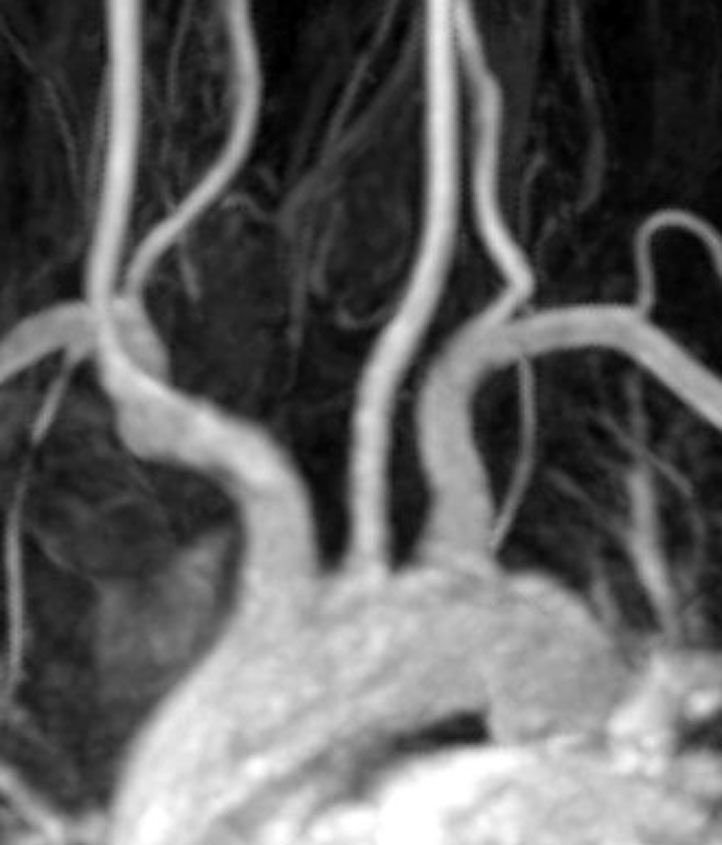
Magnetic resonance imaging can be used as a diagnostic tool to show the relationship of the aortic arches to the trachea and esophagus and also the degree of tracheal narrowing. Bronchoscopy can be useful in internally assessing the degree of tracheomalacia
Imaging
-
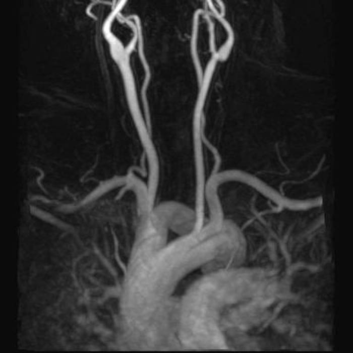 Demonstration of a double aortic arch
 -
 Normal aortic arch
(Image courtesy of Radiopaedia)

