Glioma pathophysiology: Difference between revisions
(Mahshid) |
|||
| (47 intermediate revisions by 3 users not shown) | |||
| Line 4: | Line 4: | ||
==Overview== | ==Overview== | ||
The pathogenesis of cerebral glioma involves invasion of the tumor cells into the adjacent normal brain tissue. Although in certain areas the margin of the tumor may seem to be macroscopically well defined from the brain, there are always microscopic nests of tumor cells extending well out into the brain.<ref name=aaa>Pathology of glioma. http://www.surgwiki.com/wiki/Intracranial_tumours,_infection_and_aneurysms</ref> Genes involved in the pathogenesis of glioma include ''ERCC1'', ''[[ERCC2]]'', ''[[XRCC1]]'', ''MGMT'', ''IDH1'', ''[[IDH2]]'', ''[[p53]]'', ''[[EGFR]]'', ''[[TSC1]]'', ''[[TSC2]]'', ''[[RB1]]'', ''[[APC]]'', ''hMLH1'', ''hMSH2'', ''[[PMS2]]'', ''[[PTEN]]'', ''[[NF1]]'', and ''NF2''.<ref name=ddd>Pathology of glioma. Wikipedia. https://en.wikipedia.org/wiki/Glioma</ref><ref name="pmid16932614">{{cite journal| author=Schwartzbaum JA, Fisher JL, Aldape KD, Wrensch M| title=Epidemiology and molecular pathology of glioma. | journal=Nat Clin Pract Neurol | year= 2006 | volume= 2 | issue= 9 | pages= 494-503; quiz 1 p following 516 | pmid=16932614 | doi=10.1038/ncpneuro0289 | pmc= | url=http://www.ncbi.nlm.nih.gov/entrez/eutils/elink.fcgi?dbfrom=pubmed&tool=sumsearch.org/cite&retmode=ref&cmd=prlinks&id=16932614 }} </ref> The gross and histopathological appearance of glioma varies with the tumor grade and type.<ref name=ppp>Pathology of pilocytic astrocytoma. Libre Pathology. http://librepathology.org/wiki/index.php/Pilocytic_astrocytoma</ref><ref name=fff>Pathology of gliomas. Libre Pathology. http://librepathology.org/wiki/index.php/Oligodendroglioma</ref><ref name=bbb>Pathology of anaplastic astrocytoma. Libre Pathology. http://librepathology.org/wiki/index.php?title=Neuropathology_tumours&redirect=no#Infiltrative_astrocytomas</ref><ref name=ccc>Pathology of glioblastoma. Libre Pathology. http://librepathology.org/wiki/index.php/Glioblastoma</ref><ref name=vvv>Pathology of ependymoma. Libre Pathology. http://librepathology.org/wiki/index.php/Ependymoma</ref> | |||
==Pathophysiology== | ==Pathophysiology== | ||
===Pathogenesis=== | ===Pathogenesis=== | ||
* | *The pathogenesis of cerebral glioma involves invasion of the tumor cells into the adjacent normal brain tissue. Although in certain areas the margin of the tumor may seem to be macroscopically well defined from the brain, there are always microscopic nests of tumor cells extending well out into the brain.<ref name=aaa>Pathology of glioma. http://www.surgwiki.com/wiki/Intracranial_tumours,_infection_and_aneurysms</ref> | ||
*Astrocytic projections interact with vessels and act as additional elements of the [[blood brain barrier]] (BBB) | *Astrocytic projections interact with vessels and act as additional elements of the [[blood brain barrier]] (BBB). The tumors take advantage of the blood brain barrier to ensure survival and continuous growth. | ||
*Glioma cells migrate to different regions of the brain guided by the extension of blood vessels, colonizing the healthy adjacent tissue. | *Glioma cells migrate to different regions of the brain guided by the extension of blood vessels, colonizing the healthy adjacent tissue. | ||
*Uncontrolled and fast growth also leads to the disruption of the chimeric and fragile vessels in the tumor mass resulting in peritumoral [[edema]].<ref name="pmid25565956">{{cite journal| author=Dubois LG, Campanati L, Righy C, D'Andrea-Meira I, Spohr TC, Porto-Carreiro I et al.| title=Gliomas and the vascular fragility of the blood brain barrier. | journal=Front Cell Neurosci | year= 2014 | volume= 8 | issue= | pages= 418 | pmid=25565956 | doi=10.3389/fncel.2014.00418 | pmc=PMC4264502 | url=http://www.ncbi.nlm.nih.gov/entrez/eutils/elink.fcgi?dbfrom=pubmed&tool=sumsearch.org/cite&retmode=ref&cmd=prlinks&id=25565956 }} </ref> | *Uncontrolled and fast growth also leads to the disruption of the chimeric and fragile vessels in the tumor mass resulting in peritumoral [[edema]].<ref name="pmid25565956">{{cite journal| author=Dubois LG, Campanati L, Righy C, D'Andrea-Meira I, Spohr TC, Porto-Carreiro I et al.| title=Gliomas and the vascular fragility of the blood brain barrier. | journal=Front Cell Neurosci | year= 2014 | volume= 8 | issue= | pages= 418 | pmid=25565956 | doi=10.3389/fncel.2014.00418 | pmc=PMC4264502 | url=http://www.ncbi.nlm.nih.gov/entrez/eutils/elink.fcgi?dbfrom=pubmed&tool=sumsearch.org/cite&retmode=ref&cmd=prlinks&id=25565956 }} </ref> | ||
| Line 17: | Line 16: | ||
===Genetics=== | ===Genetics=== | ||
Genes involved in the pathogenesis of glioma include:<ref name=ddd>Pathology of glioma. Wikipedia. https://en.wikipedia.org/wiki/Glioma</ref><ref name="pmid16932614">{{cite journal| author=Schwartzbaum JA, Fisher JL, Aldape KD, Wrensch M| title=Epidemiology and molecular pathology of glioma. | journal=Nat Clin Pract Neurol | year= 2006 | volume= 2 | issue= 9 | pages= 494-503; quiz 1 p following 516 | pmid=16932614 | doi=10.1038/ncpneuro0289 | pmc= | url=http://www.ncbi.nlm.nih.gov/entrez/eutils/elink.fcgi?dbfrom=pubmed&tool=sumsearch.org/cite&retmode=ref&cmd=prlinks&id=16932614 }} </ref> | Genes involved in the pathogenesis of glioma include:<ref name=ddd>Pathology of glioma. Wikipedia. https://en.wikipedia.org/wiki/Glioma</ref><ref name="pmid16932614">{{cite journal| author=Schwartzbaum JA, Fisher JL, Aldape KD, Wrensch M| title=Epidemiology and molecular pathology of glioma. | journal=Nat Clin Pract Neurol | year= 2006 | volume= 2 | issue= 9 | pages= 494-503; quiz 1 p following 516 | pmid=16932614 | doi=10.1038/ncpneuro0289 | pmc= | url=http://www.ncbi.nlm.nih.gov/entrez/eutils/elink.fcgi?dbfrom=pubmed&tool=sumsearch.org/cite&retmode=ref&cmd=prlinks&id=16932614 }} </ref> | ||
*''ERCC1'' | *''ERCC1'' | ||
*''[[ERCC2]]'' | *''[[ERCC2]]'' | ||
| Line 38: | Line 36: | ||
===Associated Conditions=== | ===Associated Conditions=== | ||
Gliomas may be associated with: | Gliomas may be associated with:<ref name="pmid16932614">{{cite journal| author=Schwartzbaum JA, Fisher JL, Aldape KD, Wrensch M| title=Epidemiology and molecular pathology of glioma. | journal=Nat Clin Pract Neurol | year= 2006 | volume= 2 | issue= 9 | pages= 494-503; quiz 1 p following 516 | pmid=16932614 | doi=10.1038/ncpneuro0289 | pmc= | url=http://www.ncbi.nlm.nih.gov/entrez/eutils/elink.fcgi?dbfrom=pubmed&tool=sumsearch.org/cite&retmode=ref&cmd=prlinks&id=16932614 }} </ref><ref name=xxx>{{cite journal|last=Reuss|first=D|author2=von Deimling, A|title=Hereditary tumor syndromes and gliomas.|journal=Recent results in cancer research. Fortschritte der Krebsforschung. Progres dans les recherches sur le cancer|year=2009|volume=171|pages=83–102|pmid=19322539|doi=10.1007/978-3-540-31206-2_5}}</ref> | ||
*[[Neurofibromatosis type 1]] | *[[Neurofibromatosis type 1]] | ||
*[[Neurofibromatosis type 2]] | *[[Neurofibromatosis type 2]] | ||
| Line 50: | Line 47: | ||
===Gross Pathology=== | ===Gross Pathology=== | ||
The gross pathological appearance of glioma varies with the tumor grade and type. Common findings are listed below:<ref name=ppp>Pathology of pilocytic astrocytoma. Libre Pathology. http://librepathology.org/wiki/index.php/Pilocytic_astrocytoma</ref><ref name=fff>Pathology of gliomas. Libre Pathology. http://librepathology.org/wiki/index.php/Oligodendroglioma</ref><ref name=bbb>Pathology of anaplastic astrocytoma. Libre Pathology. http://librepathology.org/wiki/index.php?title=Neuropathology_tumours&redirect=no#Infiltrative_astrocytomas</ref><ref name=ccc>Pathology of glioblastoma. Libre Pathology. http://librepathology.org/wiki/index.php/Glioblastoma</ref><ref name=vvv>Pathology of ependymoma. Libre Pathology. http://librepathology.org/wiki/index.php/Ependymoma</ref> | |||
{| style="border: 0px; font-size: 90%; margin: 3px; width: 600px" align=center | {| style="border: 0px; font-size: 90%; margin: 3px; width: 600px" align=center | ||
| Line 69: | Line 66: | ||
| style="padding: 5px 5px; background: #F5F5F5;" | | | style="padding: 5px 5px; background: #F5F5F5;" | | ||
:1. Poorly demarcated tumor | :1. Poorly demarcated tumor | ||
:2. Tumor causing enlargement of the involved portion of the brain and blurring of anatomical landmarks | :2. Tumor mass causing enlargement of the involved portion of the brain and blurring of anatomical landmarks | ||
:3. Commonly located in the [[cerebrum|cerebral hemisphere]] | :3. Commonly located in the [[cerebrum|cerebral hemisphere]] | ||
|- | |- | ||
| Line 106: | Line 103: | ||
====Gallery==== | |||
[http://www.peir.net Images shown below are courtesy of Professor Peter Anderson DVM PhD and published with permission © PEIR, University of Alabama at Birmingham, Department of Pathology] | [http://www.peir.net Images shown below are courtesy of Professor Peter Anderson DVM PhD and published with permission © PEIR, University of Alabama at Birmingham, Department of Pathology] | ||
<br> | <br> | ||
<div align="left"> | <div align="left"> | ||
<gallery heights="175" widths="175"> | <gallery heights="175" widths="175"> | ||
Image:Glioma Gross 1.jpg|Brain: Pontine Glioma: Gross; fixed tissue, anterior view of [[brain stem]] and [[cerebellum]] with bosselated tumor adjacent to [[basilar artery]] | Image:Glioma Gross 1.jpg|Brain: Pontine Glioma: Gross; fixed tissue, anterior view of [[brain stem]] and [[cerebellum]] with bosselated tumor adjacent to [[basilar artery]] | ||
Image:Glioma Gross 2.jpg|Brain: Pontine Glioma: Gross; fixed tissue, sagittal section [[brain stem]] and [[cerebellum]] | Image:Glioma Gross 2.jpg|Brain: Pontine Glioma: Gross; fixed tissue, sagittal section [[brain stem]] and [[cerebellum]] | ||
Image:Glioma Gross 3.jpg|Brain: Glioma: Gross; fixed tissue, horizontal section [[brain stem]] and [[cerebellum]] with obvious gelatinous appearing neoplasm a pontine glioma | Image:Glioma Gross 3.jpg|Brain: Glioma: Gross; fixed tissue, horizontal section [[brain stem]] and [[cerebellum]] with obvious gelatinous appearing neoplasm a pontine glioma | ||
Image:Glioma Gross 4.jpg|Brain: Oligodendroglioma: Gross; natural color, large, well circumscribed lesion in left [[frontal lobe]] | Image:Glioma Gross 4.jpg|Brain: Oligodendroglioma: Gross; natural color, large, well circumscribed lesion in left [[frontal lobe]] | ||
Image:Glioma Gross 5.jpg|Brain: Glioma: Gross; fixed tissue, horizontal sections [[brain stem]] and [[cerebellum]] showing large pontine glioma | Image:Glioma Gross 5.jpg|Brain: Glioma: Gross; fixed tissue, horizontal sections [[brain stem]] and [[cerebellum]] showing large pontine glioma | ||
Image:Glioma Gross 6.jpg|Brain: Pontine Glioma and Diffuse Meningeal Gliomatosis: Gross; fixed tissue, view of cerebral hemispheres from inferior with [[brain stem]] and [[cerebellum]] removed. Pontine asymmetry is easily seen due to low grade [[astrocytoma]] and meningeal gliomatosis is easily seen over [[frontal lobe]]s | Image:Glioma Gross 6.jpg|Brain: Pontine Glioma and Diffuse Meningeal Gliomatosis: Gross; fixed tissue, view of cerebral hemispheres from inferior with [[brain stem]] and [[cerebellum]] removed. Pontine asymmetry is easily seen due to low grade [[astrocytoma]] and meningeal gliomatosis is easily seen over [[frontal lobe]]s | ||
Image:Glioma Gross 7.jpg|Brain: Pontine Glioma and Diffuse Meningeal Gliomatosis in 7 yo boy: Gross; fixed tissue, view of cerebral hemispheres from vertex meningeal gliomatosis. | Image:Glioma Gross 7.jpg|Brain: Pontine Glioma and Diffuse Meningeal Gliomatosis in 7 yo boy: Gross; fixed tissue, view of cerebral hemispheres from vertex meningeal gliomatosis. | ||
Image:Glioma Gross 8.jpg|Brain: Pontine Glioma and Diffuse Meningeal Gliomatosis: Gross; in situ dural nodule | Image:Glioma Gross 8.jpg|Brain: Pontine Glioma and Diffuse Meningeal Gliomatosis: Gross; in situ dural nodule | ||
Image:Glioma Gross 9.jpg|Brain: Oligodendroglioma: Gross; fixed tissue, multiple coronal sections, cerebral hemispheres with large tumor and hemorrhage into tumor | Image:Glioma Gross 9.jpg|Brain: Oligodendroglioma: Gross; fixed tissue, multiple coronal sections, cerebral hemispheres with large tumor and hemorrhage into tumor | ||
Image:Glioma Gross 10.jpg|Brain: Oligodendroglioma: Gross; fixed tissue, coronal section, cerebral hemispheres, large hemorrhagic lesion in one hemisphere | Image:Glioma Gross 10.jpg|Brain: Oligodendroglioma: Gross; fixed tissue, coronal section, cerebral hemispheres, large hemorrhagic lesion in one hemisphere | ||
Image:Glioma Gross 11.jpg|Brain: Oligodendroglioma: Gross; fixed tissue, ischemic tissue, anterior to tumor mass | Image:Glioma Gross 11.jpg|Brain: Oligodendroglioma: Gross; fixed tissue, ischemic tissue, anterior to tumor mass | ||
Image:Glioma Gross 12.jpg|Brain: Oligodendroglioma: Gross; natural color, coronal section, cerebral hemispheres, large lesion, left parieto occipital white matter | Image:Glioma Gross 12.jpg|Brain: Oligodendroglioma: Gross; natural color, coronal section, cerebral hemispheres, large lesion, left parieto occipital white matter | ||
Image:Glioma Gross 13.jpg|Brain: Gliomatosis Cerebri: Gross; fixed tissue, coronal sections, cerebral hemispheres, lesion is in temporal lobes and hypothalamus | Image:Glioma Gross 13.jpg|Brain: Gliomatosis Cerebri: Gross; fixed tissue, coronal sections, cerebral hemispheres, lesion is in temporal lobes and hypothalamus | ||
Image:Glioma Gross 14.jpg|Brain: Ventriculitis: Gross; fixed tissue, case of glioma with meningitis, a nice view of ventriculitis in one lateral ventricle | Image:Glioma Gross 14.jpg|Brain: Ventriculitis: Gross; fixed tissue, case of glioma with meningitis, a nice view of ventriculitis in one lateral ventricle | ||
Image:Glioma Gross 15.jpg|Brain: Glioma Thalamic Grade Ii-Iii: Gross; fixed tissue, four coronal sections, cerebral hemispheres, very large hemorrhagic lesion | Image:Glioma Gross 15.jpg|Brain: Glioma Thalamic Grade Ii-Iii: Gross; fixed tissue, four coronal sections, cerebral hemispheres, very large hemorrhagic lesion | ||
Image:Glioma Gross 16.jpg|Brain: Glioma Thalamic Grade Ii-Iii: Gross; fixed tissue, coronal section, cerebral hemispheres with large hemorrhagic lesion | Image:Glioma Gross 16.jpg|Brain: Glioma Thalamic Grade Ii-Iii: Gross; fixed tissue, coronal section, cerebral hemispheres with large hemorrhagic lesion | ||
Image:Glioma Gross 17.jpg|Brain: Glioma Thalamic Grade Ii-Iii: Gross fixed tissue coronal section cerebral hemispheres lesions appears to be in choroid plexus of lateral ventricle in this picture. There is blood in fourth ventricle | Image:Glioma Gross 17.jpg|Brain: Glioma Thalamic Grade Ii-Iii: Gross fixed tissue coronal section cerebral hemispheres lesions appears to be in choroid plexus of lateral ventricle in this picture. There is blood in fourth ventricle | ||
Image:Glioma Gross 18.jpg|Brain: Cerebral Sarcoma or Microglioma: Gross; fixed tissue, coronal section, cerebral hemispheres (58 yo man) | Image:Glioma Gross 18.jpg|Brain: Cerebral Sarcoma or Microglioma: Gross; fixed tissue, coronal section, cerebral hemispheres (58 yo man) | ||
Image:Glioma Gross 19.jpg|Brain: Cerebral Sarcoma or Microglioma: Gross; fixed tissue, coronal section, cerebral hemispheres | Image:Glioma Gross 19.jpg|Brain: Cerebral Sarcoma or Microglioma: Gross; fixed tissue, coronal section, cerebral hemispheres | ||
Image:Glioma Gross 20.jpg|Brain: Cerebral Sarcoma or Microglioma: Gross; fixed tissue, coronal section, cerebral hemispheres | Image:Glioma Gross 20.jpg|Brain: Cerebral Sarcoma or Microglioma: Gross; fixed tissue, coronal section, cerebral hemispheres | ||
Image:Glioma Gross 21.jpg|Brain: Infarct Subcortical: Gross; fixed tissue, close-up view of old small subcortical infarct, a case of microglioma | Image:Glioma Gross 21.jpg|Brain: Infarct Subcortical: Gross; fixed tissue, close-up view of old small subcortical infarct, a case of microglioma | ||
Image:Glioma Gross 22.jpg|Brain: Microglioma: Gross; fixed tissue; [[cerebellum]] and fourth ventricle with periventricular tumor invasion | Image:Glioma Gross 22.jpg|Brain: Microglioma: Gross; fixed tissue; [[cerebellum]] and fourth ventricle with periventricular tumor invasion | ||
Image:Glioma Gross 23.jpg|Brain: Microglioma: Gross fixed tissue horizontal sections cerebellum and brain stem with periventricular neoplastic infiltrate | Image:Glioma Gross 23.jpg|Brain: Microglioma: Gross fixed tissue horizontal sections cerebellum and brain stem with periventricular neoplastic infiltrate | ||
Image:Glioma Gross 24.jpg|Brain: Microglioma: Gross fixed tissue horizontal section midbrain and cerebellum at mid pons level periventricular tumor infiltration | Image:Glioma Gross 24.jpg|Brain: Microglioma: Gross fixed tissue horizontal section midbrain and cerebellum at mid pons level periventricular tumor infiltration | ||
Image:Glioma Gross 25.jpg|Brain: Microglioma: Gross fixed tissue horizontal section rostral pons and cerebellum | Image:Glioma Gross 25.jpg|Brain: Microglioma: Gross fixed tissue horizontal section rostral pons and cerebellum | ||
Image:Glioma Gross 26.jpg|Brain: Microglioma: Gross fixed tissue horizontal section rostral pons and cerebellum periventricular tumor invasion | Image:Glioma Gross 26.jpg|Brain: Microglioma: Gross fixed tissue horizontal section rostral pons and cerebellum periventricular tumor invasion | ||
Image:Glioma Gross 27.jpg|Brain: Microglioma: Gross fixed tissue coronal section cerebral hemispheres with mild ventricular dilation | Image:Glioma Gross 27.jpg|Brain: Microglioma: Gross fixed tissue coronal section cerebral hemispheres with mild ventricular dilation | ||
Image:Glioma Gross 28.jpg|Glioma: Optic Nerve | Image:Glioma Gross 28.jpg|Glioma: Optic Nerve | ||
Image:Glioma Gross 29.jpg|Brain: Oligodendroglioma, Frontal Lobe | Image:Glioma Gross 29.jpg|Brain: Oligodendroglioma, Frontal Lobe | ||
Image:Glioma Gross 30.jpg|Brain: Oligodendroglioma, Mixed Astrocytoma & Oligodendroglioma | Image:Glioma Gross 30.jpg|Brain: Oligodendroglioma, Mixed Astrocytoma & Oligodendroglioma | ||
Image:Glioma Gross 31.jpg|Brain: Oligodendroglioma | Image:Glioma Gross 31.jpg|Brain: Oligodendroglioma | ||
Image:Glioma Gross 32.jpg|Brain: Oligodendroglioma | Image:Glioma Gross 32.jpg|Brain: Oligodendroglioma | ||
Image:Glioma Gross 36.jpg|Brain: Glioma, Grade II Anaplastic | Image:Glioma Gross 36.jpg|Brain: Glioma, Grade II Anaplastic | ||
Image:Glioma Gross 37.jpg|Brain: Glioma, [[Brain stem]], Low Grade | Image:Glioma Gross 37.jpg|Brain: Glioma, [[Brain stem]], Low Grade | ||
Image:Glioma Gross 38.jpg|Fundoscopy: Eye; Optic Nerve Glioma, Optic Nerve | Image:Glioma Gross 38.jpg|Fundoscopy: Eye; Optic Nerve Glioma, Optic Nerve | ||
Image:Glioma Gross 41.jpg|Brain: Glioma, Hypothalamic, Circle Around Region of Tumor | Image:Glioma Gross 41.jpg|Brain: Glioma, Hypothalamic, Circle Around Region of Tumor | ||
Image:Glioma Gross 39.jpg|CNS: Pilocytic Astrocytoma of the Spinal Cord. The fusiform expansion of the spinal cord produced by this pilocytic astrocytoma is not, on external examination alone, distinguishable from that produced by a nonresectable diffuse glioma. | Image:Glioma Gross 39.jpg|CNS: Pilocytic Astrocytoma of the Spinal Cord. The fusiform expansion of the spinal cord produced by this pilocytic astrocytoma is not, on external examination alone, distinguishable from that produced by a nonresectable diffuse glioma. | ||
Image:Glioma Gross 42.jpg|Brain: Glioma, Pontine | Image:Glioma Gross 42.jpg|Brain: Glioma, Pontine | ||
Image:Glioma Gross 43.jpg|Brain: [[Glioblastoma Multiforme]]: Gross fixed tissue close-up large necrotic tumor mass in septum pellucidum | Image:Glioma Gross 43.jpg|Brain: [[Glioblastoma Multiforme]]: Gross fixed tissue close-up large necrotic tumor mass in septum pellucidum | ||
Image:Glioma Gross 44.jpg|Brain: [[Glioblastoma Multiforme]]: Gross fixed tissue coronal section of the brain with a large necrotic tumor mass in septum pellucidum diagnosed as astrocytoma grade III | Image:Glioma Gross 44.jpg|Brain: [[Glioblastoma Multiforme]]: Gross fixed tissue coronal section of the brain with a large necrotic tumor mass in septum pellucidum diagnosed as astrocytoma grade III | ||
Image:Glioma Gross 45.jpg|Brain: [[Glioblastoma Multiforme]]: Gross natural color large hemorrhagic lesion in right centrum semiovale | Image:Glioma Gross 45.jpg|Brain: [[Glioblastoma Multiforme]]: Gross natural color large hemorrhagic lesion in right centrum semiovale | ||
Image:Pilocytic astrocytoma gross.jpg|CNS: Malignant pilocytic astrocytoma: A 29-year-old woman died 2 years after a diagnosis of "atypical pilocytic astrocytoma" of the pineal region. At autopsy, multiple tumor implants were present in the craniospinal subarachnoid spaces. | Image:Pilocytic astrocytoma gross.jpg|CNS: Malignant pilocytic astrocytoma: A 29-year-old woman died 2 years after a diagnosis of "atypical pilocytic astrocytoma" of the pineal region. At autopsy, multiple tumor implants were present in the craniospinal subarachnoid spaces. | ||
Image:Glioma Gross 33.jpg|Brain: Oligodendroglioma; Ventricular Cobblestone Effect | Image:Glioma Gross 33.jpg|Brain: Oligodendroglioma; Ventricular Cobblestone Effect | ||
</gallery> | </gallery> | ||
| Line 284: | Line 155: | ||
===Microscopic Pathology=== | ===Microscopic Pathology=== | ||
The histopathological appearance of glioma varies with the tumor grade and type, with increasing cellular [[atypia]], [[mitoses]], [[endothelial cell]] proliferation, and [[necrosis]]. Common findings are listed below:<ref name=ppp>Pathology of pilocytic astrocytoma. Libre Pathology. http://librepathology.org/wiki/index.php/Pilocytic_astrocytoma</ref><ref name=fff>Pathology of gliomas. Libre Pathology. http://librepathology.org/wiki/index.php/Oligodendroglioma</ref><ref name=bbb>Pathology of anaplastic astrocytoma. Libre Pathology. http://librepathology.org/wiki/index.php?title=Neuropathology_tumours&redirect=no#Infiltrative_astrocytomas</ref><ref name=ccc>Pathology of glioblastoma. Libre Pathology. http://librepathology.org/wiki/index.php/Glioblastoma</ref><ref name=vvv>Pathology of ependymoma. Libre Pathology. http://librepathology.org/wiki/index.php/Ependymoma</ref> | |||
{| style="border: 0px; font-size: 90%; margin: 3px; width: 600px" align=center | {| style="border: 0px; font-size: 90%; margin: 3px; width: 600px" align=center | ||
| Line 290: | Line 161: | ||
|+ | |+ | ||
! style="background: #4479BA; width: 200px;" | {{fontcolor|#FFF|Type of glioma}} | ! style="background: #4479BA; width: 200px;" | {{fontcolor|#FFF|Type of glioma}} | ||
! style="background: #4479BA; width: 400px;" | {{fontcolor|#FFF| | ! style="background: #4479BA; width: 400px;" | {{fontcolor|#FFF|Histopathological features}} | ||
|- | |- | ||
| style="padding: 5px 5px; background: #DCDCDC; font-weight: bold" | | | style="padding: 5px 5px; background: #DCDCDC; font-weight: bold" | | ||
| Line 299: | Line 170: | ||
:*Fibrillar | :*Fibrillar | ||
:* Microcystic | :* Microcystic | ||
:3. Hair-like | :3. Hair-like fibers best observed on smear or with [[GFAP]] | ||
:4. Rosenthal | :4. Rosenthal fibers | ||
:5. Eosinophilic granular bodies | :5. Eosinophilic granular bodies | ||
:6. Microvascular proliferation | :6. Microvascular proliferation | ||
| Line 358: | Line 229: | ||
====Gallery==== | |||
[http://www.peir.net Images shown below are courtesy of Professor Peter Anderson DVM PhD and published with permission © PEIR, University of Alabama at Birmingham, Department of Pathology] | [http://www.peir.net Images shown below are courtesy of Professor Peter Anderson DVM PhD and published with permission © PEIR, University of Alabama at Birmingham, Department of Pathology] | ||
<br> | <br> | ||
<div align="left"> | <div align="left"> | ||
<gallery heights="225" widths="225"> | <gallery heights="225" widths="225"> | ||
Image:Pilocytic astrocytoma 1.jpg|CNS: [[Pilocytic astrocytoma]]: variations in histologic appearance. As illustrated, many lesions are composed largely of spongy tissue rich in microcysts. Characteristic of [[pilocytic astrocytoma]]s in general, the lesion is largely a solid mass of neoplastic cells without an obvious background of infiltrated brain. | Image:Pilocytic astrocytoma 1.jpg|CNS: [[Pilocytic astrocytoma]]: variations in histologic appearance. As illustrated, many lesions are composed largely of spongy tissue rich in microcysts. Characteristic of [[pilocytic astrocytoma]]s in general, the lesion is largely a solid mass of neoplastic cells without an obvious background of infiltrated brain. | ||
Image:Pilocytic astrocytoma 2.jpg|CNS: [[Pilocytic astrocytoma]]: variations in histologic appearance. The perivascular radiating processes in some lesions can create a likeness to an [[ependymoma]]. Note the spongy background unusual for [[ependymoma]]s. | Image:Pilocytic astrocytoma 2.jpg|CNS: [[Pilocytic astrocytoma]]: variations in histologic appearance. The perivascular radiating processes in some lesions can create a likeness to an [[ependymoma]]. Note the spongy background unusual for [[ependymoma]]s. | ||
Image:Pilocytic astrocytoma 3.jpg|CNS: [[Pilocytic astrocytoma]]: variations in histologic appearance. Other [[pilocytic astrocytoma]]s are solid, rather than microcystic, and may be lobular. | Image:Pilocytic astrocytoma 3.jpg|CNS: [[Pilocytic astrocytoma]]: variations in histologic appearance. Other [[pilocytic astrocytoma]]s are solid, rather than microcystic, and may be lobular. | ||
Image:Pilocytic astrocytoma 4.jpg|CNS: [[Pilocytic astrocytoma]]: variations in histologic appearance. Rosenthal fibers, usually confined to the solid rather than spongy regions are found in many pilocytic [[astrocytoma]]s, but are not requisite for the diagnosis. | Image:Pilocytic astrocytoma 4.jpg|CNS: [[Pilocytic astrocytoma]]: variations in histologic appearance. Rosenthal fibers, usually confined to the solid rather than spongy regions are found in many pilocytic [[astrocytoma]]s, but are not requisite for the diagnosis. | ||
Image:Pilocytic astrocytoma 15.jpg|CNS: [[Pilocytic astrocytoma]]: variations in histologic appearance. Rosenthal fibers are extremely abundant in some lesions. Particularly in the [[cerebellum]], it can be difficult to distinguish such solid, paucicellular, highly fibrillar [[pilocytic astrocytoma]]s from reactive gliosis with abundant Rosenthal fiber formation. | Image:Pilocytic astrocytoma 15.jpg|CNS: [[Pilocytic astrocytoma]]: variations in histologic appearance. Rosenthal fibers are extremely abundant in some lesions. Particularly in the [[cerebellum]], it can be difficult to distinguish such solid, paucicellular, highly fibrillar [[pilocytic astrocytoma]]s from reactive gliosis with abundant Rosenthal fiber formation. | ||
Image:Pilocytic astrocytoma 16.jpg|CNS: [[Pilocytic astrocytoma]]: variations in histologic appearance. A loose array of polar cells creates an additional variant of pilocytic [[astrocytoma]]. | Image:Pilocytic astrocytoma 16.jpg|CNS: [[Pilocytic astrocytoma]]: variations in histologic appearance. A loose array of polar cells creates an additional variant of pilocytic [[astrocytoma]]. | ||
Image:Pilocytic astrocytoma 7.jpg|CNS: [[Pilocytic astrocytoma]]: variations in histologic appearance. Some pilocytic [[astrocytoma]]s are traversed by prominent collagenous septa. | Image:Pilocytic astrocytoma 7.jpg|CNS: [[Pilocytic astrocytoma]]: variations in histologic appearance. Some pilocytic [[astrocytoma]]s are traversed by prominent collagenous septa. | ||
Image:Pilocytic astrocytoma 8.jpg|CNS: [[Pilocytic astrocytoma]]: variations in histologic appearance. Unusual pilocytic [[astrocytoma]]s have an extensive mucinous background without microcysts. | Image:Pilocytic astrocytoma 8.jpg|CNS: [[Pilocytic astrocytoma]]: variations in histologic appearance. Unusual pilocytic [[astrocytoma]]s have an extensive mucinous background without microcysts. | ||
Image:Comparison.jpg|CNS: Comparison of normal [[optic nerve]] and [[pilocytic astrocytoma]] of the [[optic nerve]]. These two figures compare, at the same magnification, the normal [[optic nerve]] (left) with one containing a [[pilocytic astrocytoma]] (right). The [[neoplasm]] enlarges the compartments of the [[nerve]] and extends in collar-like fashion into the [[subarachnoid]] space. | Image:Comparison.jpg|CNS: Comparison of normal [[optic nerve]] and [[pilocytic astrocytoma]] of the [[optic nerve]]. These two figures compare, at the same magnification, the normal [[optic nerve]] (left) with one containing a [[pilocytic astrocytoma]] (right). The [[neoplasm]] enlarges the compartments of the [[nerve]] and extends in collar-like fashion into the [[subarachnoid]] space. | ||
Image:Pilocytic astrocytoma 9.jpg|CNS: [[Pilocytic astrocytoma]]; The "hair cells" for which this lesion is named are readily seen. | Image:Pilocytic astrocytoma 9.jpg|CNS: [[Pilocytic astrocytoma]]; The "hair cells" for which this lesion is named are readily seen. | ||
Image:Pilocytic astrocytoma 10.jpg|CNS: [[Pilocytic astrocytoma]]; Nuclear hyperchromasia and [[pleomorphism]] are common. Note the typical cellular elongation, and, at the center of the illustration, the [[eosinophil]]ic granular body that populates [[pilocytic astrocytoma]]s and certain other slowly growing gliomas. | Image:Pilocytic astrocytoma 10.jpg|CNS: [[Pilocytic astrocytoma]]; Nuclear hyperchromasia and [[pleomorphism]] are common. Note the typical cellular elongation, and, at the center of the illustration, the [[eosinophil]]ic granular body that populates [[pilocytic astrocytoma]]s and certain other slowly growing gliomas. | ||
Image:Pilocytic astrocytoma 11.jpg|CNS: [[Pilocytic astrocytoma]]; Intracytoplasmic Rosenthal fibers are prominent in some pilocytic [[neoplasm]]s. | Image:Pilocytic astrocytoma 11.jpg|CNS: [[Pilocytic astrocytoma]]; Intracytoplasmic Rosenthal fibers are prominent in some pilocytic [[neoplasm]]s. | ||
Image:Malignant ependymoma.jpg|Brain: [[Malignant]] [[ependymoma]]: Micro med mag H&E tumor cells. | Image:Malignant ependymoma.jpg|Brain: [[Malignant]] [[ependymoma]]: Micro med mag H&E tumor cells. | ||
Image:Oligodendroglioma 2.jpg|CNS: [[Oligodendroglioma]]; Occasional [[oligodendroglioma]]s contain cells with minute, refractile [[eosinophil]]ic bodies representing miniature Rosenthal fibers. | Image:Oligodendroglioma 2.jpg|CNS: [[Oligodendroglioma]]; Occasional [[oligodendroglioma]]s contain cells with minute, refractile [[eosinophil]]ic bodies representing miniature Rosenthal fibers. | ||
Image:Oligodendroglioma 1.jpg|CNS: [[Oligodendroglioma]]; The cells of some [[oligodendroglioma]]s acquire sufficient [[cytoplasm]] and process formation to become decidedly astrocytic, but their nuclei retain the roundness, uniformity, and chromatin distribution typical of [[oligodendroglioma]]. | Image:Oligodendroglioma 1.jpg|CNS: [[Oligodendroglioma]]; The cells of some [[oligodendroglioma]]s acquire sufficient [[cytoplasm]] and process formation to become decidedly astrocytic, but their nuclei retain the roundness, uniformity, and chromatin distribution typical of [[oligodendroglioma]]. | ||
Image:Anaplastic oligodendroglioma.jpg|[[Anaplastic]] [[oligodendroglioma]]: [[Anaplastic]] [[oligodendroglioma]]s are highly cellular and associated with vascular proliferation. | Image:Anaplastic oligodendroglioma.jpg|[[Anaplastic]] [[oligodendroglioma]]: [[Anaplastic]] [[oligodendroglioma]]s are highly cellular and associated with vascular proliferation. | ||
Image:Oligodendroglioma frozen.jpg|CNS: [[Oligodendroglioma]] (frozen section); [[Oligodendroglioma]]s in frozen sections lack the distinctive halos so often seen in permanent sections. Cellular monomorphism and infiltration of [[cerebral cortex]] with perineuronal satellitosis suggest the correct diagnosis. | Image:Oligodendroglioma frozen.jpg|CNS: [[Oligodendroglioma]] (frozen section); [[Oligodendroglioma]]s in frozen sections lack the distinctive halos so often seen in permanent sections. Cellular monomorphism and infiltration of [[cerebral cortex]] with perineuronal satellitosis suggest the correct diagnosis. | ||
Image:Clear cell ependymoma.jpg|CNS: Clear cell [[ependymoma]]; Perinuclear clearing similar to that seen in [[oligodendroglioma]]s is a prominent feature of the clear cell variant. Note the vague perivascular pseudorosettes. The lesion was a discrete occipital intraventricular mass. | Image:Clear cell ependymoma.jpg|CNS: Clear cell [[ependymoma]]; Perinuclear clearing similar to that seen in [[oligodendroglioma]]s is a prominent feature of the clear cell variant. Note the vague perivascular pseudorosettes. The lesion was a discrete occipital intraventricular mass. | ||
Image:Glioblastoma multiforme 1.jpg|CNS: [[Glioblastoma multiforme]]; Brain: [[Glioblastoma multiforme]]. Grade I-Ii: Micro med mag with H&E, [[tumor]] well shown | Image:Glioblastoma multiforme 1.jpg|CNS: [[Glioblastoma multiforme]]; Brain: [[Glioblastoma multiforme]]. Grade I-Ii: Micro med mag with H&E, [[tumor]] well shown | ||
Image:Glioblastoma multiforme 2.jpg|CNS: [[Glioblastoma multiforme]] arising in an [[astrocytoma]]. At higher magnification, gemistocytic [[astrocytoma]] with microcystic change is apparent at the bottom of the illustration and cellular nodules of [[glioblastoma multiforme]] are seen at the top. The 6-year history of symptoms attested to the initially low-grade nature of this astrocytic tumor. | Image:Glioblastoma multiforme 2.jpg|CNS: [[Glioblastoma multiforme]] arising in an [[astrocytoma]]. At higher magnification, gemistocytic [[astrocytoma]] with microcystic change is apparent at the bottom of the illustration and cellular nodules of [[glioblastoma multiforme]] are seen at the top. The 6-year history of symptoms attested to the initially low-grade nature of this astrocytic tumor. | ||
Image:Glioblastoma multiforme 103.jpg|CNS: [[Glioblastoma multiforme]]; Characteristic of most [[glioblastoma]]s are small cells with elongated nuclei and bipolar processes. As here, the [[chromatin]] is generally not markedly dense nor are nucleoli usually prominent. | Image:Glioblastoma multiforme 103.jpg|CNS: [[Glioblastoma multiforme]]; Characteristic of most [[glioblastoma]]s are small cells with elongated nuclei and bipolar processes. As here, the [[chromatin]] is generally not markedly dense nor are nucleoli usually prominent. | ||
Image:Glioblastoma multiforme 104.jpg|CNS: [[Glioblastoma multiforme]]; Vascular proliferation, a common feature of glioblastoma, produces tufts which often grow directionally. Here, as is often the case, they are oriented toward a focus of necrosis (top right). | Image:Glioblastoma multiforme 104.jpg|CNS: [[Glioblastoma multiforme]]; Vascular proliferation, a common feature of glioblastoma, produces tufts which often grow directionally. Here, as is often the case, they are oriented toward a focus of necrosis (top right). | ||
Image:Glioblastoma multiforme 105.jpg|CNS: [[Glioblastoma multiforme]]; At high magnification, the neovascular tuft is a mass which, as can be confirmed by immunohistochemistry, is formed of both endothelial cells and smooth muscle cells (pericytes). | Image:Glioblastoma multiforme 105.jpg|CNS: [[Glioblastoma multiforme]]; At high magnification, the neovascular tuft is a mass which, as can be confirmed by immunohistochemistry, is formed of both endothelial cells and smooth muscle cells (pericytes). | ||
Image:Glioblastoma multiforme 106.jpg|CNS: [[Glioblastoma multiforme]]; In many instances, [[necrosis]] is surrounded by a distinctive collar of cells, which are often smaller than those in surrounding neoplastic tissue. The phenomenon is referred to as pseudopalisading. | Image:Glioblastoma multiforme 106.jpg|CNS: [[Glioblastoma multiforme]]; In many instances, [[necrosis]] is surrounded by a distinctive collar of cells, which are often smaller than those in surrounding neoplastic tissue. The phenomenon is referred to as pseudopalisading. | ||
Image:Cerebrospinal dissemination of glioblastoma multiforme 1.jpg|CNS: Cerebrospinal dissemination of glioblastoma multiforme; As seen at low (left) and high (right) magnification, the small undifferentiated-appearing cells of this glioblastoma are drop metastases colonizing the nerve roots of the cauda equina. | Image:Cerebrospinal dissemination of glioblastoma multiforme 1.jpg|CNS: Cerebrospinal dissemination of glioblastoma multiforme; As seen at low (left) and high (right) magnification, the small undifferentiated-appearing cells of this glioblastoma are drop metastases colonizing the nerve roots of the cauda equina. | ||
Image:Cerebrospinal dissemination of glioblastoma multiforme 2.jpg|CNS: Cerebrospinal dissemination of glioblastoma multiforme; As seen at low (left) and high (right) magnification, the small undifferentiated-appearing cells of this glioblastoma are drop metastases colonizing the nerve roots of the cauda equina. | Image:Cerebrospinal dissemination of glioblastoma multiforme 2.jpg|CNS: Cerebrospinal dissemination of glioblastoma multiforme; As seen at low (left) and high (right) magnification, the small undifferentiated-appearing cells of this glioblastoma are drop metastases colonizing the nerve roots of the cauda equina. | ||
Image:Glioblastoma multiforme 116.jpg|CNS: [[Glioblastoma multiforme]]; Higher magnification reveals the small cell nature of such tumors. | Image:Glioblastoma multiforme 116.jpg|CNS: [[Glioblastoma multiforme]]; Higher magnification reveals the small cell nature of such tumors. | ||
Image:Glioblastoma multiforme 117.jpg|CNS: [[Glioblastoma multiforme]]; Some glioblastomas are especially infiltrative of the cerebral cortex where subpial, perivascular, and perineuronal accumulations are prominent. | Image:Glioblastoma multiforme 117.jpg|CNS: [[Glioblastoma multiforme]]; Some glioblastomas are especially infiltrative of the cerebral cortex where subpial, perivascular, and perineuronal accumulations are prominent. | ||
Image:Glioblastoma multiforme 126.jpg|CNS: [[Glioblastoma multiforme]]; Although this densely cellular and largely undifferentiated lesion technically merits a diagnosis of [[anaplastic]] [[astrocytoma]], it is, for practical purposes, a glioblastoma. | Image:Glioblastoma multiforme 126.jpg|CNS: [[Glioblastoma multiforme]]; Although this densely cellular and largely undifferentiated lesion technically merits a diagnosis of [[anaplastic]] [[astrocytoma]], it is, for practical purposes, a glioblastoma. | ||
Image:Glioblastoma multiforme malignant astrocytoma 1.jpg|Brain: [[Glioblastoma multiforme]] [[malignant]] [[astrocytoma]] | Image:Glioblastoma multiforme malignant astrocytoma 1.jpg|Brain: [[Glioblastoma multiforme]] [[malignant]] [[astrocytoma]] | ||
Image:Glioblastoma multiforme pallisading.jpg|Brain: [[Glioblastoma multiforme]]; pallisading | Image:Glioblastoma multiforme pallisading.jpg|Brain: [[Glioblastoma multiforme]]; pallisading | ||
Image:Glioblastoma multiforme vascular proliferation.jpg|Brain: [[Glioblastoma multiforme]]; vascular proliferation | Image:Glioblastoma multiforme vascular proliferation.jpg|Brain: [[Glioblastoma multiforme]]; vascular proliferation | ||
Image:Glioblastoma multiforme extravasated blood.jpg|Brain: [[Glioblastoma multiforme]]; Plump and juicy [[endothelial cell]]s, extravasated blood | Image:Glioblastoma multiforme extravasated blood.jpg|Brain: [[Glioblastoma multiforme]]; Plump and juicy [[endothelial cell]]s, extravasated blood | ||
Image:Glioblastoma multiforme perivascular lymphocytes.jpg|Brain: [[Glioblastoma multiforme]]; perivascular [[lymphocyte]]s | Image:Glioblastoma multiforme perivascular lymphocytes.jpg|Brain: [[Glioblastoma multiforme]]; perivascular [[lymphocyte]]s | ||
Image:Glioblastoma multiforme thrombosed vessel.jpg|Brain: [[Glioblastoma multiforme]]; thrombosed vessel | Image:Glioblastoma multiforme thrombosed vessel.jpg|Brain: [[Glioblastoma multiforme]]; thrombosed vessel | ||
Image:Hemosiderin in glioblastoma.jpg|Brain: [[Glioblastoma multiforme]]; [[Hemosiderin]] in glioblastoma | Image:Hemosiderin in glioblastoma.jpg|Brain: [[Glioblastoma multiforme]]; [[Hemosiderin]] in glioblastoma | ||
| Line 513: | Line 283: | ||
{{WikiDoc Help Menu}} | {{WikiDoc Help Menu}} | ||
{{WikiDoc Sources}} | {{WikiDoc Sources}} | ||
[[Category:Up-To-Date]] | |||
[[Category:Oncology]] | |||
[[Category:Medicine]] | |||
[[Category:Neurology]] | |||
[[Category:Neurosurgery]] | |||
[[Category:Up-To-Date]] | |||
[[Category:Oncology]] | |||
[[Category:Medicine]] | |||
[[Category:Neurology]] | |||
[[Category:Neurosurgery]] | |||
Latest revision as of 01:45, 27 November 2017
|
Glioma Microchapters |
|
Diagnosis |
|---|
|
Treatment |
|
Case Studies |
|
Glioma pathophysiology On the Web |
|
American Roentgen Ray Society Images of Glioma pathophysiology |
|
Risk calculators and risk factors for Glioma pathophysiology |
Editor-In-Chief: C. Michael Gibson, M.S., M.D. [1]; Associate Editor-In-Chief: Cafer Zorkun, M.D., Ph.D. [2], Sujit Routray, M.D. [3]
Overview
The pathogenesis of cerebral glioma involves invasion of the tumor cells into the adjacent normal brain tissue. Although in certain areas the margin of the tumor may seem to be macroscopically well defined from the brain, there are always microscopic nests of tumor cells extending well out into the brain.[1] Genes involved in the pathogenesis of glioma include ERCC1, ERCC2, XRCC1, MGMT, IDH1, IDH2, p53, EGFR, TSC1, TSC2, RB1, APC, hMLH1, hMSH2, PMS2, PTEN, NF1, and NF2.[2][3] The gross and histopathological appearance of glioma varies with the tumor grade and type.[4][5][6][7][8]
Pathophysiology
Pathogenesis
- The pathogenesis of cerebral glioma involves invasion of the tumor cells into the adjacent normal brain tissue. Although in certain areas the margin of the tumor may seem to be macroscopically well defined from the brain, there are always microscopic nests of tumor cells extending well out into the brain.[1]
- Astrocytic projections interact with vessels and act as additional elements of the blood brain barrier (BBB). The tumors take advantage of the blood brain barrier to ensure survival and continuous growth.
- Glioma cells migrate to different regions of the brain guided by the extension of blood vessels, colonizing the healthy adjacent tissue.
- Uncontrolled and fast growth also leads to the disruption of the chimeric and fragile vessels in the tumor mass resulting in peritumoral edema.[9]
Genetics
Genes involved in the pathogenesis of glioma include:[2][3]
Associated Conditions
Gliomas may be associated with:[3][10]
- Neurofibromatosis type 1
- Neurofibromatosis type 2
- Tuberous sclerosis
- Li-Fraumeni syndrome
- Turcot syndrome
- Maffucci syndrome
- Von Hippel-Lindau disease
- Retinoblastoma
Gross Pathology
The gross pathological appearance of glioma varies with the tumor grade and type. Common findings are listed below:[4][5][6][7][8]
| Type of glioma | Gross pathological features |
|---|---|
| |
| |
| |
| |
| |
|
Gallery
-
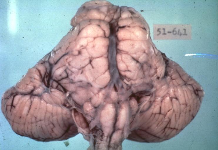
Brain: Pontine Glioma: Gross; fixed tissue, anterior view of brain stem and cerebellum with bosselated tumor adjacent to basilar artery
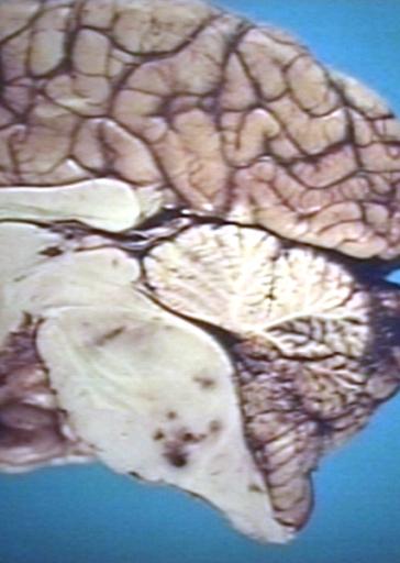
-
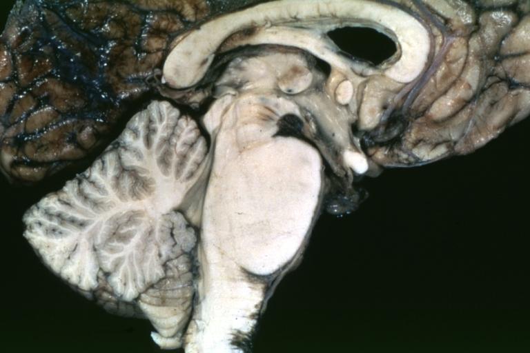
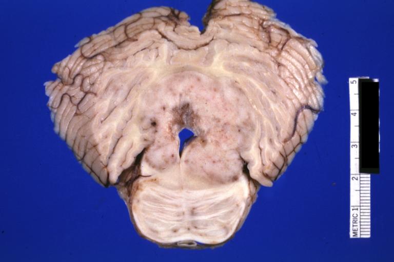
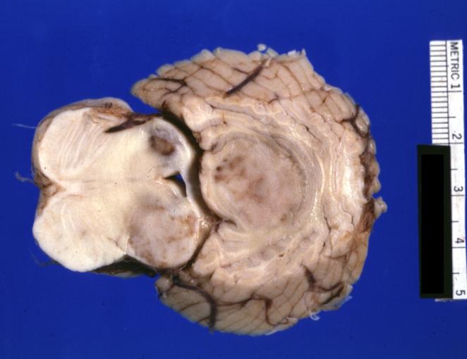
Brain: Pontine Glioma: Gross; fixed tissue, sagittal section brain stem and cerebellum
-
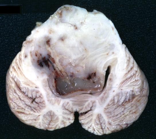
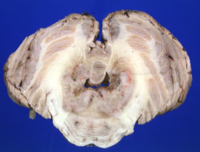
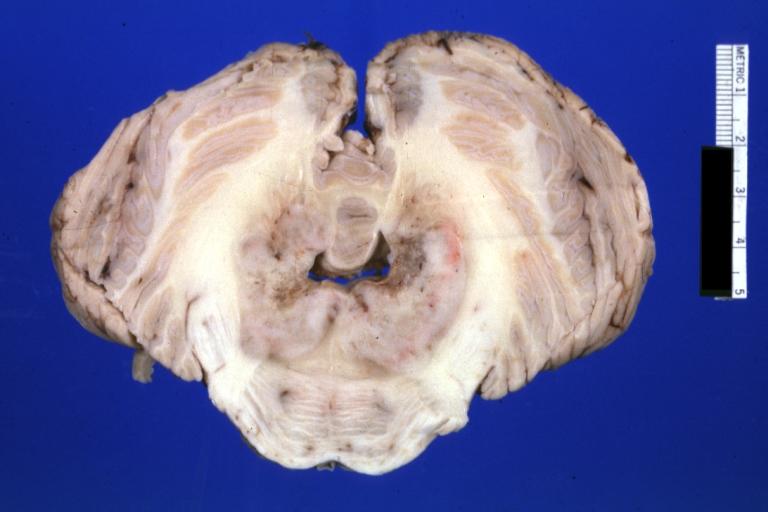
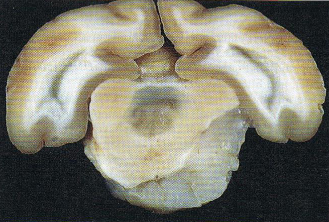
Brain: Glioma: Gross; fixed tissue, horizontal section brain stem and cerebellum with obvious gelatinous appearing neoplasm a pontine glioma
-
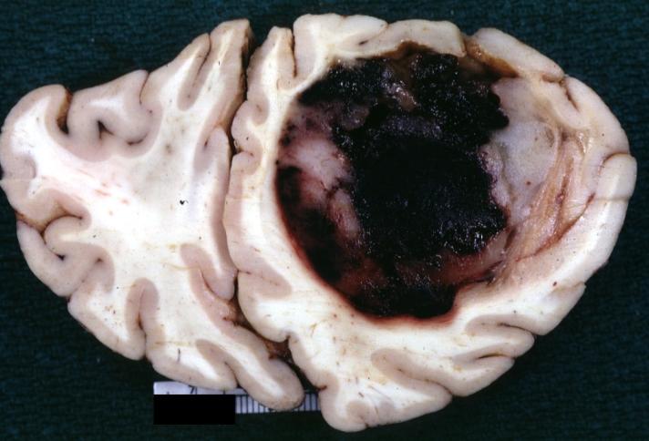
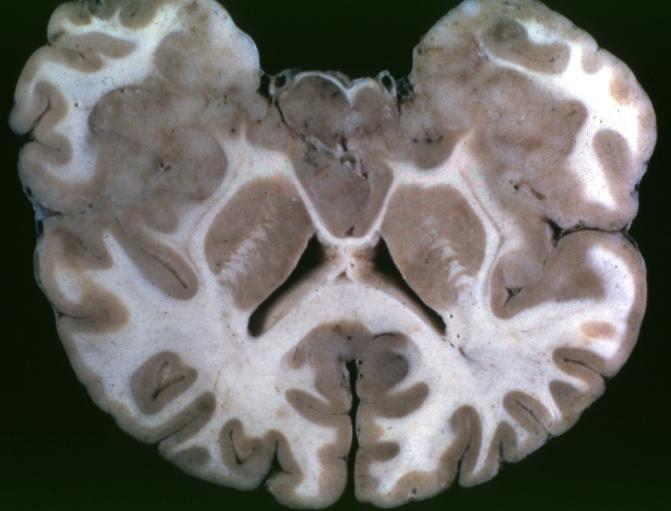
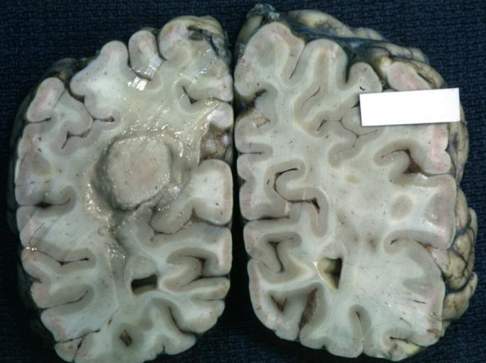
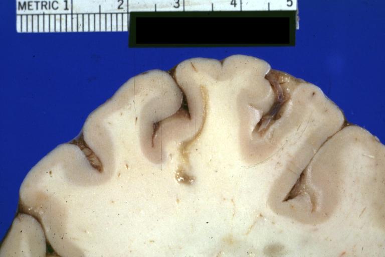
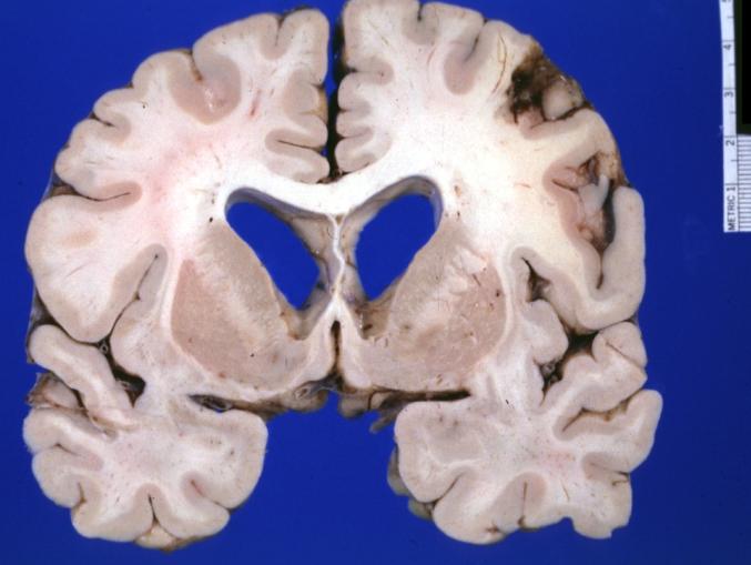
Brain: Oligodendroglioma: Gross; natural color, large, well circumscribed lesion in left frontal lobe
-
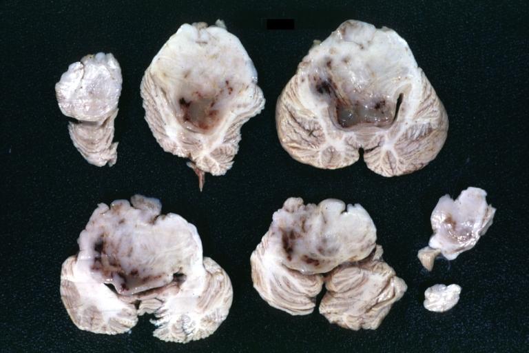
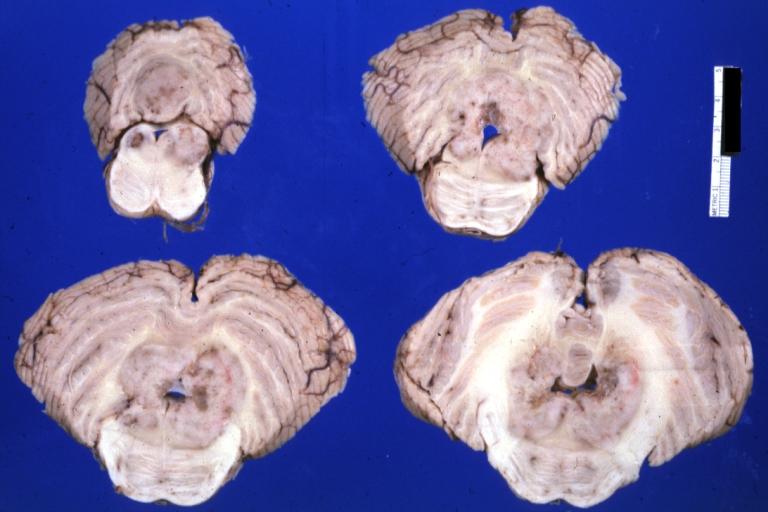
Brain: Glioma: Gross; fixed tissue, horizontal sections brain stem and cerebellum showing large pontine glioma
-
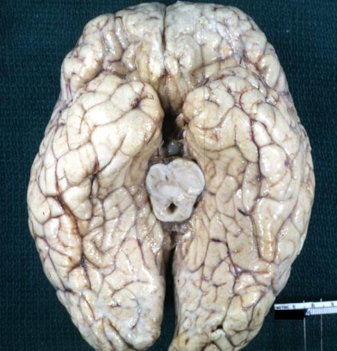
Brain: Pontine Glioma and Diffuse Meningeal Gliomatosis: Gross; fixed tissue, view of cerebral hemispheres from inferior with brain stem and cerebellum removed. Pontine asymmetry is easily seen due to low grade astrocytoma and meningeal gliomatosis is easily seen over frontal lobes
-
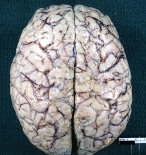
Brain: Pontine Glioma and Diffuse Meningeal Gliomatosis in 7 yo boy: Gross; fixed tissue, view of cerebral hemispheres from vertex meningeal gliomatosis.
-
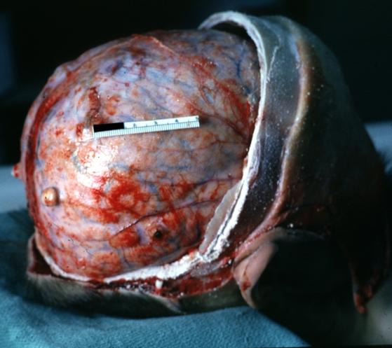
Brain: Pontine Glioma and Diffuse Meningeal Gliomatosis: Gross; in situ dural nodule
-
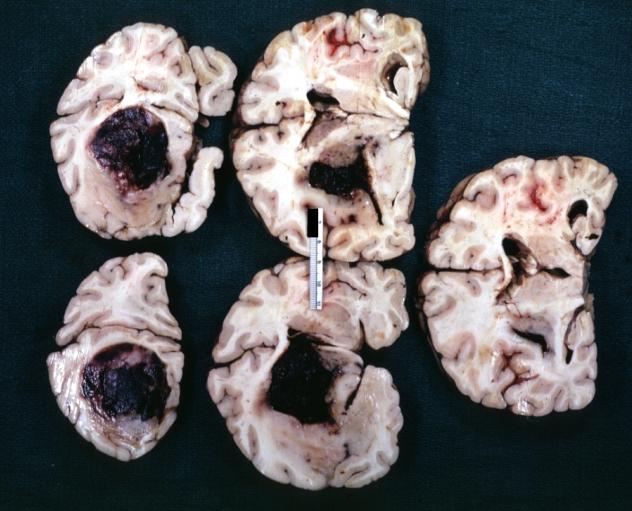
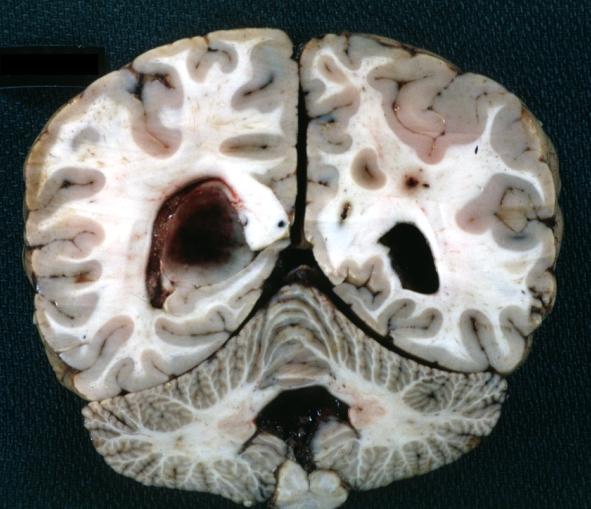
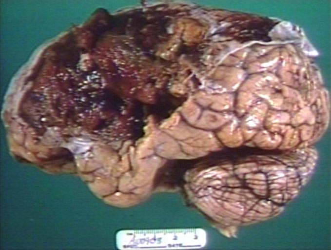
Brain: Oligodendroglioma: Gross; fixed tissue, multiple coronal sections, cerebral hemispheres with large tumor and hemorrhage into tumor
-
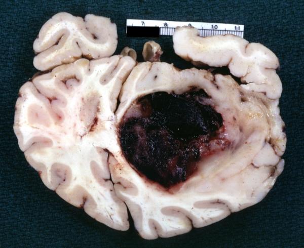
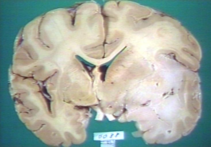
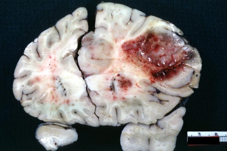
Brain: Oligodendroglioma: Gross; fixed tissue, coronal section, cerebral hemispheres, large hemorrhagic lesion in one hemisphere
-
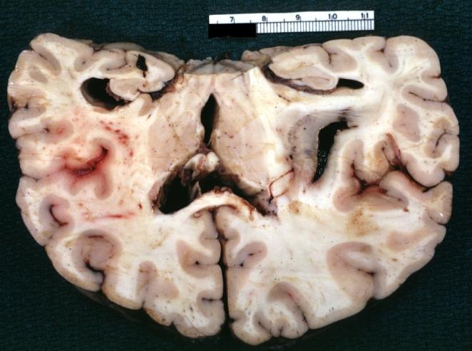
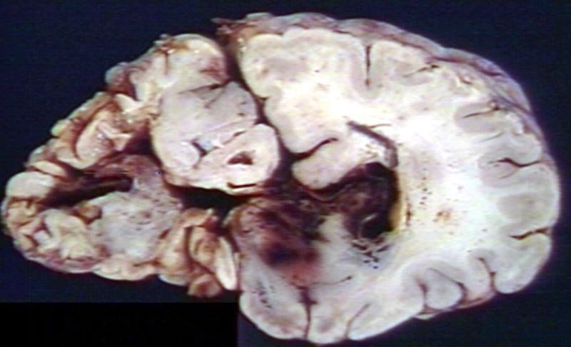
Brain: Oligodendroglioma: Gross; fixed tissue, ischemic tissue, anterior to tumor mass
-
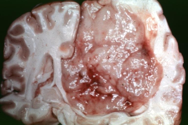
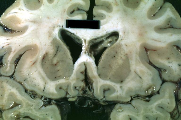
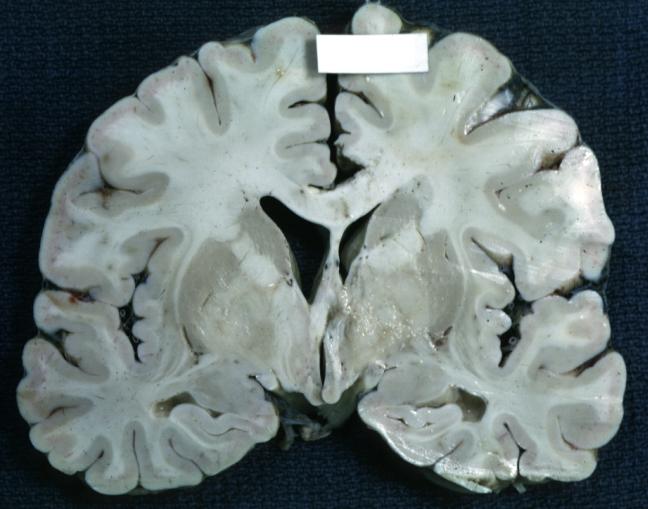
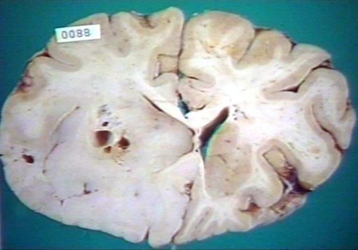
Brain: Oligodendroglioma: Gross; natural color, coronal section, cerebral hemispheres, large lesion, left parieto occipital white matter
-
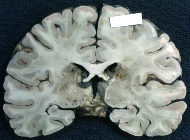
Brain: Gliomatosis Cerebri: Gross; fixed tissue, coronal sections, cerebral hemispheres, lesion is in temporal lobes and hypothalamus
-
Brain: Ventriculitis: Gross; fixed tissue, case of glioma with meningitis, a nice view of ventriculitis in one lateral ventricle
-
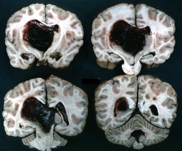
Brain: Glioma Thalamic Grade Ii-Iii: Gross; fixed tissue, four coronal sections, cerebral hemispheres, very large hemorrhagic lesion
-
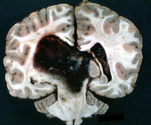
Brain: Glioma Thalamic Grade Ii-Iii: Gross; fixed tissue, coronal section, cerebral hemispheres with large hemorrhagic lesion
-
Brain: Glioma Thalamic Grade Ii-Iii: Gross fixed tissue coronal section cerebral hemispheres lesions appears to be in choroid plexus of lateral ventricle in this picture. There is blood in fourth ventricle
-
Brain: Cerebral Sarcoma or Microglioma: Gross; fixed tissue, coronal section, cerebral hemispheres (58 yo man)
-
Brain: Cerebral Sarcoma or Microglioma: Gross; fixed tissue, coronal section, cerebral hemispheres
-
Brain: Cerebral Sarcoma or Microglioma: Gross; fixed tissue, coronal section, cerebral hemispheres
-
Brain: Infarct Subcortical: Gross; fixed tissue, close-up view of old small subcortical infarct, a case of microglioma
-
Brain: Microglioma: Gross; fixed tissue; cerebellum and fourth ventricle with periventricular tumor invasion
-
Brain: Microglioma: Gross fixed tissue horizontal sections cerebellum and brain stem with periventricular neoplastic infiltrate
-
Brain: Microglioma: Gross fixed tissue horizontal section midbrain and cerebellum at mid pons level periventricular tumor infiltration
-
Brain: Microglioma: Gross fixed tissue horizontal section rostral pons and cerebellum
-
Brain: Microglioma: Gross fixed tissue horizontal section rostral pons and cerebellum periventricular tumor invasion
-
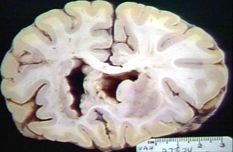
Brain: Microglioma: Gross fixed tissue coronal section cerebral hemispheres with mild ventricular dilation
-
Glioma: Optic Nerve
-
Brain: Oligodendroglioma, Frontal Lobe
-
Brain: Oligodendroglioma, Mixed Astrocytoma & Oligodendroglioma
-
Brain: Oligodendroglioma
-
Brain: Oligodendroglioma
-
Brain: Glioma, Grade II Anaplastic
-
Brain: Glioma, Brain stem, Low Grade
-
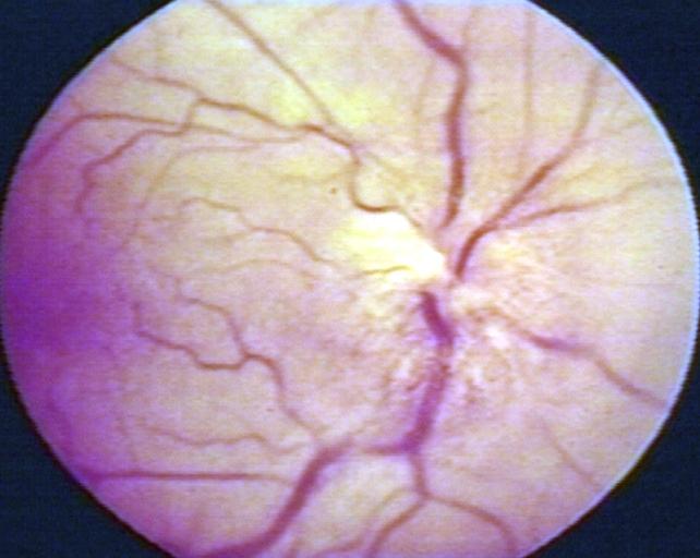
Fundoscopy: Eye; Optic Nerve Glioma, Optic Nerve
-
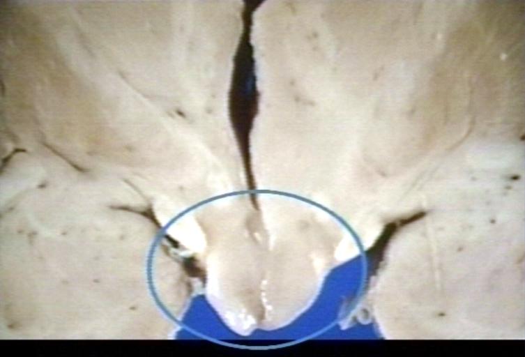
Brain: Glioma, Hypothalamic, Circle Around Region of Tumor
-
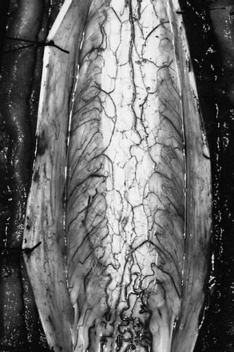
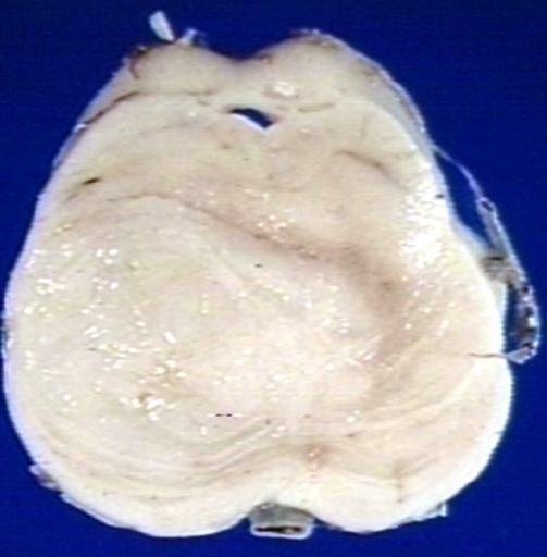
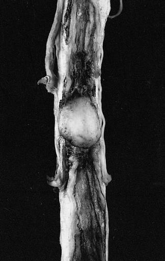
CNS: Pilocytic Astrocytoma of the Spinal Cord. The fusiform expansion of the spinal cord produced by this pilocytic astrocytoma is not, on external examination alone, distinguishable from that produced by a nonresectable diffuse glioma.
-
Brain: Glioma, Pontine
-
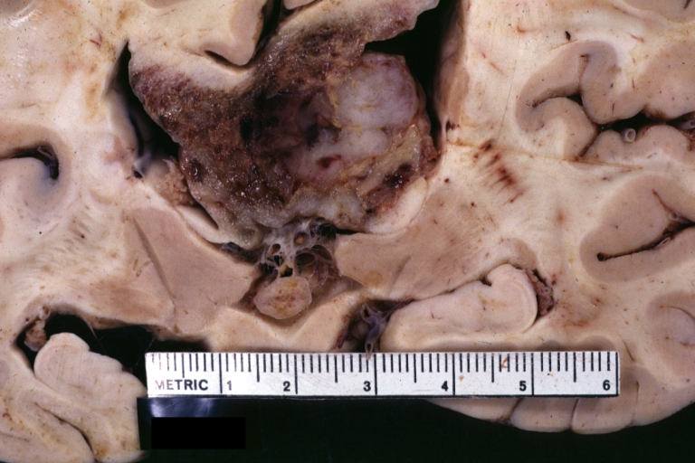
Brain: Glioblastoma Multiforme: Gross fixed tissue close-up large necrotic tumor mass in septum pellucidum
-
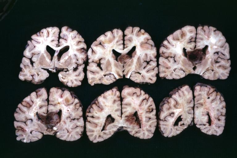
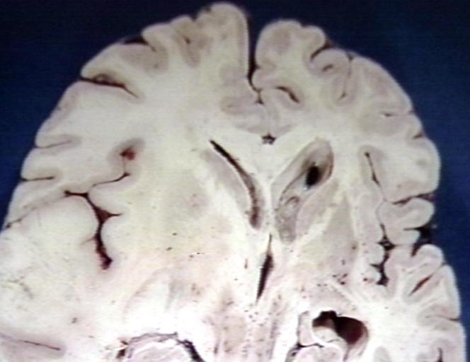
Brain: Glioblastoma Multiforme: Gross fixed tissue coronal section of the brain with a large necrotic tumor mass in septum pellucidum diagnosed as astrocytoma grade III
-
Brain: Glioblastoma Multiforme: Gross natural color large hemorrhagic lesion in right centrum semiovale
-
CNS: Malignant pilocytic astrocytoma: A 29-year-old woman died 2 years after a diagnosis of "atypical pilocytic astrocytoma" of the pineal region. At autopsy, multiple tumor implants were present in the craniospinal subarachnoid spaces.
-
Brain: Oligodendroglioma; Ventricular Cobblestone Effect


Microscopic Pathology
The histopathological appearance of glioma varies with the tumor grade and type, with increasing cellular atypia, mitoses, endothelial cell proliferation, and necrosis. Common findings are listed below:[4][5][6][7][8]
| Type of glioma | Histopathological features |
|---|---|
| |
| |
| |
| |
|
Gallery
-
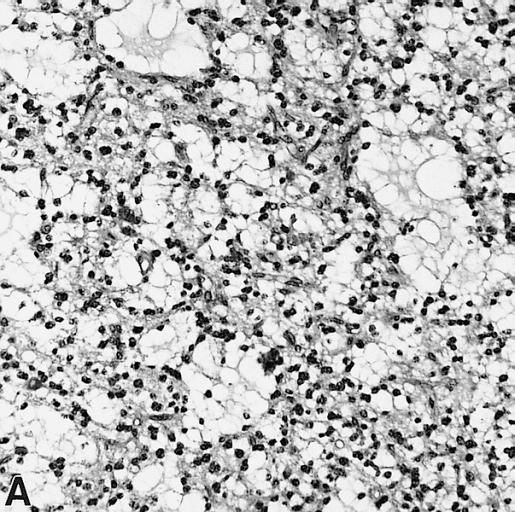
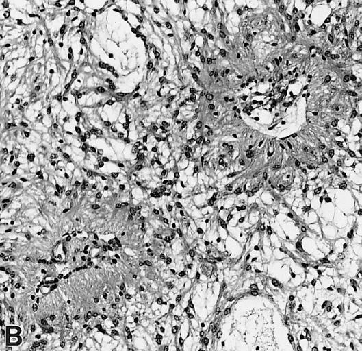
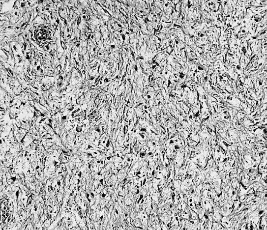
CNS: Pilocytic astrocytoma: variations in histologic appearance. As illustrated, many lesions are composed largely of spongy tissue rich in microcysts. Characteristic of pilocytic astrocytomas in general, the lesion is largely a solid mass of neoplastic cells without an obvious background of infiltrated brain.
-
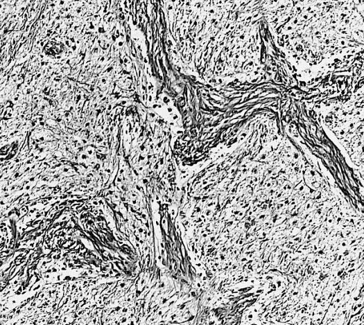
CNS: Pilocytic astrocytoma: variations in histologic appearance. The perivascular radiating processes in some lesions can create a likeness to an ependymoma. Note the spongy background unusual for ependymomas.
-
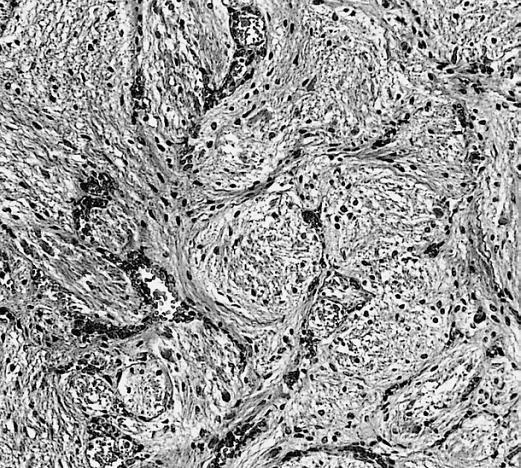
CNS: Pilocytic astrocytoma: variations in histologic appearance. Other pilocytic astrocytomas are solid, rather than microcystic, and may be lobular.
-
CNS: Pilocytic astrocytoma: variations in histologic appearance. Rosenthal fibers, usually confined to the solid rather than spongy regions are found in many pilocytic astrocytomas, but are not requisite for the diagnosis.
-
CNS: Pilocytic astrocytoma: variations in histologic appearance. Rosenthal fibers are extremely abundant in some lesions. Particularly in the cerebellum, it can be difficult to distinguish such solid, paucicellular, highly fibrillar pilocytic astrocytomas from reactive gliosis with abundant Rosenthal fiber formation.
-
CNS: Pilocytic astrocytoma: variations in histologic appearance. A loose array of polar cells creates an additional variant of pilocytic astrocytoma.
-
CNS: Pilocytic astrocytoma: variations in histologic appearance. Some pilocytic astrocytomas are traversed by prominent collagenous septa.
-
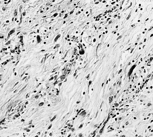
CNS: Pilocytic astrocytoma: variations in histologic appearance. Unusual pilocytic astrocytomas have an extensive mucinous background without microcysts.
-
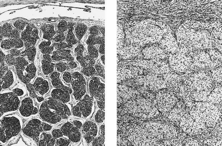
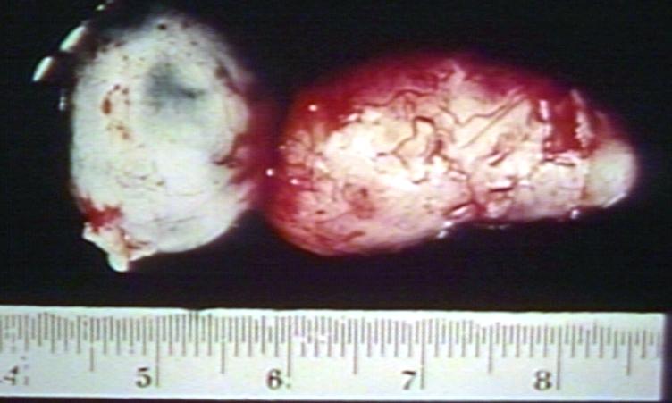
CNS: Comparison of normal optic nerve and pilocytic astrocytoma of the optic nerve. These two figures compare, at the same magnification, the normal optic nerve (left) with one containing a pilocytic astrocytoma (right). The neoplasm enlarges the compartments of the nerve and extends in collar-like fashion into the subarachnoid space.
-
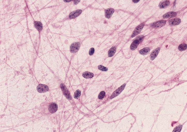
CNS: Pilocytic astrocytoma; The "hair cells" for which this lesion is named are readily seen.
-
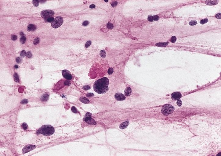
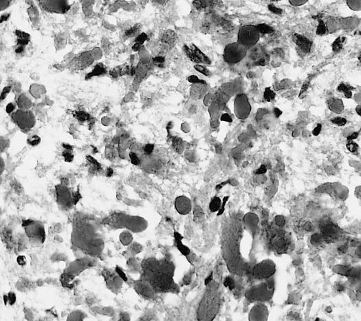
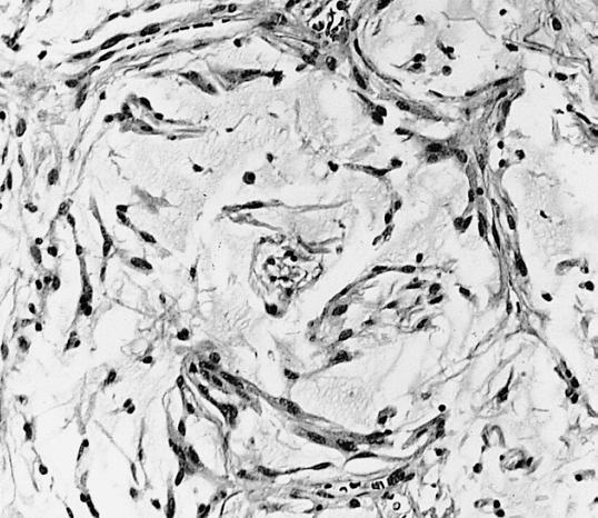
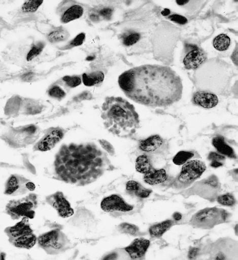
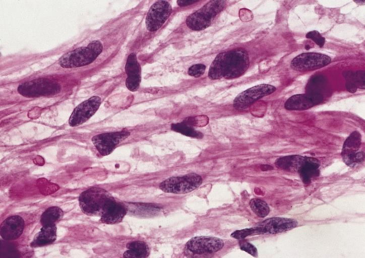
CNS: Pilocytic astrocytoma; Nuclear hyperchromasia and pleomorphism are common. Note the typical cellular elongation, and, at the center of the illustration, the eosinophilic granular body that populates pilocytic astrocytomas and certain other slowly growing gliomas.
-
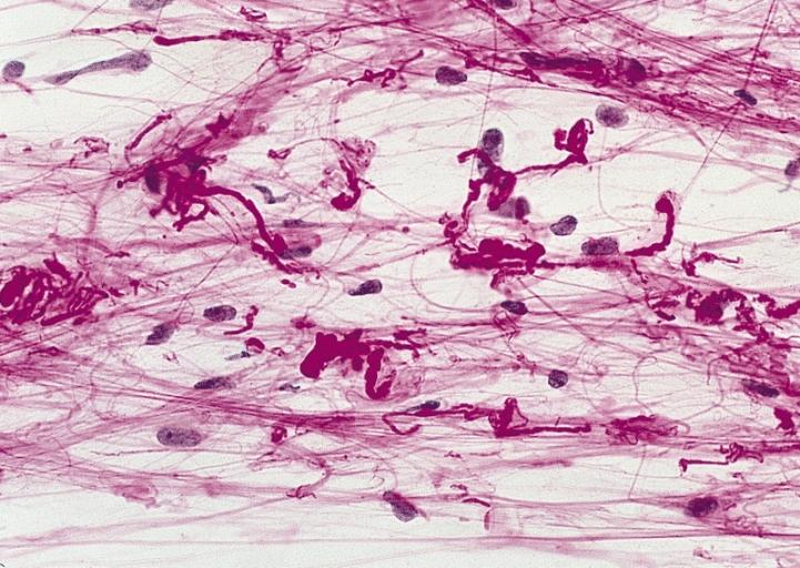
CNS: Pilocytic astrocytoma; Intracytoplasmic Rosenthal fibers are prominent in some pilocytic neoplasms.
-
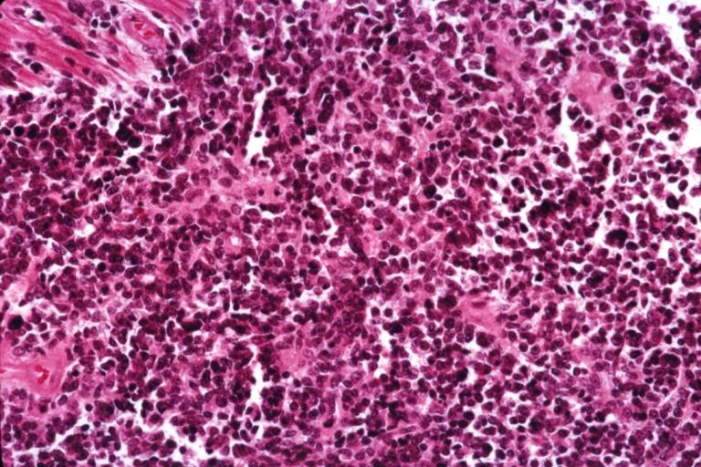
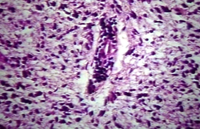
Brain: Malignant ependymoma: Micro med mag H&E tumor cells.
-
CNS: Oligodendroglioma; Occasional oligodendrogliomas contain cells with minute, refractile eosinophilic bodies representing miniature Rosenthal fibers.
-
CNS: Oligodendroglioma; The cells of some oligodendrogliomas acquire sufficient cytoplasm and process formation to become decidedly astrocytic, but their nuclei retain the roundness, uniformity, and chromatin distribution typical of oligodendroglioma.
-
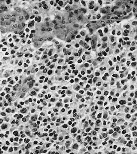
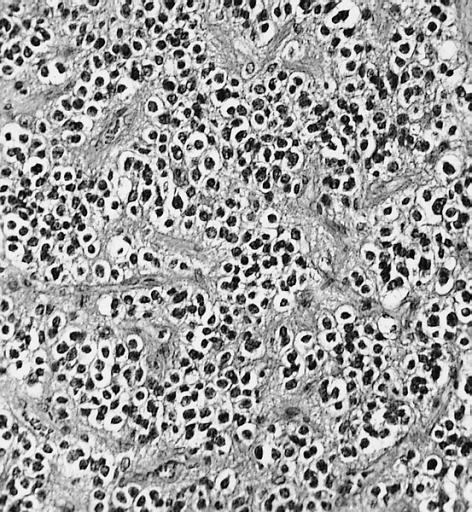
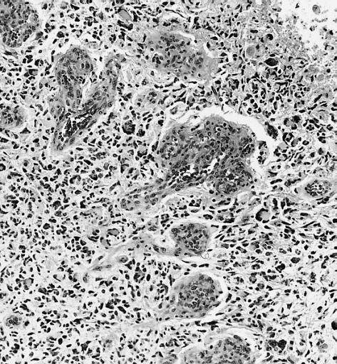
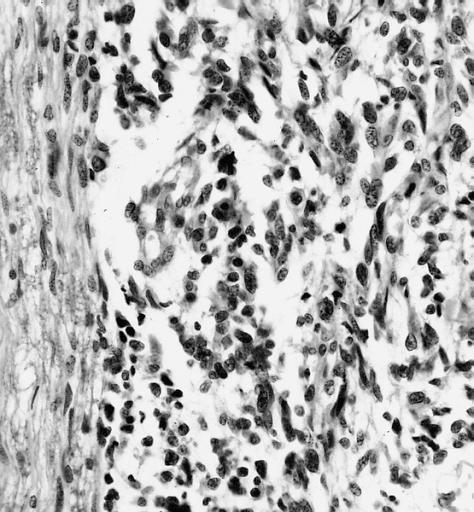
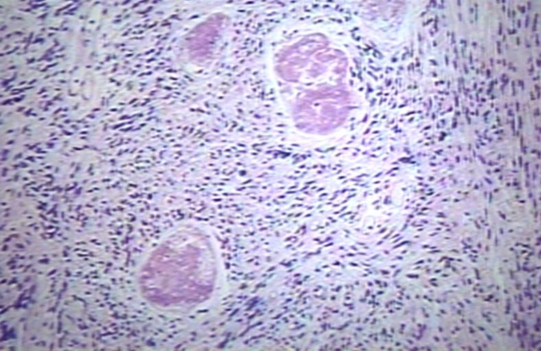
Anaplastic oligodendroglioma: Anaplastic oligodendrogliomas are highly cellular and associated with vascular proliferation.
-
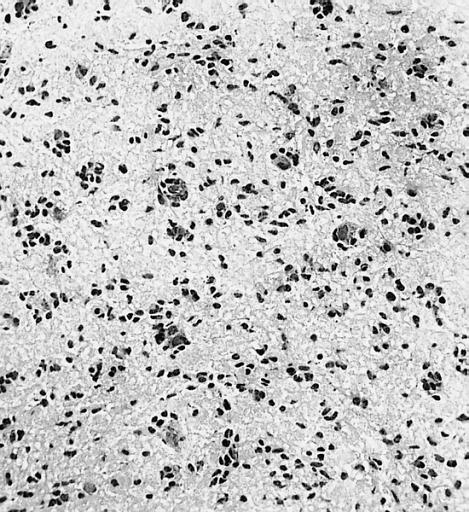
CNS: Oligodendroglioma (frozen section); Oligodendrogliomas in frozen sections lack the distinctive halos so often seen in permanent sections. Cellular monomorphism and infiltration of cerebral cortex with perineuronal satellitosis suggest the correct diagnosis.
-
CNS: Clear cell ependymoma; Perinuclear clearing similar to that seen in oligodendrogliomas is a prominent feature of the clear cell variant. Note the vague perivascular pseudorosettes. The lesion was a discrete occipital intraventricular mass.
-
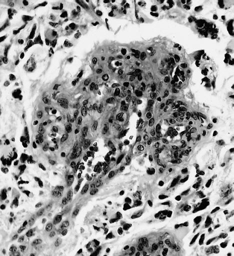
CNS: Glioblastoma multiforme; Brain: Glioblastoma multiforme. Grade I-Ii: Micro med mag with H&E, tumor well shown
-
CNS: Glioblastoma multiforme arising in an astrocytoma. At higher magnification, gemistocytic astrocytoma with microcystic change is apparent at the bottom of the illustration and cellular nodules of glioblastoma multiforme are seen at the top. The 6-year history of symptoms attested to the initially low-grade nature of this astrocytic tumor.
-
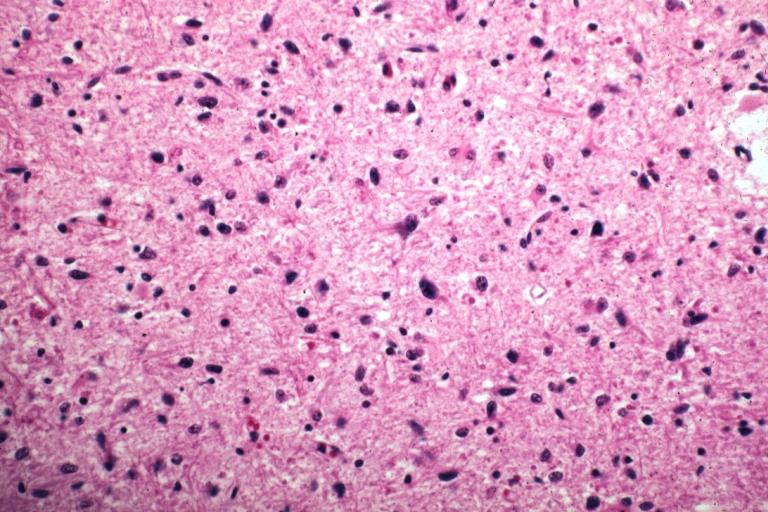
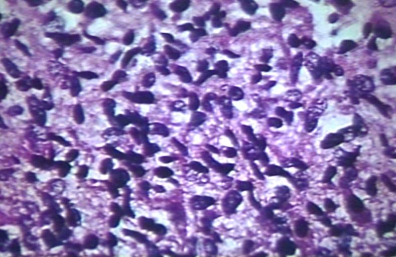
CNS: Glioblastoma multiforme; Characteristic of most glioblastomas are small cells with elongated nuclei and bipolar processes. As here, the chromatin is generally not markedly dense nor are nucleoli usually prominent.
-
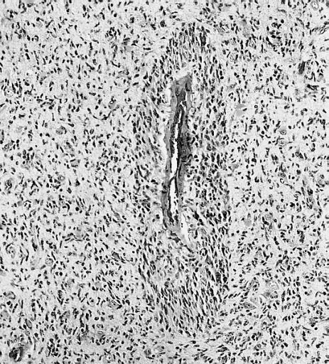
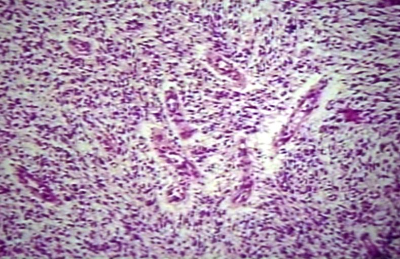
CNS: Glioblastoma multiforme; Vascular proliferation, a common feature of glioblastoma, produces tufts which often grow directionally. Here, as is often the case, they are oriented toward a focus of necrosis (top right).
-
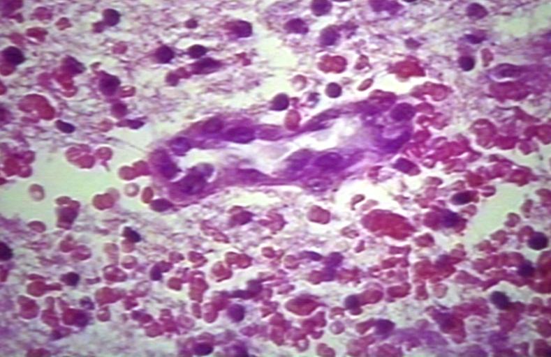
CNS: Glioblastoma multiforme; At high magnification, the neovascular tuft is a mass which, as can be confirmed by immunohistochemistry, is formed of both endothelial cells and smooth muscle cells (pericytes).
-
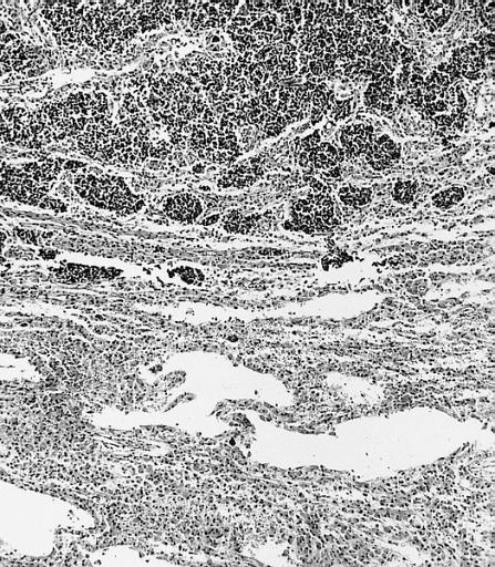
CNS: Glioblastoma multiforme; In many instances, necrosis is surrounded by a distinctive collar of cells, which are often smaller than those in surrounding neoplastic tissue. The phenomenon is referred to as pseudopalisading.
-
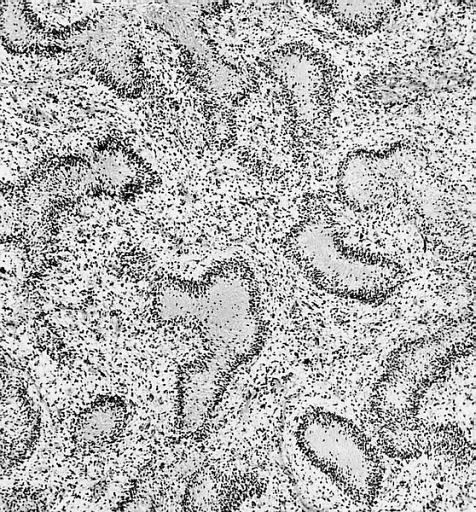
CNS: Cerebrospinal dissemination of glioblastoma multiforme; As seen at low (left) and high (right) magnification, the small undifferentiated-appearing cells of this glioblastoma are drop metastases colonizing the nerve roots of the cauda equina.
-
CNS: Cerebrospinal dissemination of glioblastoma multiforme; As seen at low (left) and high (right) magnification, the small undifferentiated-appearing cells of this glioblastoma are drop metastases colonizing the nerve roots of the cauda equina.
-
CNS: Glioblastoma multiforme; Higher magnification reveals the small cell nature of such tumors.
-
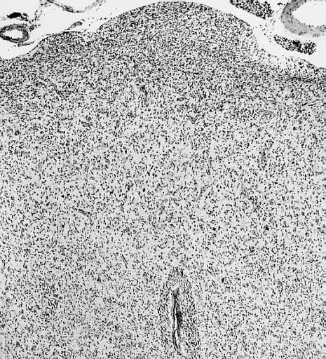
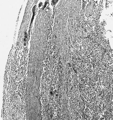
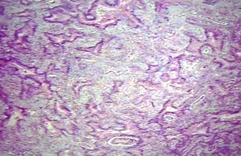
CNS: Glioblastoma multiforme; Some glioblastomas are especially infiltrative of the cerebral cortex where subpial, perivascular, and perineuronal accumulations are prominent.
-
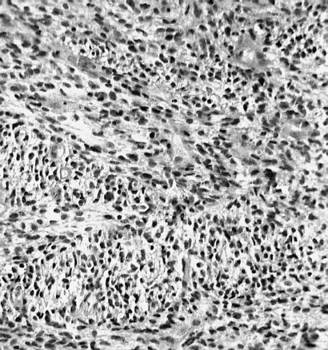
CNS: Glioblastoma multiforme; Although this densely cellular and largely undifferentiated lesion technically merits a diagnosis of anaplastic astrocytoma, it is, for practical purposes, a glioblastoma.
-
-
Brain: Glioblastoma multiforme; pallisading
-
Brain: Glioblastoma multiforme; vascular proliferation
-
Brain: Glioblastoma multiforme; Plump and juicy endothelial cells, extravasated blood
-
Brain: Glioblastoma multiforme; perivascular lymphocytes
-
Brain: Glioblastoma multiforme; thrombosed vessel
-
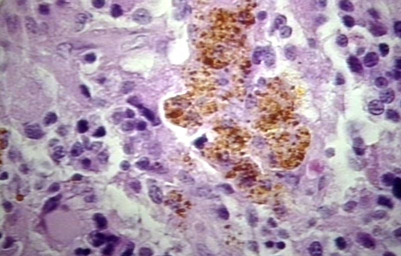
Brain: Glioblastoma multiforme; Hemosiderin in glioblastoma


References
- ↑ 1.0 1.1 Pathology of glioma. http://www.surgwiki.com/wiki/Intracranial_tumours,_infection_and_aneurysms
- ↑ 2.0 2.1 Pathology of glioma. Wikipedia. https://en.wikipedia.org/wiki/Glioma
- ↑ 3.0 3.1 3.2 Schwartzbaum JA, Fisher JL, Aldape KD, Wrensch M (2006). "Epidemiology and molecular pathology of glioma". Nat Clin Pract Neurol. 2 (9): 494–503, quiz 1 p following 516. doi:10.1038/ncpneuro0289. PMID 16932614.
- ↑ 4.0 4.1 4.2 Pathology of pilocytic astrocytoma. Libre Pathology. http://librepathology.org/wiki/index.php/Pilocytic_astrocytoma
- ↑ 5.0 5.1 5.2 Pathology of gliomas. Libre Pathology. http://librepathology.org/wiki/index.php/Oligodendroglioma
- ↑ 6.0 6.1 6.2 Pathology of anaplastic astrocytoma. Libre Pathology. http://librepathology.org/wiki/index.php?title=Neuropathology_tumours&redirect=no#Infiltrative_astrocytomas
- ↑ 7.0 7.1 7.2 Pathology of glioblastoma. Libre Pathology. http://librepathology.org/wiki/index.php/Glioblastoma
- ↑ 8.0 8.1 8.2 Pathology of ependymoma. Libre Pathology. http://librepathology.org/wiki/index.php/Ependymoma
- ↑ Dubois LG, Campanati L, Righy C, D'Andrea-Meira I, Spohr TC, Porto-Carreiro I; et al. (2014). "Gliomas and the vascular fragility of the blood brain barrier". Front Cell Neurosci. 8: 418. doi:10.3389/fncel.2014.00418. PMC 4264502. PMID 25565956.
- ↑ Reuss, D; von Deimling, A (2009). "Hereditary tumor syndromes and gliomas". Recent results in cancer research. Fortschritte der Krebsforschung. Progres dans les recherches sur le cancer. 171: 83–102. doi:10.1007/978-3-540-31206-2_5. PMID 19322539.