Fourth ventricle
Editor-In-Chief: C. Michael Gibson, M.S., M.D. [1]
Overview
The fourth ventricle is one of the four connected fluid-filled cavities within the human brain. These cavities, known collectively as the ventricular system, consist of the left and right lateral ventricles, the third ventricle, and the fourth ventricle. The fourth ventricle extends from the cerebral aqueduct (aqueduct of Sylvius) to the obex, and is filled with cerebrospinal fluid (CSF).
The fourth ventricle has a characteristic diamond shape in cross-sections of the human brain. It is located within the pons or in the upper part of the medulla. CSF entering the fourth ventricle through the cerebral aqueduct can exit to the subarachnoid space of the spinal cord through two lateral foramina of Luschka (singular: foramen of Luschka) and a single, midline foramen of Magendie (see List of human anatomical parts named after people).
Roof and floor
The fourth ventricle has a "roof" dorsally and a "floor" ventrally. The roof of the fourth ventricle is formed by the cerebellum (superior and inferior medullary velums), the floor by the rhomboid fossa, and the side "walls" formed by the cerebellar peduncles. Among the prominent features of the floor of the fourth ventricle are the:
- facial colliculus: formed by the internal part of the facial nerve as it loops around the abducens nucleus in the lower pons;
- sulcus limitans: which represents the border between the alar plate and the basal plate of the developing neural tube;
- obex: represents the caudal tip of the fourth ventricle; the obex is also a marker for the level of the foramen magnum of the skull and therefore is a marker for the imaginary dividing line between the medulla and spinal cord.
Additional images
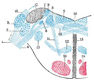
-
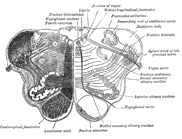
Transverse section of medulla oblongata below the middle of the olive.
-
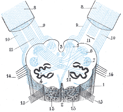
Diagram showing the course of the arcuate fibers.
-
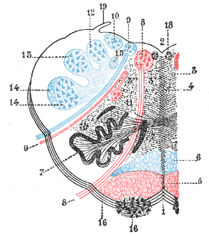
The formatio reticularis of the medulla oblongata, shown by a transverse section passing through the middle of the olive.
-
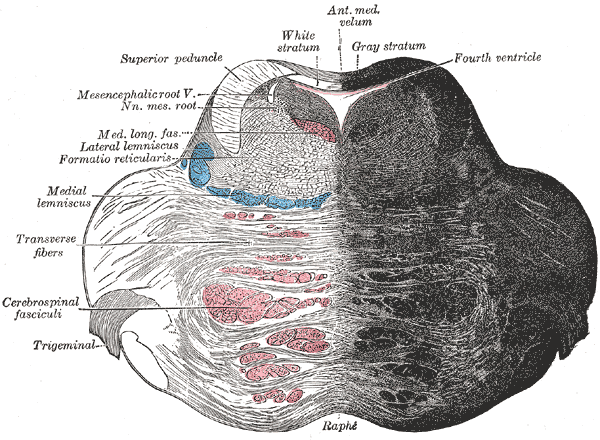
Coronal section of the pons, at its upper part.
-
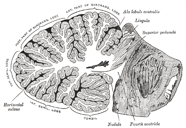
Sagittal section of the cerebellum, near the junction of the vermis with the hemisphere.
-
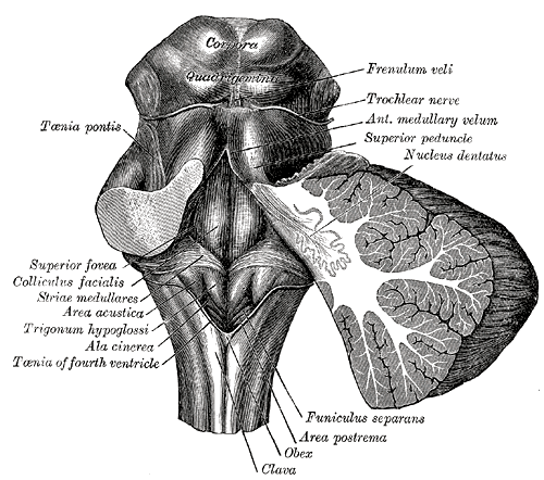
Rhomboid fossa.
-
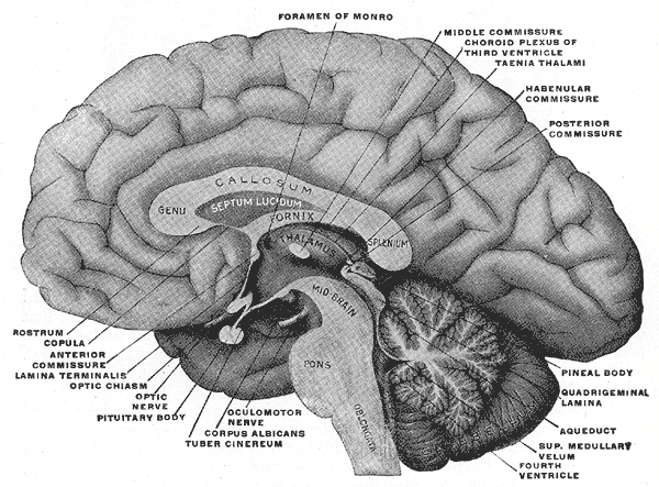
Mesal aspect of a brain sectioned in the median sagittal plane.
-
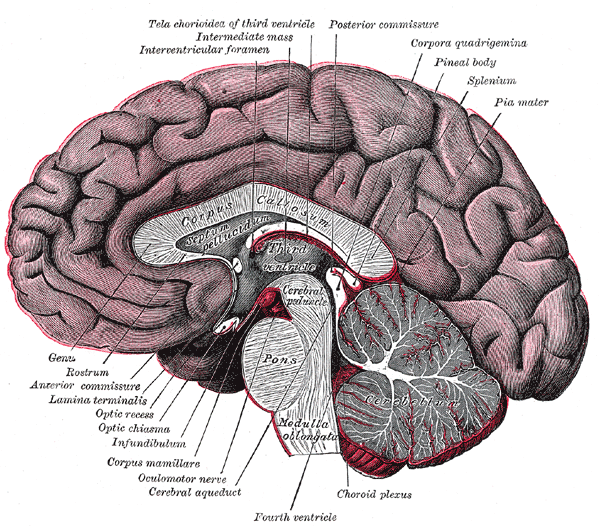
Median sagittal section of brain.
-
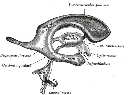
Drawing of a cast of the ventricular cavities, viewed from the side.
-
Terminal nuclei of the vestibular nerve, with their upper connections.
-
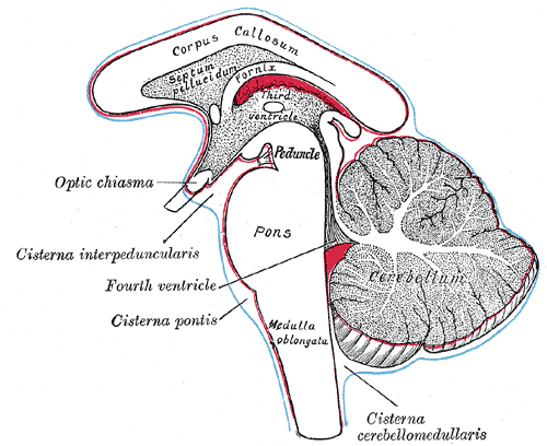
Diagram showing the positions of the three principal subarachnoid cisternæ.


External links
- Template:UMichAtlas - "Fourth Ventricle, Sagittal Section, Medial View"
- Template:BrainMaps
- Template:EMedicineDictionary
- Template:RocheLexicon
de:Hirnventrikel#Vierter Ventrikel nl:Vierde ventrikel no:Fjerde ventrikkel sk:Štvrtá mozgová komora