Subarachnoid space

Jump to navigation Jump to search

Template:Infobox Anatomy

WikiDoc Resources for Subarachnoid space

Articles

Most recent articles on Subarachnoid space

Most cited articles on Subarachnoid space

Review articles on Subarachnoid space

Articles on Subarachnoid space in N Eng J Med, Lancet, BMJ

Media

Powerpoint slides on Subarachnoid space

Images of Subarachnoid space

Photos of Subarachnoid space

Podcasts & MP3s on Subarachnoid space

Videos on Subarachnoid space

Evidence Based Medicine

Cochrane Collaboration on Subarachnoid space

Bandolier on Subarachnoid space

TRIP on Subarachnoid space

Clinical Trials

Ongoing Trials on Subarachnoid space at Clinical Trials.gov

Trial results on Subarachnoid space

Clinical Trials on Subarachnoid space at Google

Guidelines / Policies / Govt

US National Guidelines Clearinghouse on Subarachnoid space

NICE Guidance on Subarachnoid space

NHS PRODIGY Guidance

FDA on Subarachnoid space

CDC on Subarachnoid space

Books

Books on Subarachnoid space

News

Subarachnoid space in the news

Be alerted to news on Subarachnoid space

News trends on Subarachnoid space

Commentary

Blogs on Subarachnoid space

Definitions

Definitions of Subarachnoid space

Patient Resources / Community

Patient resources on Subarachnoid space

Discussion groups on Subarachnoid space

Patient Handouts on Subarachnoid space

Directions to Hospitals Treating Subarachnoid space

Risk calculators and risk factors for Subarachnoid space

Healthcare Provider Resources

Symptoms of Subarachnoid space

Causes & Risk Factors for Subarachnoid space

Diagnostic studies for Subarachnoid space

Treatment of Subarachnoid space

Continuing Medical Education (CME)

CME Programs on Subarachnoid space

International

Subarachnoid space en Espanol

Subarachnoid space en Francais

Business

Subarachnoid space in the Marketplace

Patents on Subarachnoid space

Experimental / Informatics

List of terms related to Subarachnoid space


Overview

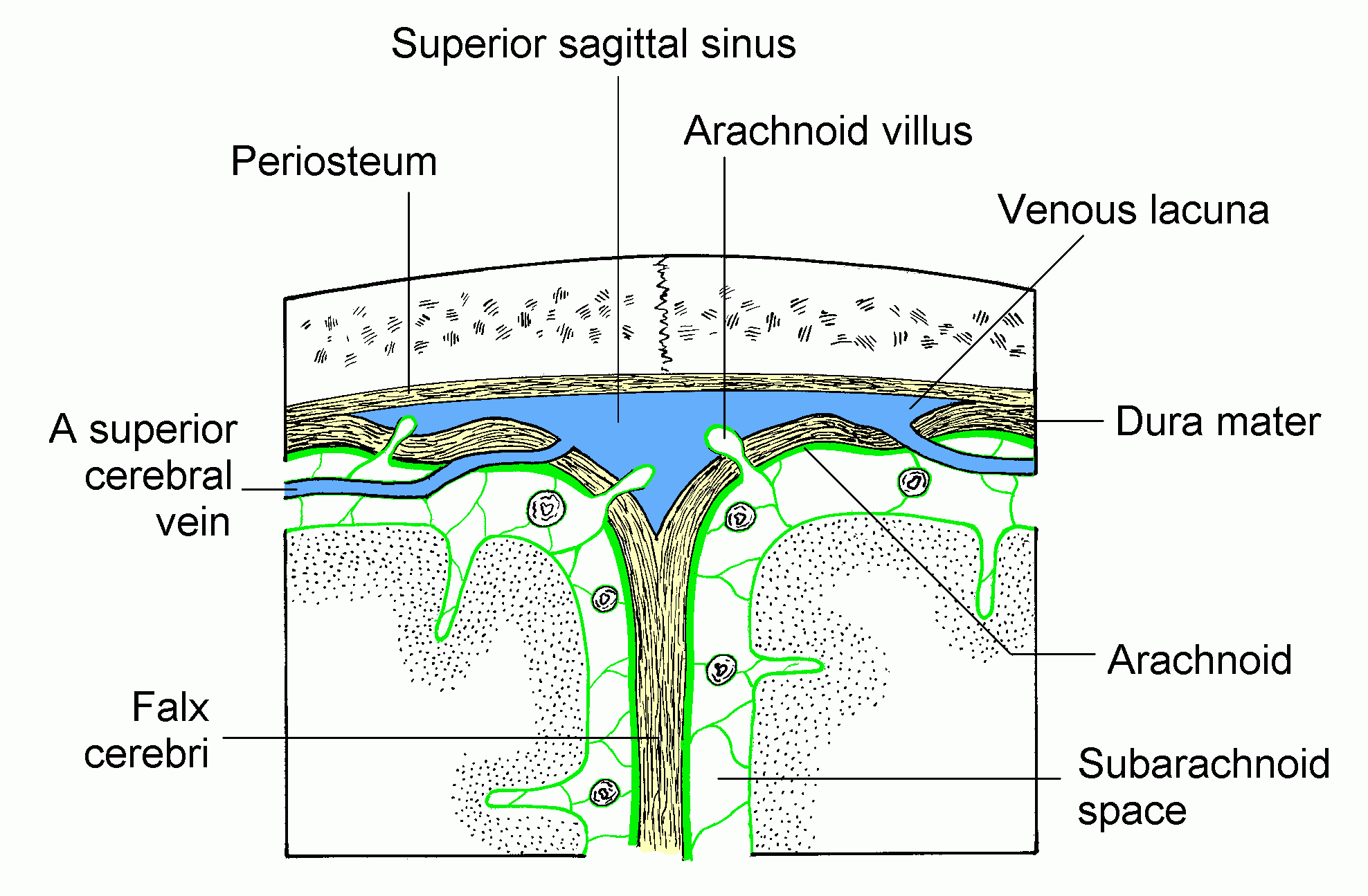
In the brain, the subarachnoid cavity (subarachnoid space) is the interval between the arachnoid mater and pia mater.

It is occupied by a spongy tissue consisting of trabeculæ (delicate connective tissue filaments that extend from the arachnoid mater and blend into the pia mater) and intercommunicating channels in which the cerebrospinal fluid is contained.

This cavity is small on the surface of the hemispheres of the brain. On the summit of each gyrus the pia mater and the arachnoid are in close contact, but in the sulci between the gyri, triangular spaces are left, in which the subarachnoid trabecular tissue is found. Whilst the pia mater closely follows the surface of the brain and dips into the sulci, the arachnoid bridges across them from gyrus to gyrus.

At certain parts of the base of the brain, the arachnoid is separated from the pia mater by wide intervals, which communicate freely with each other and are named subarachnoid cisternæ; in these the subarachnoid tissue is less abundant. The subarachnoid space is the location of the interface between the vascular tissue and the cerebrospinal fluid and is active in the blood brain barrier.

The arachnoid mater continues down the spinal cord too, and the subarachnoid layer with it. It serves a similar function in the spinal cord as it does in the brain.

See also

Subarachnoid hemorrhage

External links

Template:Gray's

Template:Meninges Template:Spinal cord

Template:WH Template:WS