Pulmonic regurgitation electrocardiogram
|
Pulmonic regurgitation Microchapters |
|
Diagnosis |
|---|
Editor-In-Chief: C. Michael Gibson, M.S., M.D. [1], Associate Editor(s)-in-Chief: Aravind Kuchkuntla, M.B.B.S[2], Aysha Anwar, M.B.B.S[3], Javaria Anwer M.D.[4]
Overview
EKG findings among patients wit chronic Pulmonic regurgitation (PR) may be non-specific. Ventricular tachycardia is demonstrated on EKG among patients with PR and RV dilatation. Patients may develop atrial flutter/fibrillation after years of PR development. Among patients with tetralogy of Fallot (TOF), increased QRS duration with widened QRS complex reflects the severity of PR and right ventricular dilation predisposes the patients to develop malignant arrythmias.
Electrocardiogram
Key EKG Findings in Pulmonic regurgitation
EKG findings among patients with pulmonary regurgitation (PR) may include the following:
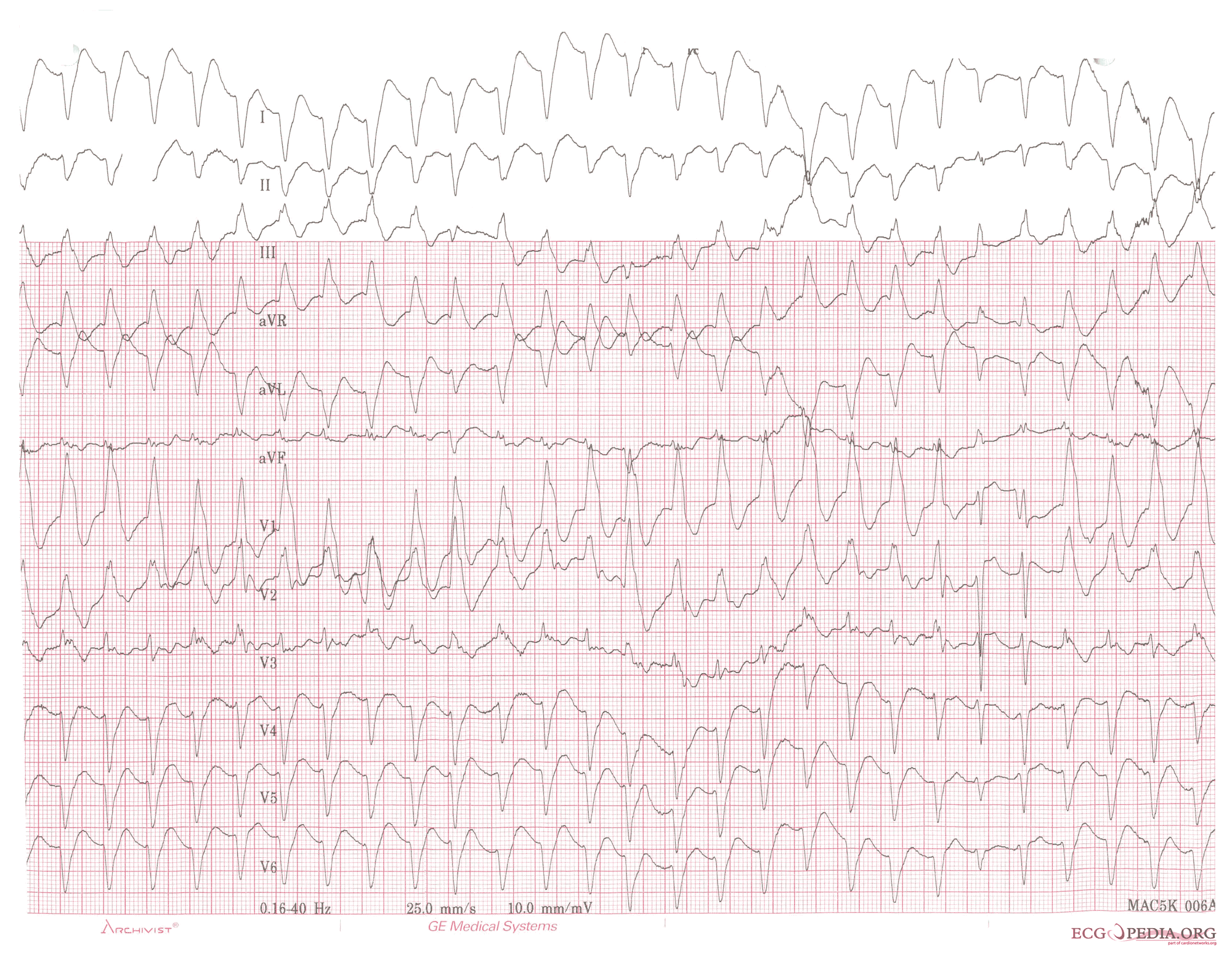
- Ventricular tachycardia is demonstrated on EKG among patients with PR and RV dilatation.[1] (EKG 1)
- Mild PR: Signs of RV hypertrophy may be demonstrated on EKG such as tall P waves, increased R to S ratio in the right precordial leads and right axis deviation [2]:
- Severity of PR[3]: A strong correlation between QRS duration and PR index has been demonstrated. QRS duration ≥160 ms predicted severe PR with 100% sensitivity and 87% specificity among patients with repaired TOF in a cohort study.
- Chronic PR[1][4]:
- EKG findings demonstrated in chronic PR are non-specific.
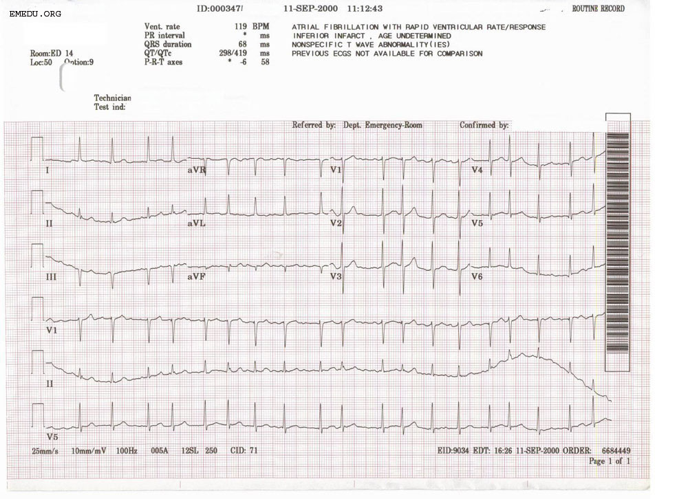
- Patients may develop atrial flutter/fibrillation after years of PR development. (EKG 2)
- Due to PR, chronic RV volume overload has been associated with a prolonged QRS. All patients with ventricular tachycardia or sudden death have demonstrated QRS duration of ⩾180 ms.
- Isolated PR: Among patients with RV volume overload and isolated PR, QRS prolongation with rSr morphology can be seen in right precordial leads.
- TOF and Post-TOF repair[4][5]:
- Among patients with tetralogy of Fallot (TOF) increased QRS duration with widened QRS complex reflects the severity of PR and right ventricular dilation predisposes the patients to develop malignant arrythmias.
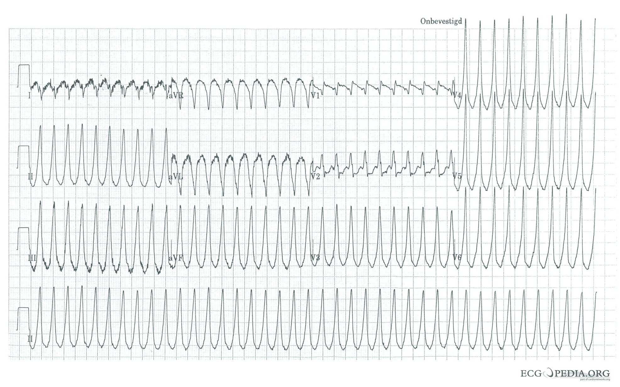
- RBB is common among the majority of patients who have tetralogy of Fallot repair with right ventriculotomy. (EKG 3)
- Pulmonary hypertension (PAH): Among patients with PAH, RV hypertrophy may be demonstrated on EKG as[2]:
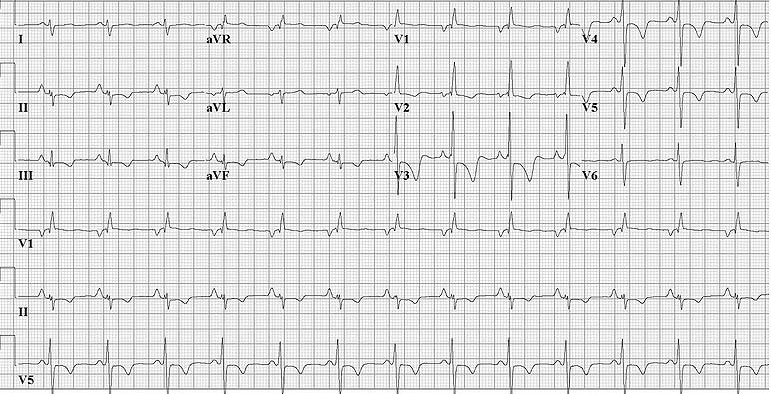
- P pulmonale (tall P waves- demonstrating right atrial enlargement, (EKG 4)
- Increased R to S ratio in the right precordial leads
- Right axis deviation
EKG examples
EKG1: The EKG demonstrates ventricular tachycardia with a rate of 150 bpm, and a right bundle branch block pattern with right heart axis. The 5th and 6th complexes from the right side are fusion complexes. Moreover, this EKG shows baseline drift, which is a technical artifact.

Copyleft image obtained courtesy of ECGpedia,http://en.ecgpedia.org/wiki/Main_Page
EKG 2: The EKG demonstrates irregularly irregular rhythm with no P waves, suggestive of atrial fibrillation.

Copyleft image obtained courtesy of ECGpedia,http://en.ecgpedia.org/wiki/Main_Page
EKG 3: The EKG demonstrates ventricular tachycardia with a rate of 250 bpm, and a right bundle branch block pattern with a right heart axis.

Copyleft image obtained courtesy of ECGpedia,http://en.ecgpedia.org/wiki/Main_Page
EKG 4: The EKG demonstrates right ventricular hypertrophy and right atrial enlargement in a patient with chronic PAH. Note P pulmonale that is a P wave amplitude >2.5mm among inferior leads (II, III, and AVF) and T wave inversion among leads II, III, aVF, V2, V3, V4, V5.

Copyleft image obtained courtesy of ECGpedia,http://en.ecgpedia.org/wiki/Main_Page
References
- ↑ 1.0 1.1 Chaturvedi RR, Redington AN (July 2007). "Pulmonary regurgitation in congenital heart disease". Heart. 93 (7): 880–9. doi:10.1136/hrt.2005.075234. PMC 1994453. PMID 17569817.
- ↑ 2.0 2.1 Glancy DL, Jain N, Jaligam VR, Ilie CC, Atluri P (July 2011). "Electrocardiogram in a woman with cor pulmonale". Proc (Bayl Univ Med Cent). 24 (3): 255–6. doi:10.1080/08998280.2011.11928728. PMC 3124915. PMID 21738303.
- ↑ Tanasan A, Kocharian A, Zanjani KS, Payravian FK, Torabian S (October 2013). "Correlation between QRS Duration, Pulmonary Insufficiency and Right Ventricle Performance in Totally Corrected Tetralogy of Fallot". Iran J Pediatr. 23 (5): 593–6. PMC 4006512. PMID 24800023.
- ↑ 4.0 4.1 Gatzoulis MA, Till JA, Somerville J, Redington AN (July 1995). "Mechanoelectrical interaction in tetralogy of Fallot. QRS prolongation relates to right ventricular size and predicts malignant ventricular arrhythmias and sudden death". Circulation. 92 (2): 231–7. doi:10.1161/01.cir.92.2.231. PMID 7600655.
- ↑ Abd El Rahman MY, Abdul-Khaliq H, Vogel M, Alexi-Meskishvili V, Gutberlet M, Lange PE (2000). "Relation between right ventricular enlargement, QRS duration, and right ventricular function in patients with tetralogy of Fallot and pulmonary regurgitation after surgical repair". Heart. 84 (4): 416–20. PMC 1729453. PMID 10995413.