Osteogenesis imperfecta classification
|
Osteogenesis imperfecta Microchapters |
|
Diagnosis |
|---|
|
Treatment |
|
Case Studies |
|
Osteogenesis imperfecta classification On the Web |
|
American Roentgen Ray Society Images of Osteogenesis imperfecta classification |
|
Risk calculators and risk factors for Osteogenesis imperfecta classification |
Editor-In-Chief: C. Michael Gibson, M.S., M.D. [1]
Overview
Classification
There are eight types of OI, Type I being the most common, though the symptoms range from person to person.
| Type | Description | OMIM |
| Type I | mild | 166240 (IA), 166200 (IB) |
| Type II | severe and usually lethal in the perinatal period | 166210 |
| Type III | considered progressive and deforming | 259420 |
| Type IV | deforming, but with normal scleras | 166220 |
| Type V | shares the same clinical features of IV, but has unique histologic findings ("mesh-like") | 610967 |
| Type VI | shares the same clinical features of IV, but has unique histologic findings ("fish scale") | 610968 |
| Type VII | associated with cartilage associated protein | 610682 |
| Type VIII | associated with leprecan | 610915 |
Type I
Collagen is of normal quality but is produced in insufficient quantities:
- Bones fracture easily
- Slight spinal curvature
- Loose joints
- Poor muscle tone
- Discolouration of the sclera (whites of the eyes), usually giving them a blue-gray color. The blue-gray color of the sclera is due to the reflection of underlying choroidal veins. The underlying choroidal veins reflect through the sclera because there is defective synthesis of type 1 collagen.
- Early loss of hearing in some children
- Slight protrusion of the eyes
IA and IB are defined to be distinguished by the absence/presence of dentinogenesis imperfecta (characterized by opalescent teeth.) (Absent in IA, present in IB.)
Type II
Collagen is not of a sufficient quality or quantity
- Most cases die within the first year of life due to respiratory failure or intracerebral hemorrhage
- Severe respiratory problems due to underdeveloped lungs
- Severe bone deformity and small stature
Type II can be further subclassified into groups A, B, C, which are distinguished by radiographic evaluation of the long bones and ribs. Type IIA demonstrates broad and short long bones with broad and beaded ribs. Type IIB demonstrates broad and short long bones with thin ribs that have little or no beading. Type IIC demonstrates thin and longer long bones with thin and beaded ribs.
Type III
Collagen quantity is sufficient but is not of a high enough quality
- Bones fracture easily, sometimes even before birth
- Bone deformity, often severe
- Respiratory problems possible
- Short stature, spinal curvature and sometimes barrel-shaped rib cage
- Loose joints
- Poor muscle tone in arms and legs
- Discolouration of the sclera (whites of the eyes)
- Early loss of hearing, sometimes
Type III is distinguished among the other classifications as being the "Progressive Deforming" type, wherein a neonate presents with mild symptoms at birth and develops the aforementioned symptoms throughout life. Lifespan may be normal, albeit with severe physical handicapping.
Type IV
Collagen quantity is sufficient but is not of a high enough quality
- Bones fracture easily, especially before puberty
- Short stature, spinal curvature and barrel-shaped rib cage
- Bone deformity is mild to moderate
- Early loss of hearing
Similar to Type I, Type IV can be further subclassified into types IVA and IVB characterized by absence (IVA) or presence (IVB) of dentinogenesis imperfecta.
Type V


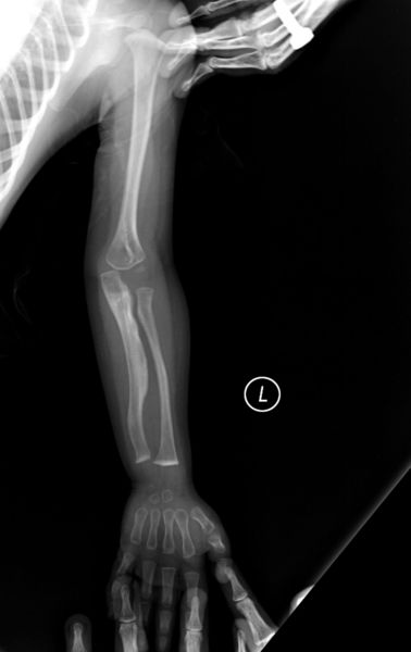
Same clinical features as Type IV. Distinguished histologically by "mesh-like" bone appearance. Further characterized by the "V Triad" consisting of a) radio-opaque band adjacent to growth plates, b) hypertrophic calluses at fracture sites, and c) calcification of the radio-ulnarinterosseous membrane.
- As per Doctors Francis Glorieux, Frank Rauch, and Leanne Ward in the Shriners Hospital for Children in Quebec[1]
OI Type V leads to calcification of the membrane between the two forearm bones, making it difficult to turn the wrist. Another symptom is abnormally large amounts of repair tissue (hyperplasic callus) at the site of fractures. At the present time, the cause for Type V is unknown, though doctors have determined that it is inherited.
X-Ray OI Type V in Adult X-Ray OI Type V Kid
More on Type V Research More on OI Study
Type VI
Same clinical features as Type IV. Distinguished histologically by "fish-scale" bone appearance.
Type VII
- In 2005 a recessive form called "Type VII" was discovered. Thus far it seems to be limited to a First Nations people in Quebec.