Knee
|
WikiDoc Resources for Knee |
|
Articles |
|---|
|
Media |
|
Evidence Based Medicine |
|
Clinical Trials |
|
Ongoing Trials on Knee at Clinical Trials.gov Clinical Trials on Knee at Google
|
|
Guidelines / Policies / Govt |
|
US National Guidelines Clearinghouse on Knee
|
|
Books |
|
News |
|
Commentary |
|
Definitions |
|
Patient Resources / Community |
|
Directions to Hospitals Treating Knee Risk calculators and risk factors for Knee
|
|
Healthcare Provider Resources |
|
Continuing Medical Education (CME) |
|
International |
|
|
|
Business |
|
Experimental / Informatics |
Editor-In-Chief: C. Michael Gibson, M.S., M.D. [1]
Overview
In human anatomy, the knee is the lower extremity joint connecting the femur and the tibia. Since in humans the knee supports nearly the entire weight of the body, it is the joint most vulnerable both to acute injury and to the development of osteoarthritis.
Function of the knee
The knee functions as a living, self-maintaining, biologic transmission, the purpose of which is to accept and transfer biomechanical loads between the femur, tibia, patella, and fibula. In this analogy the ligaments represent non-rigid adaptable sensate linkages within the biologic transmission. The articular cartilages act as bearing surfaces, and the menisci as mobile bearings. The muscles function as living cellular engines that in concentric contraction provide motive forces across the joint, and in eccentric contraction act as brakes and dampening systems, absorbing loads.
Human anatomy
Upon birth, a baby will not have a conventional knee cap, but a knee cap formed of cartilege. In Human females this turns to a normal bone knee cap by the age of 3, in males the age of 5.
The knee is a complex, compound, condyloid variety of a synovial joint which hovers. It actually comprises two separate joints.
- The femoro-patellar joint consists of the patella, or "kneecap", a so-called sesamoid bone which sits within the tendon of the anterior thigh muscle (m. quadriceps femoris), and the patellar groove on the front of the femur through which it slides.
- The femoro-tibial joint links the femur, or thigh bone, with the tibia, the main bone of the (lower) leg. The joint is bathed in a viscous (synovial) fluid which is contained inside the "synovial" membrane, or joint capsule.
The recess behind the knee is called the popliteal fossa it can also be called a "knee pit."
Ligaments
| Name | Capsule | From | To | Description |
| anterior cruciate ligament (ACL) | inside | lateral condyle of femur | anterior intercondylar area | The critically important ACL prevents the tibia from being pushed too far anterior relative to the femur. |
| posterior cruciate ligament (PCL) | inside | medial condyle of femur | posterior intercondylar area | Injury to this ligament is uncommon but can occur as a direct result of forced trauma to the ligament. This ligament prevents posterior displacment of the tibia relative to the femur. |
| capsular ligament | - | - | - | - |
| patellar ligament | outside | patella | tuberosity of the tibia | - Also occasionally called the patellar tendon because there is no definite separation between the quadriceps tendon (which surrounds the patella) and the area connecting the patella to the tibia. This very strong ligament helps give the patella its mechanical leverage and also functions as a cap for the condyles of the femur. |
| medial collateral ligament (MCL a.k.a. "tibial") | outside | medial epicondyle of the femur | medial tibial condyle | The MCL protects the medial side of the knee from being bent open by a stress applied to the lateral side of the knee (a valgus force). |
| lateral collateral ligament (LCL a.k.a. "fibular") | outside | lateral epicondyle of the femur | head of fibula | The LCL protects the lateral side from an inside bending force (a varus force). |
| oblique popliteal ligament | outside | medial condyle of tibia | - | Tendinous expansion of the semimembranosus muscle. Strengthens the back of the capsule |
| arcuate popliteal ligament | outside | Intercondylar area of tibia, and lateral condyle of femur, to below head of fibula | Connects to the medial portion of the fibular head | -This ligament strengthes the knee posterolaterally and usually when injured is in combination with a PCL and popliteus tendon injury. |
Menisci
These are cartilaginous elements within the knee joint which serve to protect the ends of the bones from rubbing on each other and to effectively deepen the tibial sockets into which the femur attaches. They also play a role in shock absorption. There are two menisci in each knee, the medial meniscus and the lateral meniscus. Either or both may be cracked, or torn, when the knee is forcefully rotated and/or bent.
Movements
The knee permits the following movements: flexion, extension, as well as slight medial and lateral rotation. Also, the knee has special locking and unlocking mechanisms, related to movement by the femoral condyles on the tibial plateau. The ligaments and menisci, along with the muscles which traverse the joint, prevent movement beyond the knee's intended range of motion. It is also classified as a hinge joint.
The range of movement is as follows: Flexion is permitted up to 120º when the hip is extended, 140º when the hip is flexed and 160º when the knee is flexed passively. Medial rotation is limited to 10º and lateral rotation to 30º .
Blood Supply of Knee Joint
The femoral artery and the popliteal artery help form the arterial network surrounding the knee joint (articular rete). There are 6 main branches:
1. Superior medial genicular artery
2. Superior lateral genicular artery
3. Inferior medial genicular artery
4. Inferior lateral genicular artery
5. Descending genicular artery
6. Recurrent branch of anterior tibial artery
It is important to note that the medial genicular arteries penetrate the knee joint
Injury

In sports that place great stress on the knees, especially with twisting forces, it is common to tear one or more ligaments or cartilages. The anterior cruciate ligament is often torn as a result of a rapid direction change while running or as a result of some other type of violent twisting motion. It can also be torn by being extended forcefully beyond its normal range, or as a result of being forced sideways. In such cases, other structures will incur damage as well. Especially debilitating is the unfortunately common "unhappy triad" of torn medial collateral and anterior cruciate ligaments and a torn medial meniscus. This typically arises from a combination of inwards forcing and twisting.
Before the advent of arthroscopy and arthroscopic surgery, patients having surgery for a torn ACL required at least nine months of rehabilitation. With current techniques, such patients may be walking without crutches in two weeks, and playing some sports in but a few months. In Australian rules football, knee injuries are among the most common, with a great deal of controversy caused in ruck contests, where the crashing of two knees during the leap has caused injuries to numerous players. This forced new rule changes in the Australian Football League for the 2005 season.
In addition to developing new surgical procedures, ongoing research is looking into underlying problems which may increase the likelihood of an athlete suffering a severe knee injury. These findings may lead to effective preventive measures. Techniques to minimize the risk of an ACL injury while skiing are published by Vermont Safety Research
See also
Additional images
-
 Knee MR
 -
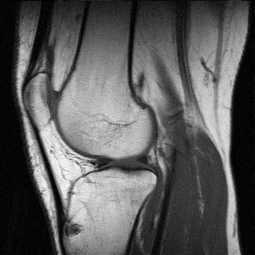 Knee MR
 -
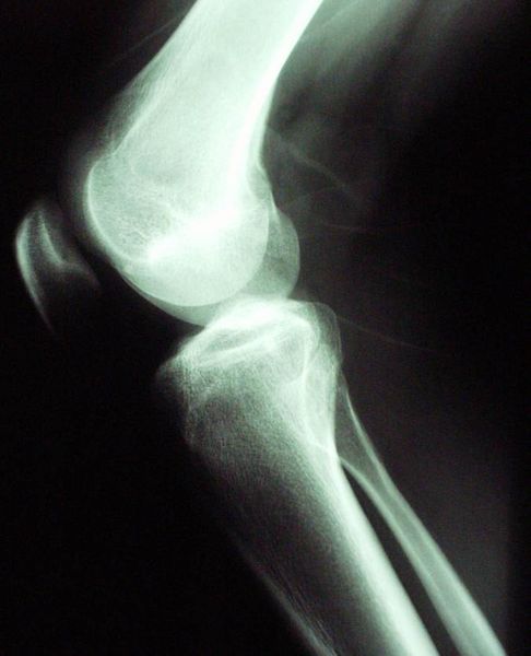 Knee X-ray
 -
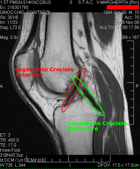 Cruciate ligaments
 -
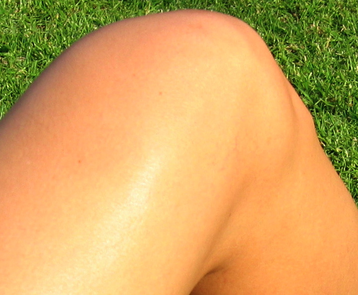 Female knee
 -
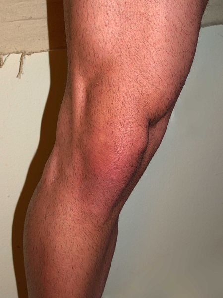 Male knee
 -
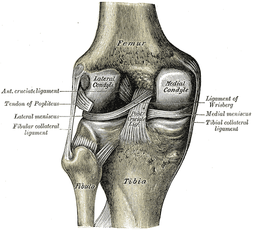 Left knee-joint from behind, showing interior ligaments.
 -
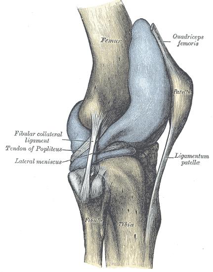 Capsule of right knee-joint (distended). Lateral aspect.


-
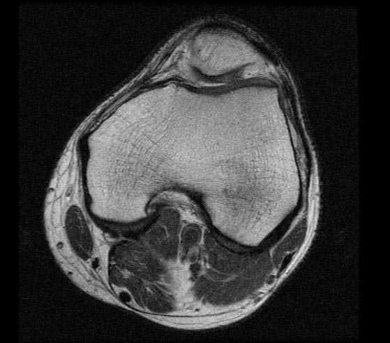 MRI: Normal knee
 -
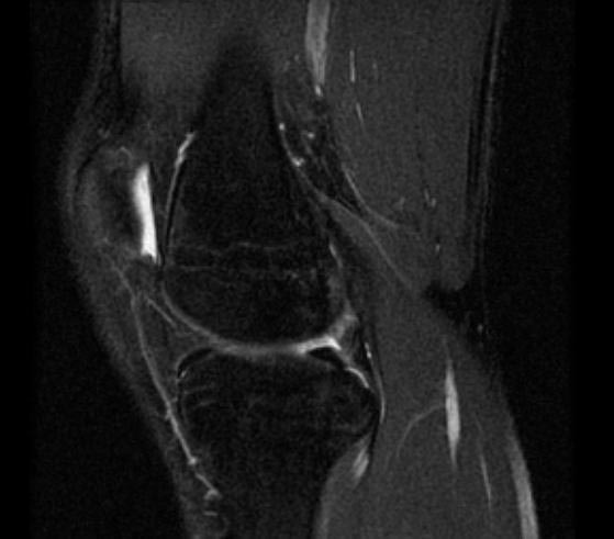 MRI: Normal knee
 -
MRI: Normal knee


-
MRI: Normal knee
-
MRI: Normal knee
External links
- treatment by Alimorad Farshchian, MD
- London Knee Clinic
- The Knee - useful resource from the University of Glasgow
- Patient Information for all knee issues
- The Knee Library
- Knee1
- Template:DartmouthAnatomy
- Total Knee Replacement
Template:Joints of lower limbs Template:Human anatomical features
cs:Koleno de:Kniegelenk eo:Genuo id:Lutut it:Ginocchio he:ברך la:Genu nl:Knie oc:Genolh ug:تىز scn:Dinocchiu simple:Knee sl:Kolenski sklep fi:Polvi sv:Knä