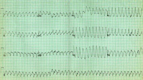
Ventricular tachycardia pathophysiology
|
Ventricular tachycardia Microchapters |
|
Differentiating Ventricular Tachycardia from other Disorders |
|---|
|
Diagnosis |
|
Treatment |
|
Case Studies |
|
Ventricular tachycardia pathophysiology On the Web |
|
to Hospitals Treating Ventricular tachycardia pathophysiology |
|
Risk calculators and risk factors for Ventricular tachycardia pathophysiology |
Editor-In-Chief: C. Michael Gibson, M.S., M.D. [1]; Associate Editor-In-Chief: Cafer Zorkun, M.D., Ph.D. [2]
Overview
The underlying mechanism of VT is due to automaticity arising in either the myocardium or in the distal conduction system. The most common underlying substrate for ventricular tachycardia is ischemic heart disease. The morphology of ventricular tachycardia often depends on its cause.
Monomorphic Ventricular Tachycardia
There are two reasons the morphology of the QRS does not vary in monomorphic ventricular tachycardia:
- A single site that generates automaticity of a single point in either the left or right ventricle
- A reentry circuit within the ventricle
Causes
Common
- Scar from a prior MI (the most common cause of monomorphic ventricular tachycardia)
Rare
Polymorphic Ventricular Tachycardia
Polymorphic ventricular tachycardia, on the other hand, is most commonly caused by abnormalities of ventricular muscle repolarization. The predisposition to this problem usually manifests on the EKG as a prolongation of the QT interval. QT prolongation may be congenital or acquired. Congenital problems include Long QT syndrome and Catecholaminergic polymorphic ventricular tachycardia. Acquired problems are usually related to drug toxicity or electrolyte abnormalities, but can occur as a result of myocardial ischaemia. Class III anti-arrhythmic drugs such as sotalol and amiodarone prolong the QT interval and may in some circumstances be pro-arrhythmic. Other relatively common drugs including some antibiotics and antihistamines may also be a danger, particularly in combination with one another. Problems with blood levels of potassium, magnesium and calcium may also contribute. High dose magnesium is often used as an antidote in cardiac arrest protocols.

Bundle Branch Re-entrant Ventricular Tachycardia
Bundle branch reentry ventricular tachycardia usually occurs either in patients with structural heart disease or in patients with conduction disturbances with a structurally normal heart. Bundle branch reentry is a macro-reentrant tachycardia that incorporates the His-Purkinje system, the bundle branches, and transseptal myocardial conduction in the circuit. Typical Bundle Branch reentry tachycardia uses the right bundle as the anterograde limb and the left bundle as the retrograde limb. Atypical Bundle Branch reentry uses the left bundle (anterior fascicle, posterior fascicle or both) as the antegrade limb and the right bundle as the retrograde limb. The tachycardia appears as a typical Left Bundle Branch Block or Right Bundle Branch Block.