Tricuspid regurgitation chest x ray: Difference between revisions
(→X ray) |
(→X ray) |
||
| Line 4: | Line 4: | ||
==Overview== | ==Overview== | ||
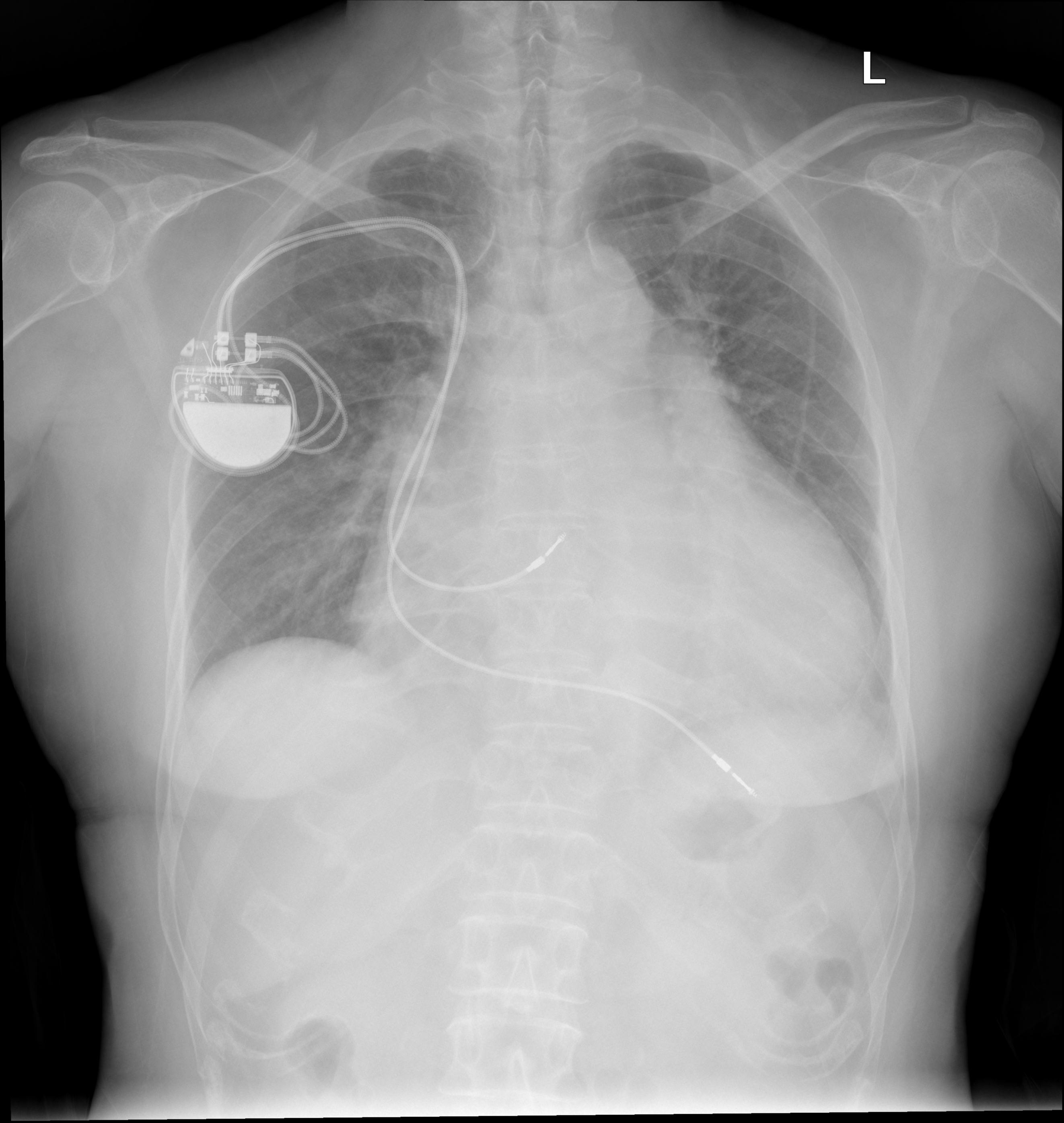
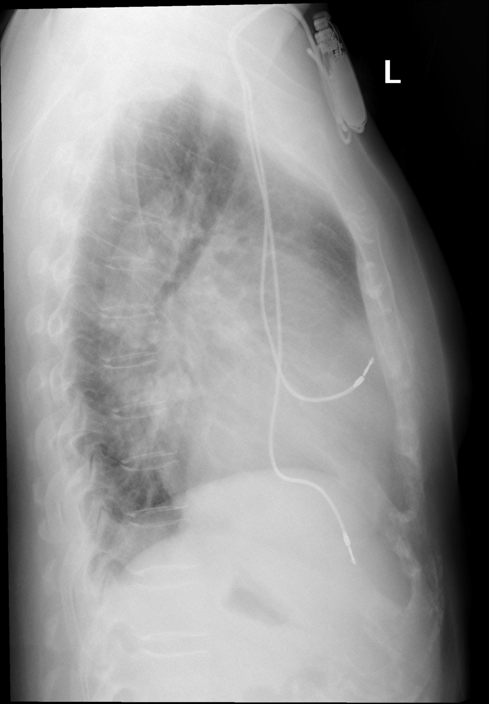
An [[Chest X-ray|chest x-ray]] may be helpful in the diagnosis of [[tricuspid regurgitation]] during the initial evaluation as well as during follow-up among adolescent and young adult patients. Findings on an [[x-ray]] suggestive of [[tricuspid regurgitation]] include [[cardiomegaly]], prominent cardiac silhouette, [[Right atrium|right atrial]] enlargement and [[pleural effusion]]<nowiki/>s. | An [[Chest X-ray|chest x-ray]] may be helpful in the diagnosis of [[tricuspid regurgitation]] during the initial evaluation as well as during follow-up among adolescent and young adult patients. Findings on an [[x-ray]] suggestive of [[tricuspid regurgitation]] include [[cardiomegaly]], prominent cardiac silhouette, [[Right atrium|right atrial]] enlargement and [[pleural effusion]]<nowiki/>s. | ||
== X ray == | == X ray == | ||
An [[Chest X-ray|chest x-ray]] may be helpful in the diagnosis of [[tricuspid regurgitation]]. Findings on an [[Chest X-ray|chest x-ray]] suggestive [[tricuspid regurgitation]] include:<ref name="pmid26503944">{{cite journal| author=Tornos Mas P, Rodríguez-Palomares JF, Antunes MJ| title=Secondary tricuspid valve regurgitation: a forgotten entity. | journal=Heart | year= 2015 | volume= 101 | issue= 22 | pages= 1840-8 | pmid=26503944 | doi=10.1136/heartjnl-2014-307252 | pmc=4680164 | url=https://www.ncbi.nlm.nih.gov/entrez/eutils/elink.fcgi?dbfrom=pubmed&tool=sumsearch.org/cite&retmode=ref&cmd=prlinks&id=26503944 }}</ref> | An [[Chest X-ray|chest x-ray]] may be helpful in the diagnosis of [[tricuspid regurgitation]]. Findings on an [[Chest X-ray|chest x-ray]] suggestive [[tricuspid regurgitation]] include:<ref name="pmid26503944">{{cite journal| author=Tornos Mas P, Rodríguez-Palomares JF, Antunes MJ| title=Secondary tricuspid valve regurgitation: a forgotten entity. | journal=Heart | year= 2015 | volume= 101 | issue= 22 | pages= 1840-8 | pmid=26503944 | doi=10.1136/heartjnl-2014-307252 | pmc=4680164 | url=https://www.ncbi.nlm.nih.gov/entrez/eutils/elink.fcgi?dbfrom=pubmed&tool=sumsearch.org/cite&retmode=ref&cmd=prlinks&id=26503944 }}</ref> | ||
Revision as of 16:40, 20 April 2020
|
Tricuspid Regurgitation Microchapters |
|
Diagnosis |
|---|
|
Treatment |
|
Case Studies |
|
Tricuspid regurgitation chest x ray On the Web |
|
American Roentgen Ray Society Images of Tricuspid regurgitation chest x ray |
|
Risk calculators and risk factors for Tricuspid regurgitation chest x ray |
Editor-In-Chief: C. Michael Gibson, M.S., M.D. [1] Associate Editor(s)-in-Chief: Rim Halaby, M.D. [2] Fatimo Biobaku M.B.B.S [3]
Overview
An chest x-ray may be helpful in the diagnosis of tricuspid regurgitation during the initial evaluation as well as during follow-up among adolescent and young adult patients. Findings on an x-ray suggestive of tricuspid regurgitation include cardiomegaly, prominent cardiac silhouette, right atrial enlargement and pleural effusions.
X ray
An chest x-ray may be helpful in the diagnosis of tricuspid regurgitation. Findings on an chest x-ray suggestive tricuspid regurgitation include:[1]
- Cardiomegaly can be noticed when the tricuspid regurgitation is severe due to right ventricle enlargement
- Cardiac silhouette can be noticed on the right in the x-ray along with the pulmonary artery view
- Right atrial enlargement can be noticed
- A view of azygos vein can be seen when the pressure is elevated in the right atrium
- Pleural effusions can be noticed
- An upward displacement of the diaphragm can be noticed due to ascites
- Prominent right and left pulmonary artery hilar segments can be noticed on the x-ray
2008 ACC/AHA Guidelines for the Management of Patients with Valvular Heart Disease - Evaluation of Tricuspid Valve Disease in Adolescents and Young Adults (DO NOT EDIT)[2]
| Class I |
| "1. Chest X-ray is indicated for the initial evaluation of adolescent and young adult patients with TR, and serially every 1 to 3 years, depending on severity. (Level C)" |


References
- ↑ Tornos Mas P, Rodríguez-Palomares JF, Antunes MJ (2015). "Secondary tricuspid valve regurgitation: a forgotten entity". Heart. 101 (22): 1840–8. doi:10.1136/heartjnl-2014-307252. PMC 4680164. PMID 26503944.
- ↑ Bonow RO, Carabello BA, Chatterjee K; et al. (2008). "2008 Focused update incorporated into the ACC/AHA 2006 guidelines for the management of patients with valvular heart disease: a report of the American College of Cardiology/American Heart Association Task Force on Practice Guidelines (Writing Committee to Revise the 1998 Guidelines for the Management of Patients With Valvular Heart Disease): endorsed by the Society of Cardiovascular Anesthesiologists, Society for Cardiovascular Angiography and Interventions, and Society of Thoracic Surgeons". Circulation. 118 (15): e523–661. doi:10.1161/CIRCULATIONAHA.108.190748. PMID 18820172. Unknown parameter
|month=ignored (help)