Nasopharyngeal carcinoma pathophysiology
|
Nasopharyngeal carcinoma Microchapters |
|
Differentiating Nasopharyngeal carcinoma from other Diseases |
|---|
|
Diagnosis |
|
Treatment |
|
Case Studies |
|
Nasopharyngeal carcinoma pathophysiology On the Web |
|
American Roentgen Ray Society Images of Nasopharyngeal carcinoma pathophysiology |
|
Risk calculators and risk factors for Nasopharyngeal carcinoma pathophysiology |
Editor-In-Chief: C. Michael Gibson, M.S., M.D. [1] Associate Editor(s)-in-Chief: Faizan Sheraz, M.D. [2]
Pathophysiology
Gross
- Nasal cavity involvement - common in early disease.[1]
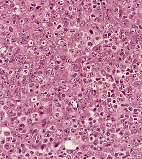
Microscopic
Features:[2]
- Prominent lymphoid component - key feature.
- Features of squamous cell carcinoma:
- Cohesive cells with:
- Abundant dense eosinophilic cytoplasm.
- Central nuclei +/- small/indistinct nucleoli.
Nasopharyngeal carcinoma
- Cohesive cells with:

Histologic subclassification
World Health Classification (2005) for NPC:[2]
| Type | Histology | Description | EBV | Prevalence | Prognosis |
|---|---|---|---|---|---|
| 1 | Keratinizing SCC | graded poorly-well-diff. | -ve | bad | |
| 2a | Nonkeratinizing carcinoma, differentiated | well defined cell borders & tumour nest borders | +ve | good | |
| 2b | Nonkeratinizing carcinoma, undifferentiated | sheets/syncytial, vescicular nuclei, prominent nucleoli, pink cytoplasm | most common | ||
| 3 | Basaloid SCC | mimics basal cell carcinoma | least common |
Immunohistochemistry
- EBER +ve.
- p16 -ve.[3]
References
- ↑ Abdel Khalek Abdel Razek, A.; King, A. (2012). "MRI and CT of nasopharyngeal carcinoma". AJR Am J Roentgenol. 198 (1): 11–8. doi:10.2214/AJR.11.6954. PMID 22194474. Unknown parameter
|month=ignored (help) - ↑ 2.0 2.1 Nasopharyngeal carcinoma http://librepathology.org/wiki/index.php/Nasopharyngeal_carcinoma
- ↑ Gulley ML, Nicholls JM, Schneider BG, Amin MB, Ro JY, Geradts J (1998). "Nasopharyngeal carcinomas frequently lack the p16/MTS1 tumor suppressor protein but consistently express the retinoblastoma gene product". Am. J. Pathol. 152 (4): 865–9. PMC 1858242. PMID 9546345. Unknown parameter
|month=ignored (help)