Mononucleosis pathophysiology: Difference between revisions
(Created page with "{{Mononucleosis}} {{CMG}} ==Overview== ==References== {{reflist|2}} {{WikiDoc Help Menu}} {{WikiDoc Sources}} Category:Disease Category:Infectious disease [[Catego...") |
m (Changes made per Mahshid's request) |
||
| (16 intermediate revisions by 4 users not shown) | |||
| Line 1: | Line 1: | ||
__NOTOC__ | |||
{{Mononucleosis}} | {{Mononucleosis}} | ||
{{CMG}} | {{CMG}} | ||
==Overview== | ==Overview== | ||
Epstein-Barr virus, frequently referred to as [[EBV]], is a member of the [[herpes|herpesvirus family]] that targets oro-pharyngeal epithelium and [[B cells]]. Transmission of the [[EBV]] through the air or blood does not normally occur. The incubation period, or the time from infection to appearance of symptoms, ranges from 4 to 6 weeks. Persons with infectious mononucleosis may be able to spread the infection to others for a period of weeks. However, no special precautions or isolation procedures are recommended, since the virus is also found frequently in the saliva of healthy people. In fact, many healthy people can carry and spread the virus intermittently for life. These people are usually the primary reservoir for person-to-person transmission. For this reason, transmission of the virus is almost impossible to prevent. | |||
==Pathophysiology== | |||
*Following intimate contact with infected saliva, the [[EBV|virus]] infects [[B cells]] located in the oropharyngeal epithelium and subsequently spreads to involve the [[lymph nodes]], [[liver]] and [[spleen]]. | |||
*Mononucleosis was so-named because the count of mononuclear leucocytes ([[white blood cells]] with a one-lobed nucleus) rises significantly. | |||
:*There are two main types of mononuclear leucocytes: [[monocyte]]s and [[lymphocyte]]s. | |||
:*Mononuclear leucocytes normally account for about 35% of all white blood cells and in patients infected with mononucleosis, this count can rise to 50-70%. | |||
:*In addition, the total white blood count may increase to 10,000-20,000 per cubic millimeter. | |||
*Humoral response: As with many viral infections, such as [[chicken pox]], [[antibody|antibodies]] to the viral antigens are developed with resultant recovery from acute illness. | |||
:*In addition, these [[antibody|antibodies]] remain in the system for most individuals, creating a lifelong immunity to further infections. | |||
:*Also, assessment of these specific antibodies forms the basis to diagnose mononucleosis in patients with atypical presentation or in heterophile negative cases. | |||
*Cellular response: | |||
:*Is required to control the proliferation of infected [[B cells]]. | |||
:*This in turn, helps to terminate active EBV infection and also suppress future infections with [[EBV]]. | |||
:*Ineffective cellular response results in excessive proliferation of [[B cells]] with resultant EBV-associated malignancies such as [[Burkitt's lymphoma]] and [[nasopharyngeal carcinoma]]. | |||
===Transmission=== | |||
*Transmission of [[EBV]] requires intimate contact with the saliva of an infected person. | |||
*Typically, the disease is transmitted from asymptomatic individuals through [[blood]] or [[saliva]] (hence the kissing disease), or by sharing a drink, or sharing eating utensils. The disease is far less contagious than is commonly thought. In rare cases, a person may have a high resistance to infection. | |||
*Modes of transmission include: | |||
:* Saliva | |||
::* Epstein-Barr virus (EBV) shed for up to 18 months after primary infection | |||
::* Intermittent viral shedding thereafter in asymptomatic sero-positive patients | |||
::* Increased viral shedding in immunocompromised patients | |||
:* Blood transfusion is rare. | |||
*Individuals in close living arrangements nearly always pass the infection onto each other, although symptoms may not present for months or even years. | |||
===Electron Microscopy=== | |||
[[image:Epstein Barr Virus virions EM 10.1371 journal.pbio.0030430.g001-L.JPG|Two Epstein-Barr [[Virion#Structure|virions]]|250px|left]] | |||
<br clear="left"/> | |||
===Microscopic Pathology=== | |||
Images shown below are courtesy of Professor Peter Anderson DVM PhD and published with permission. [http://www.peir.net © PEIR, University of Alabama at Birmingham, Department of Pathology] | |||
<div align="left"> | |||
<gallery heights="175" widths="175"> | |||
Image:IM 1.jpg|BONE MARROW: INFECTION-ASSOCIATED HEMOPHAGOCYTIC SYNDROME A bone marrow aspirate smear from a child with infection-associated hemophagocytic syndrome secondary to an Epstein-Barr virus infection. On this field there are several large histiocytes. Phagocytosis of nucleated red blood cells, neutrophils, and platelets is evident. The histiocytes have the appearance of reactive cells and should be readily distinguishable from neoplastic histiocytes. (Wright-Giemsa stain) | |||
Image:IM 2.jpg|LOWER RESPIRATORY TRACT: (Supplement) AIL/LYG CD20 staining (for B cells) in the viable tissue shows positive staining of scattered large lymphoid cells (C) which were also the cells that were positive for Epstein-Barr virus by in situ hybridization | |||
Image:IM 3.jpg|LOWER RESPIRATORY TRACT: (Supplement) AIL/LYG CD20 staining (for B cells) in the viable tissue shows positive staining of scattered large lymphoid cells (C) which were also the cells that were positive for Epstein-Barr virus by in situ hybridization | |||
</gallery> | |||
</div> | |||
<div align="left"> | |||
<gallery heights="175" widths="175"> | |||
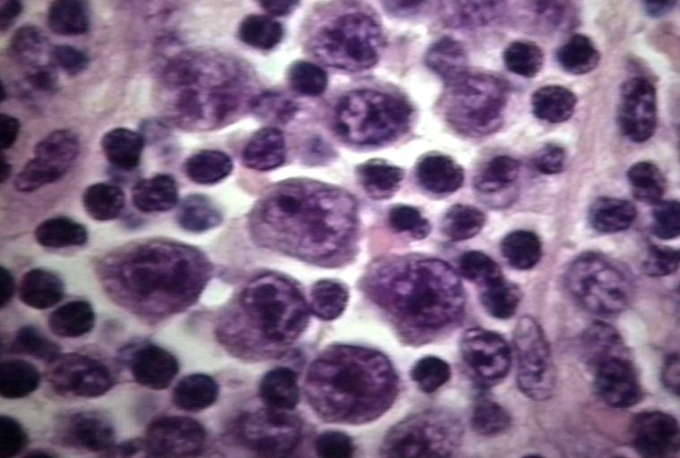
Image:IM 4.jpg|LYMPH NODES-SPLEEN: EPSTEIN-BARR VIRUS-ASSOCIATED INFECTIOUS MONONUCLEOSIS A heterogeneous population is present, including cells mimicking Hodgkin cells. However, the spectrum of cell types and the extensive apoptosis present would not be found in Hodgkin's disease. | |||
Image:IM 5.jpg|LYMPH NODES-SPLEEN: METASTATIC NASOPHARYNGEAL CARCINOMA IN LYMPH NODE PRESENTING AS METASTATIC CARCINOMA OF UNKNOWN PRIMARY Nuclear labeling of the carcinoma cells for Epstein-Barr virus-encoded RNA strongly suggests that the primary is nasopharyngeal. | |||
Image:IM 6.jpg|BONE MARROW: REACTIVE (ATYPICAL) LYMPHOCYTES This blood smear is from a 19-year-old male college student with infectious mononucleosis. The two reactive (atypical) lymphocytes are large, with abundant cytoplasm and coarse nuclear chromatin, and lack a nucleolus. Cytoplasmic basophilia is radial in distribution and accentuated at the cell margin. In contrast, lymphoblasts are generally smaller, have less cytoplasm with uniform basophilia and more dispersed nuclear chromatin, and may contain a nucleolus. (Wright-Giemsa stain) | |||
</gallery> | |||
</div> | |||
<div align="left"> | |||
<gallery heights="175" widths="175"> | |||
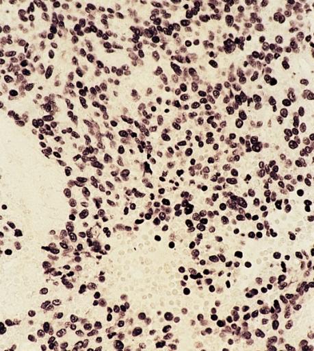
Image:IM 7.jpg|LYMPH NODE: INFECTIOUS MONONUCLEOSIS, LYMPH NODE | |||
Image:IM 8.jpg|BLOOD: INFECTIOUS MONONUCLEOSIS; PERIPHERAL BLOOD | |||
Image:IM 9.jpg|EYE AND OCULAR ADNEXA: BURKITT LYMPHOMA Bilateral involvement. | |||
Image:IM 10.jpg|EYE AND OCULAR ADNEXA: BURKITT LYMPHOMA Lymphoblastic tumor with numerous mitotic figures. | |||
</gallery> | |||
</div> | |||
==References== | ==References== | ||
{{ | {{Reflist|2}} | ||
{{WikiDoc Help Menu}} | {{WikiDoc Help Menu}} | ||
| Line 11: | Line 79: | ||
[[Category:Disease]] | [[Category:Disease]] | ||
[[Category:Pediatrics]] | [[Category:Pediatrics]] | ||
[[Category:Medicine]] | |||
[[Category:Otolaryngology]] | |||
[[Category:Lymphocytes]] | |||
[[Category:Viral diseases]] | |||
Latest revision as of 18:06, 18 September 2017
|
Mononucleosis Microchapters |
|
Diagnosis |
|---|
|
Treatment |
|
Case Studies |
|
Mononucleosis pathophysiology On the Web |
|
American Roentgen Ray Society Images of Mononucleosis pathophysiology |
|
Risk calculators and risk factors for Mononucleosis pathophysiology |
Editor-In-Chief: C. Michael Gibson, M.S., M.D. [1]
Overview
Epstein-Barr virus, frequently referred to as EBV, is a member of the herpesvirus family that targets oro-pharyngeal epithelium and B cells. Transmission of the EBV through the air or blood does not normally occur. The incubation period, or the time from infection to appearance of symptoms, ranges from 4 to 6 weeks. Persons with infectious mononucleosis may be able to spread the infection to others for a period of weeks. However, no special precautions or isolation procedures are recommended, since the virus is also found frequently in the saliva of healthy people. In fact, many healthy people can carry and spread the virus intermittently for life. These people are usually the primary reservoir for person-to-person transmission. For this reason, transmission of the virus is almost impossible to prevent.
Pathophysiology
- Following intimate contact with infected saliva, the virus infects B cells located in the oropharyngeal epithelium and subsequently spreads to involve the lymph nodes, liver and spleen.
- Mononucleosis was so-named because the count of mononuclear leucocytes (white blood cells with a one-lobed nucleus) rises significantly.
- There are two main types of mononuclear leucocytes: monocytes and lymphocytes.
- Mononuclear leucocytes normally account for about 35% of all white blood cells and in patients infected with mononucleosis, this count can rise to 50-70%.
- In addition, the total white blood count may increase to 10,000-20,000 per cubic millimeter.
- Humoral response: As with many viral infections, such as chicken pox, antibodies to the viral antigens are developed with resultant recovery from acute illness.
- In addition, these antibodies remain in the system for most individuals, creating a lifelong immunity to further infections.
- Also, assessment of these specific antibodies forms the basis to diagnose mononucleosis in patients with atypical presentation or in heterophile negative cases.
- Cellular response:
- Is required to control the proliferation of infected B cells.
- This in turn, helps to terminate active EBV infection and also suppress future infections with EBV.
- Ineffective cellular response results in excessive proliferation of B cells with resultant EBV-associated malignancies such as Burkitt's lymphoma and nasopharyngeal carcinoma.
Transmission
- Transmission of EBV requires intimate contact with the saliva of an infected person.
- Typically, the disease is transmitted from asymptomatic individuals through blood or saliva (hence the kissing disease), or by sharing a drink, or sharing eating utensils. The disease is far less contagious than is commonly thought. In rare cases, a person may have a high resistance to infection.
- Modes of transmission include:
- Saliva
- Epstein-Barr virus (EBV) shed for up to 18 months after primary infection
- Intermittent viral shedding thereafter in asymptomatic sero-positive patients
- Increased viral shedding in immunocompromised patients
- Blood transfusion is rare.
- Individuals in close living arrangements nearly always pass the infection onto each other, although symptoms may not present for months or even years.
Electron Microscopy

Microscopic Pathology
Images shown below are courtesy of Professor Peter Anderson DVM PhD and published with permission. © PEIR, University of Alabama at Birmingham, Department of Pathology
-
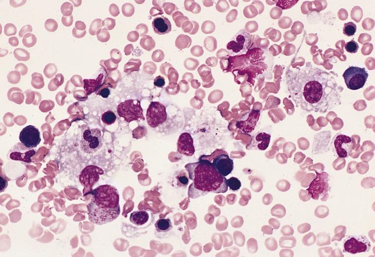
BONE MARROW: INFECTION-ASSOCIATED HEMOPHAGOCYTIC SYNDROME A bone marrow aspirate smear from a child with infection-associated hemophagocytic syndrome secondary to an Epstein-Barr virus infection. On this field there are several large histiocytes. Phagocytosis of nucleated red blood cells, neutrophils, and platelets is evident. The histiocytes have the appearance of reactive cells and should be readily distinguishable from neoplastic histiocytes. (Wright-Giemsa stain)
-
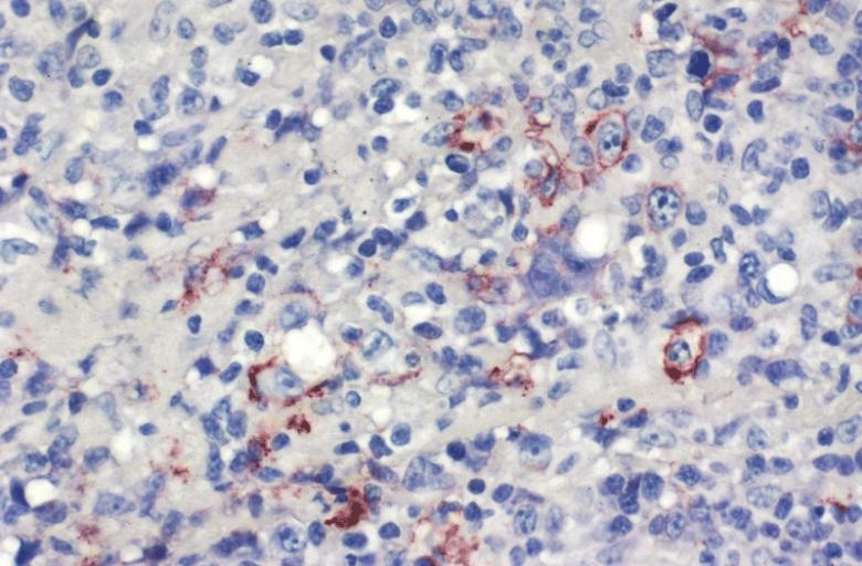
LOWER RESPIRATORY TRACT: (Supplement) AIL/LYG CD20 staining (for B cells) in the viable tissue shows positive staining of scattered large lymphoid cells (C) which were also the cells that were positive for Epstein-Barr virus by in situ hybridization
-
LOWER RESPIRATORY TRACT: (Supplement) AIL/LYG CD20 staining (for B cells) in the viable tissue shows positive staining of scattered large lymphoid cells (C) which were also the cells that were positive for Epstein-Barr virus by in situ hybridization


-
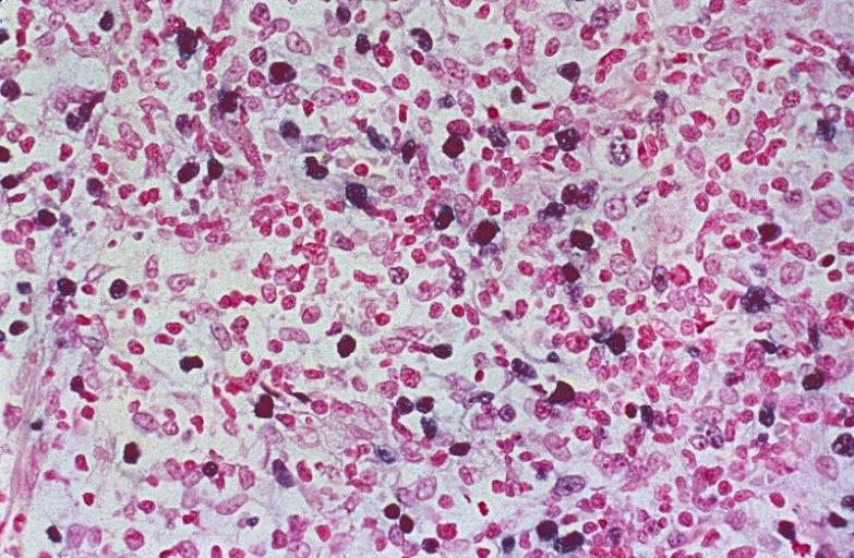
LYMPH NODES-SPLEEN: EPSTEIN-BARR VIRUS-ASSOCIATED INFECTIOUS MONONUCLEOSIS A heterogeneous population is present, including cells mimicking Hodgkin cells. However, the spectrum of cell types and the extensive apoptosis present would not be found in Hodgkin's disease.
-
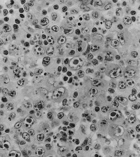
LYMPH NODES-SPLEEN: METASTATIC NASOPHARYNGEAL CARCINOMA IN LYMPH NODE PRESENTING AS METASTATIC CARCINOMA OF UNKNOWN PRIMARY Nuclear labeling of the carcinoma cells for Epstein-Barr virus-encoded RNA strongly suggests that the primary is nasopharyngeal.
-
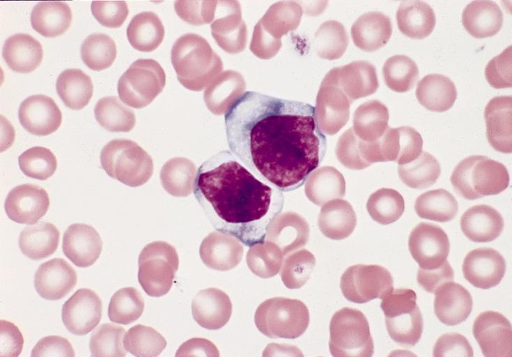
BONE MARROW: REACTIVE (ATYPICAL) LYMPHOCYTES This blood smear is from a 19-year-old male college student with infectious mononucleosis. The two reactive (atypical) lymphocytes are large, with abundant cytoplasm and coarse nuclear chromatin, and lack a nucleolus. Cytoplasmic basophilia is radial in distribution and accentuated at the cell margin. In contrast, lymphoblasts are generally smaller, have less cytoplasm with uniform basophilia and more dispersed nuclear chromatin, and may contain a nucleolus. (Wright-Giemsa stain)


-
LYMPH NODE: INFECTIOUS MONONUCLEOSIS, LYMPH NODE
-
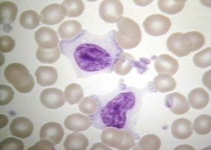
BLOOD: INFECTIOUS MONONUCLEOSIS; PERIPHERAL BLOOD
-
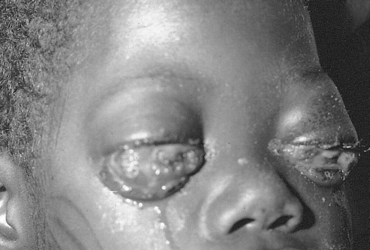
EYE AND OCULAR ADNEXA: BURKITT LYMPHOMA Bilateral involvement.
-
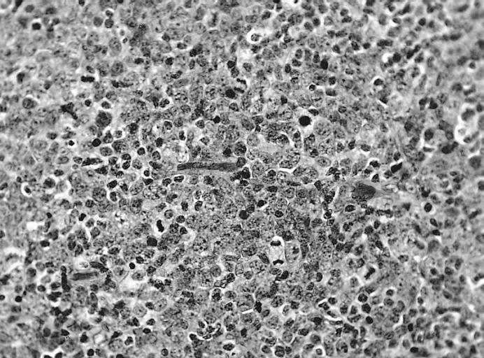
EYE AND OCULAR ADNEXA: BURKITT LYMPHOMA Lymphoblastic tumor with numerous mitotic figures.


