Merkel cell cancer physical examination: Difference between revisions
Jump to navigation
Jump to search
No edit summary |
Sargun Walia (talk | contribs) No edit summary |
||
| Line 3: | Line 3: | ||
==Overview== | ==Overview== | ||
Physical exam findings of | Physical exam findings of merkel cell cancer include red/violaceous skin [[mass]] that appear in [[Sun exposure|sun exposed]] areas. | ||
==Physical examination== | ==Physical examination== | ||
[[Physical examination]] of patients with | [[Physical examination]] of patients with merkel cell cancer is usually remarkable for flesh-colored or bluish-red, intracutaneous [[Nodule (medicine)|nodules]]. | ||
=== Appearance of the Patient === | === Appearance of the Patient === | ||
* [[Patient|Patients]] with | * [[Patient|Patients]] with merkel cell cancer usually appear normal. | ||
=== Vital Signs === | === Vital Signs === | ||
| Line 14: | Line 14: | ||
=== Skin === | === Skin === | ||
* Skin examination of patients with | {|align="right" | ||
**A mass that appear in sun exposed areas of the skin | | | ||
**Mass that usually 1-2 cm | [[File:Merkel Cell Cancer.gif|alt=Merkel Cell Cancer|center|thumb|'''Merkel Cell Cancer''' [https://commons.wikimedia.org/wiki/File:Merkel_cell_carcinoma_arm.jpg#/media/File:Merkel_cell_carcinoma_arm.jpg Source:Wikimedia Commons]]] | ||
**Non-tender, firm, shiny, flesh-colored or bluish-red or pink colour intracutaneous [[Nodule (medicine)|nodule]] | |} | ||
**Presence of a polymorphous [[vascular]] pattern is very characteristic to | * Skin examination of patients with merkel cell cancer is significant for:<ref name="pmid18280333">{{cite journal |vauthors=Heath M, Jaimes N, Lemos B, Mostaghimi A, Wang LC, Peñas PF, Nghiem P |title=Clinical characteristics of Merkel cell carcinoma at diagnosis in 195 patients: the AEIOU features |journal=J. Am. Acad. Dermatol. |volume=58 |issue=3 |pages=375–81 |date=March 2008 |pmid=18280333 |pmc=2335370 |doi=10.1016/j.jaad.2007.11.020 |url=}}</ref><ref name="pmid29085720">{{cite journal |vauthors=Geller S, Pulitzer M, Brady MS, Myskowski PL |title=Dermoscopic assessment of vascular structures in solitary small pink lesions-differentiating between good and evil |journal=Dermatol Pract Concept |volume=7 |issue=3 |pages=47–50 |date=July 2017 |pmid=29085720 |doi=10.5826/dpc.0703a10 |url=}}</ref><ref name="pmid23574613">{{cite journal |vauthors=Jalilian C, Chamberlain AJ, Haskett M, Rosendahl C, Goh M, Beck H, Keir J, Varghese P, Mar A, Hosking S, Hussain I, Rich M, McLean C, Kelly JW |title=Clinical and dermoscopic characteristics of Merkel cell carcinoma |journal=Br. J. Dermatol. |volume=169 |issue=2 |pages=294–7 |date=August 2013 |pmid=23574613 |doi=10.1111/bjd.12376 |url=}}</ref> | ||
**A mass that appear in sun exposed areas of the skin. | |||
**Mass that usually 1-2 cm. | |||
**Non-tender, firm, shiny, flesh-colored or bluish-red or pink colour intracutaneous [[Nodule (medicine)|nodule]]. | |||
**Presence of a polymorphous [[vascular]] pattern is very characteristic to merkel cell cancer. | |||
**Dome-shaped or raised nodule | **Dome-shaped or raised nodule | ||
**Red/violaceous skin mass | **Red/violaceous skin mass | ||
| Line 27: | Line 31: | ||
**#[[Lower limbs]](LL) and [[Hip (anatomy)|hip]] | **#[[Lower limbs]](LL) and [[Hip (anatomy)|hip]] | ||
**#[[Trunk]] | **#[[Trunk]] | ||
=== HEENT === | === HEENT === | ||
Revision as of 02:47, 31 January 2019
|
Merkel cell cancer Microchapters |
|
Diagnosis |
|---|
|
Treatment |
|
Case Studies |
|
Merkel cell cancer physical examination On the Web |
|
American Roentgen Ray Society Images of Merkel cell cancer physical examination |
|
Risk calculators and risk factors for Merkel cell cancer physical examination |
Editor-In-Chief: C. Michael Gibson, M.S., M.D. [1]; Associate Editor(s)-in-Chief: Vamsikrishna Gunnam M.B.B.S [2]
Overview
Physical exam findings of merkel cell cancer include red/violaceous skin mass that appear in sun exposed areas.
Physical examination
Physical examination of patients with merkel cell cancer is usually remarkable for flesh-colored or bluish-red, intracutaneous nodules.
Appearance of the Patient
- Patients with merkel cell cancer usually appear normal.
Vital Signs
- Mostly all vitals are within normal limits.
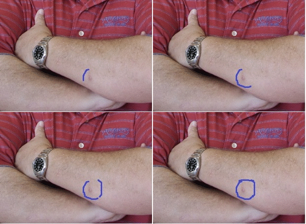
Skin
 |
- Skin examination of patients with merkel cell cancer is significant for:[1][2][3]
- A mass that appear in sun exposed areas of the skin.
- Mass that usually 1-2 cm.
- Non-tender, firm, shiny, flesh-colored or bluish-red or pink colour intracutaneous nodule.
- Presence of a polymorphous vascular pattern is very characteristic to merkel cell cancer.
- Dome-shaped or raised nodule
- Red/violaceous skin mass
- +/- Crusting and ulceration
- According to National Cancer Database the following are the most common locations of merkel cell cancer in decreasing order:
- Head and neck
- Upper limbs(UL) and shoulder
- Lower limbs(LL) and hip
- Trunk
HEENT
- HEENT examination of patients with merkel cell cancer is usually shows the following:[4][5]
- Mass measuring 2 X 3 cm most commonly in the right nasal ala.
- Due to the mass effect patients with merkel cell cancer might experience nasal obstruction.
Lungs
- Pulmonary examination of patients with merkel cell cancer is usually normal.
Heart
- Cardiovascular examination of patients with merkel cell cancer is usually normal.
Abdomen
- Abdominal examination of patients with merkel cell cancer is usually normal.
Back
- Back examination of patients with merkel cell cancer is usually normal.
Genitourinary
- Genitourinary examination of patients with merkel cell cancer is usually normal.
Neuromuscular
- Neuromuscular examination of patients with merkel cell cancer shows the following:[6][7]
- Clonus may be present
- Sudden onset of severe headache
- Gait disturbance
- Confusion
- Brain metastasis from merkel cell carcinoma
Extremities
- Extremities examination of patients with merkel cell carcinoma is usually normal.
References
- ↑ Heath M, Jaimes N, Lemos B, Mostaghimi A, Wang LC, Peñas PF, Nghiem P (March 2008). "Clinical characteristics of Merkel cell carcinoma at diagnosis in 195 patients: the AEIOU features". J. Am. Acad. Dermatol. 58 (3): 375–81. doi:10.1016/j.jaad.2007.11.020. PMC 2335370. PMID 18280333.
- ↑ Geller S, Pulitzer M, Brady MS, Myskowski PL (July 2017). "Dermoscopic assessment of vascular structures in solitary small pink lesions-differentiating between good and evil". Dermatol Pract Concept. 7 (3): 47–50. doi:10.5826/dpc.0703a10. PMID 29085720.
- ↑ Jalilian C, Chamberlain AJ, Haskett M, Rosendahl C, Goh M, Beck H, Keir J, Varghese P, Mar A, Hosking S, Hussain I, Rich M, McLean C, Kelly JW (August 2013). "Clinical and dermoscopic characteristics of Merkel cell carcinoma". Br. J. Dermatol. 169 (2): 294–7. doi:10.1111/bjd.12376. PMID 23574613.
- ↑ Becker JC, Stang A, DeCaprio JA, Cerroni L, Lebbé C, Veness M, Nghiem P (October 2017). "Merkel cell carcinoma". Nat Rev Dis Primers. 3: 17077. doi:10.1038/nrdp.2017.77. PMC 6054450. PMID 29072302.
- ↑ Schmerling RA, Casas JG, Cinat G, Ospina F, Kassuga L, Tlahuel J, Mazzuoccolo LD (July 2018). "Burden of Disease, Early Diagnosis, and Treatment of Merkel Cell Carcinoma in Latin America". J Glob Oncol (4): 1–11. doi:10.1200/JGO.18.00041. PMC 6223512. PMID 30085832. Vancouver style error: initials (help)
- ↑ Honeybul, S. (2016). "Cerebral metastases from Merkel cell carcinoma: long-term survival". Journal of Surgical Case Reports. 2016 (10): rjw165. doi:10.1093/jscr/rjw165. ISSN 2042-8812.
- ↑ Eggers SD, Salomao DR, Dinapoli RP, Vernino S (March 2001). "Paraneoplastic and metastatic neurologic complications of Merkel cell carcinoma". Mayo Clin. Proc. 76 (3): 327–30. doi:10.4065/76.3.327. PMID 11243282.