Atrial fibrillation differential diagnosis
| Resident Survival Guide |
 |
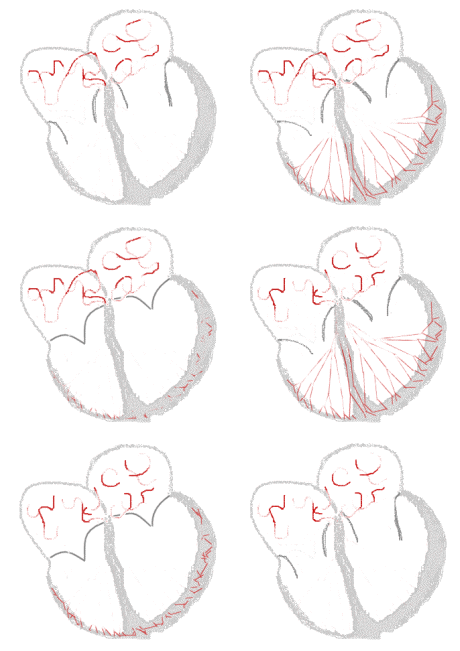
Sinus rhythm  |
Atrial fibrillation  |
|
Atrial Fibrillation Microchapters | |
|
Special Groups | |
|---|---|
|
Diagnosis | |
|
Treatment | |
|
Cardioversion | |
|
Anticoagulation | |
|
Surgery | |
|
Case Studies | |
|
Atrial fibrillation differential diagnosis On the Web | |
|
Directions to Hospitals Treating Atrial fibrillation differential diagnosis | |
|
Risk calculators and risk factors for Atrial fibrillation differential diagnosis | |
Editor-In-Chief: C. Michael Gibson, M.S., M.D. [1]
Overview
Atrial fibrillation must be distinguished from other common atrial arrhythmias, which include atrial flutter, atrial tachycardia, paroxysmal supraventricular tachycardia, Wolff-Parkinson-White syndrome, and atrioventricular nodal reentry tachycardia.
Differentiating Atrial Fibrillation from other Diseases
Atrial fibrillation has to be differnetiated from other diseases like:
- Atrial flutter
- Atrial tachycardia
- Atrioventricular nodal reentry tachycardia (AVNRT)
- Multifocal atrial tachycardia
- Paroxysmal supraventricular tachycardia
- Wolff-Parkinson-White syndrome
The differentiating features are largely based on both EKG findings and cardiovascular examination.
- Atrial fibrillation is irregularly irregular, while the other rhythms such as atrial flutter, sinus tachycardia, AV nodal reentry tachycardia and paroxysmal supraventricular tachycardia are all much more regular.
- An atrioventricular nodal reentry tachycardia will often break with either carotid sinus massage or AV nodal blocking agents.
- If the patient has Wolff-Parkinson-White syndrome there may be much more rapid conduction. The presence of the delta wave on EKG is characteristic.
| Arrhythmia | Rhythm | Rate | P wave | PR Interval | QRS Complex | Response to Maneuvers | Epidemiology | Co-existing Conditions |
|---|---|---|---|---|---|---|---|---|
| Atrial Fibrillation[1][2] | Irregularly irregular | On a 10-second 12-lead EKG strip, multiply number of QRS complexes by 6 | Absent, fibrillatory waves | Absent | Less than 0.12 seconds, consistent, and normal in morphology in the absence of aberrant conduction | Does not break with adenosine or vagal maneuvers |
|
|
| Atrial Flutter | Regular or Irregular | 75 (4:1 block), 100 (3:1 block) and 150 (2:1 block) bpm, but 150 is more common | Sawtooth pattern of P waves at 250 to 350 beats per minute | Varies depending upon the magnitude of the block, but is short | Less than 0.12 seconds, consistent, and normal in morphology | Conduction may vary in response to drugs and maneuvers dropping the rate from 150 to 100 or to 75 bpm |
|
|
| Atrioventricular nodal reentry tachycardia (AVNRT)[3][4] | Inverted, superimposed on or buried within the QRS complex (pseudo R prime in V1/pseudo S wave in inferior leads) | Absent (P wave can appear after the QRS complex and before the T wave, and in atypical AVNRT, the P wave can appear just before the QRS complex) | Less than 0.12 seconds, consistent, and normal in morphology in the absence of aberrant conduction, QRS alternans may be present | May break with adenosine or vagal maneuvers | 60%-70% of all SVTs | |||
| Multifocal Atrial Tachycardia[5][6] | Irregular | Atrial rate is > 100 beats per minute | Varying morphology from at least three different foci, absence of one dominant atrial pacemaker, can be mistaken for atrial fibrillation if the P waves are of low amplitude | Variable PR intervals, RR intervals, and PP intervals | Less than 0.12 seconds, consistent, and normal in morphology | Does not terminate with adenosine or vagal maneuvers | High incidence in the elderly and in those with COPD | |
| Paroxysmal Supraventricular Tachycardia | ||||||||
| Wolff-Parkinson-White Syndrome[7][8] | Regular | Atrial rate is nearly 300 bpm and ventricular rate is at 150 bpm | With orthodromic conduction due to a bypass tract, the P wave generally follows the QRS complex, whereas in AVNRT, the P wave is generally buried in the QRS complex. | Less than 0.12 seconds | A delta wave and evidence of ventricular pre-excitation if there is conduction to the ventricle via ante-grade conduction down an accessory pathway. It should be noted, however, that in some patients with WPW, a delta wave and pre-excitation may not be present because bypass tracts do not conduct ante-grade. | May break in response to procainamide, adenosine, vagal maneuvers | Worldwide prevalence of WPW syndrome is 100 - 300 per 100,000 | |
| Ventricular Fibrillation | Irregular | 150 to 500 bpm | Absent | Absent | Absent (R on T phenomenon in the setting of ischemia) | Myocardial ischemia / infarction,
cardiomyopathy, channelopathies e.g. Long QT (acquired / congenital), aortic stenosis, aortic dissection, myocarditis, cardiac tamponade, blunt trauma (Commotio Cordis), sepsis, hypothermia, pneumothroax, seizures, stroke | ||
| Ventricular Tacycardia |
References
- ↑ Lankveld TA, Zeemering S, Crijns HJ, Schotten U (July 2014). "The ECG as a tool to determine atrial fibrillation complexity". Heart. 100 (14): 1077–84. doi:10.1136/heartjnl-2013-305149. PMID 24837984.
- ↑ Harris K, Edwards D, Mant J (2012). "How can we best detect atrial fibrillation?". J R Coll Physicians Edinb. 42 Suppl 18: 5–22. doi:10.4997/JRCPE.2012.S02. PMID 22518390.
- ↑ Katritsis DG, Josephson ME (August 2016). "Classification, Electrophysiological Features and Therapy of Atrioventricular Nodal Reentrant Tachycardia". Arrhythm Electrophysiol Rev. 5 (2): 130–5. doi:10.15420/AER.2016.18.2. PMC 5013176. PMID 27617092.
- ↑ Letsas KP, Weber R, Siklody CH, Mihas CC, Stockinger J, Blum T, Kalusche D, Arentz T (April 2010). "Electrocardiographic differentiation of common type atrioventricular nodal reentrant tachycardia from atrioventricular reciprocating tachycardia via a concealed accessory pathway". Acta Cardiol. 65 (2): 171–6. doi:10.2143/AC.65.2.2047050. PMID 20458824.
- ↑ Scher DL, Arsura EL (September 1989). "Multifocal atrial tachycardia: mechanisms, clinical correlates, and treatment". Am. Heart J. 118 (3): 574–80. doi:10.1016/0002-8703(89)90275-5. PMID 2570520.
- ↑ Goodacre S, Irons R (March 2002). "ABC of clinical electrocardiography: Atrial arrhythmias". BMJ. 324 (7337): 594–7. doi:10.1136/bmj.324.7337.594. PMC 1122515. PMID 11884328.
- ↑ Rao AL, Salerno JC, Asif IM, Drezner JA (July 2014). "Evaluation and management of wolff-Parkinson-white in athletes". Sports Health. 6 (4): 326–32. doi:10.1177/1941738113509059. PMC 4065555. PMID 24982705.
- ↑ Rosner MH, Brady WJ, Kefer MP, Martin ML (November 1999). "Electrocardiography in the patient with the Wolff-Parkinson-White syndrome: diagnostic and initial therapeutic issues". Am J Emerg Med. 17 (7): 705–14. doi:10.1016/s0735-6757(99)90167-5. PMID 10597097.