Atrial fibrillation differential diagnosis: Difference between revisions
No edit summary |
No edit summary |
||
| Line 48: | Line 48: | ||
|- | |- | ||
|Atrial Fibrillation | |Atrial Fibrillation | ||
|Irregularly irregular | |||
| | | | ||
|Absent, fibrillatory waves | |||
|Absent | |||
|Less than 0.12 seconds, consistent, and normal in morphology in the absence of aberrant conduction | |||
|Does not break with adenosine or vagal maneuvers | |||
| | | | ||
| | |Old age, following bypass surgery, in mitral valve disease, hyperthyroidism | ||
|- | |- | ||
|Atrial Flutter | |Atrial Flutter | ||
| | | | ||
|75 (4:1 block), 100 (3:1 block) and 150 (2:1 block) bpm, but 150 is more common | |||
|Sawtooth pattern of P waves at 250 to 350 beats per minute | |||
|Varies depending upon the magnitude of the block, but is short | |||
|Less than 0.12 seconds, consistent, and normal in morphology | |||
|Conduction may vary in response to drugs and maneuvers dropping the rate from 150 to 100 or to 75 bpm | |||
| | | | ||
| | |More common in the elderly, after alcohol | ||
|- | |- | ||
|Atrioventricular nodal reentry tachycardia (AVNRT) | |Atrioventricular nodal reentry tachycardia (AVNRT) | ||
Revision as of 21:00, 14 November 2019
| Resident Survival Guide |
 |
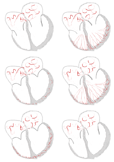
Sinus rhythm  |
Atrial fibrillation  |
|
Atrial Fibrillation Microchapters | |
|
Special Groups | |
|---|---|
|
Diagnosis | |
|
Treatment | |
|
Cardioversion | |
|
Anticoagulation | |
|
Surgery | |
|
Case Studies | |
|
Atrial fibrillation differential diagnosis On the Web | |
|
Directions to Hospitals Treating Atrial fibrillation differential diagnosis | |
|
Risk calculators and risk factors for Atrial fibrillation differential diagnosis | |
Editor-In-Chief: C. Michael Gibson, M.S., M.D. [1]
Overview
Atrial fibrillation must be distinguished from other common atrial arrhythmias, which include atrial flutter, atrial tachycardia, paroxysmal supraventricular tachycardia, Wolff-Parkinson-White syndrome, and atrioventricular nodal reentry tachycardia.
Differentiating Atrial Fibrillation from other Diseases
Atrial fibrillation has to be differnetiated from other diseases like:
- Atrial flutter
- Atrial tachycardia
- Atrioventricular nodal reentry tachycardia (AVNRT)
- Multifocal atrial tachycardia
- Paroxysmal supraventricular tachycardia
- Wolff-Parkinson-White syndrome
The differentiating features are largely based on both EKG findings and cardiovascular examination.
- Atrial fibrillation is irregularly irregular, while the other rhythms such as atrial flutter, sinus tachycardia, AV nodal reentry tachycardia and paroxysmal supraventricular tachycardia are all much more regular.
- An atrioventricular nodal reentry tachycardia will often break with either carotid sinus massage or AV nodal blocking agents.
- If the patient has Wolff-Parkinson-White syndrome there may be much more rapid conduction. The presence of the delta wave on EKG is characteristic.
| Arrhythmia | Rhythm | Rate | P wave | PR Interval | QRS Complex | Response to Maneuvers | Epidemiology | Co-existing Conditions |
|---|---|---|---|---|---|---|---|---|
| Atrial Fibrillation | Irregularly irregular | Absent, fibrillatory waves | Absent | Less than 0.12 seconds, consistent, and normal in morphology in the absence of aberrant conduction | Does not break with adenosine or vagal maneuvers | Old age, following bypass surgery, in mitral valve disease, hyperthyroidism | ||
| Atrial Flutter | 75 (4:1 block), 100 (3:1 block) and 150 (2:1 block) bpm, but 150 is more common | Sawtooth pattern of P waves at 250 to 350 beats per minute | Varies depending upon the magnitude of the block, but is short | Less than 0.12 seconds, consistent, and normal in morphology | Conduction may vary in response to drugs and maneuvers dropping the rate from 150 to 100 or to 75 bpm | More common in the elderly, after alcohol | ||
| Atrioventricular nodal reentry tachycardia (AVNRT) | ||||||||
| Multifocal Atrial Tachycardia | ||||||||
| Paroxysmal Supraventricular Tachycardia | ||||||||
| Wolff-Parkinson-White Syndrome | ||||||||
| Ventricular Fibrillation | ||||||||
| Ventricular Tacycardia |