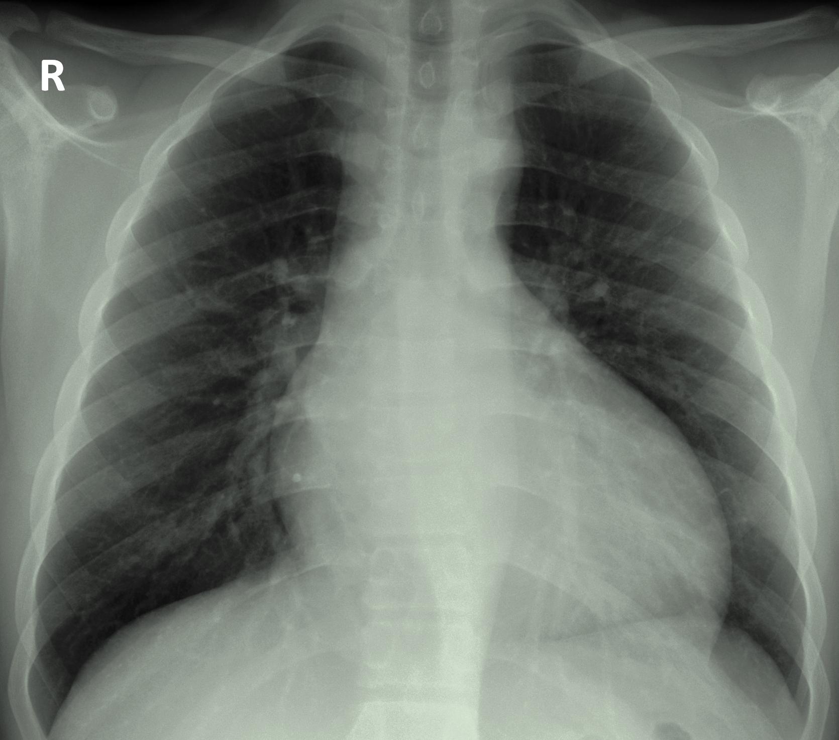
Aortic regurgitation chest x-ray
|
Aortic Regurgitation Microchapters |
|
Diagnosis |
|---|
|
Treatment |
|
Acute Aortic regurgitation |
|
Chronic Aortic regurgitation |
|
Special Scenarios |
|
Case Studies |
|
Aortic regurgitation chest x-ray On the Web |
|
American Roentgen Ray Society Images of Aortic regurgitation chest x-ray |
|
Risk calculators and risk factors for Aortic regurgitation chest x-ray |
Editor-In-Chief: C. Michael Gibson, M.S., M.D. [1]; Associate Editor(s)-in-Chief: Cafer Zorkun, M.D., Ph.D. [2]; Varun Kumar, M.B.B.S.; Lakshmi Gopalakrishnan, M.B.B.S.
Overview
Chest x ray findings associated with aortic regurgitation may include left ventricular enlargement, cardiomegaly, prominent aortic root with valvular calcification, prosthetic valve dis-lodgement, or aortic dilation. If aortic regurgitation is severe, signs of pulmonary edema may also be present.
Chest X Ray
In patients with aortic regurgitation, chest radiograph may demonstrate any of the following findings:
- Cardiomegaly
- Aortic dilation
- Increased cardiac silhouette (suggestive of aortic dissection)
- Widened mediastinum (suggestive of aortic root dilation)
- Pulmonary congestion (suggestive of pulmonary edema or pulmonary hypertension in severe AR)
- Prominent aortic root
- Aortic valve calcification
- Prosthetic valve dislodgement
Below is the chest radiograph demonstrating left ventricular enlargement secondary to chronic aortic regurgitation as a result of increased left ventricular systolic pressure and volume overload.
