Aortic regurgitation chest x-ray: Difference between revisions
No edit summary |
No edit summary |
||
| (19 intermediate revisions by 8 users not shown) | |||
| Line 1: | Line 1: | ||
__NOTOC__ | |||
{| class="infobox" style="float:right;" | |||
|- | |||
| [[File:Siren.gif|30px|link=Aortic regurgitation resident survival guide]]|| <br> || <br> | |||
| [[Aortic regurgitation resident survival guide|'''Resident'''<br>'''Survival'''<br>'''Guide''']] | |||
|} | |||
{{Aortic insufficiency}} | {{Aortic insufficiency}} | ||
{{CMG}}; {{AE}} {{CZ}}; [[Varun Kumar]], M.B.B.S.; [[Lakshmi Gopalakrishnan]], M.B.B.S; {{USAMA}} | |||
==Overview== | |||
[[Chest X-ray]] findings associated with aortic regurgitation may include [[left ventricular enlargement]], [[cardiomegaly]], prominent [[aortic root]] with valvular [[calcification]], [[prosthetic valve]] dislodgement, or aortic dilation. If aortic regurgitation is severe, signs of [[pulmonary edema]] may also be present.<ref name="pmid9870202">{{cite journal| author=Bonow RO, Carabello B, de Leon AC, Edmunds LH, Fedderly BJ, Freed MD et al.| title=ACC/AHA Guidelines for the Management of Patients With Valvular Heart Disease. Executive Summary. A report of the American College of Cardiology/American Heart Association Task Force on Practice Guidelines (Committee on Management of Patients With Valvular Heart Disease). | journal=J Heart Valve Dis | year= 1998 | volume= 7 | issue= 6 | pages= 672-707 | pmid=9870202 | doi= | pmc= | url=https://www.ncbi.nlm.nih.gov/entrez/eutils/elink.fcgi?dbfrom=pubmed&tool=sumsearch.org/cite&retmode=ref&cmd=prlinks&id=9870202 }} </ref> | |||
== | ==Chest X-Ray== | ||
In patients with aortic regurgitation, chest radiograph may demonstrate any of the following findings:<ref name="pmid9870202">{{cite journal| author=Bonow RO, Carabello B, de Leon AC, Edmunds LH, Fedderly BJ, Freed MD et al.| title=ACC/AHA Guidelines for the Management of Patients With Valvular Heart Disease. Executive Summary. A report of the American College of Cardiology/American Heart Association Task Force on Practice Guidelines (Committee on Management of Patients With Valvular Heart Disease). | journal=J Heart Valve Dis | year= 1998 | volume= 7 | issue= 6 | pages= 672-707 | pmid=9870202 | doi= | pmc= | url=https://www.ncbi.nlm.nih.gov/entrez/eutils/elink.fcgi?dbfrom=pubmed&tool=sumsearch.org/cite&retmode=ref&cmd=prlinks&id=9870202 }} </ref><ref name="pmid27515955">{{cite journal| author=McWilliams E, Zehr K, Alshehri A| title=An unusual shadow above the aortic valve. | journal=Heart | year= 2016 | volume= 102 | issue= 24 | pages= 1942 | pmid=27515955 | doi=10.1136/heartjnl-2016-309932 | pmc= | url=https://www.ncbi.nlm.nih.gov/entrez/eutils/elink.fcgi?dbfrom=pubmed&tool=sumsearch.org/cite&retmode=ref&cmd=prlinks&id=27515955 }} </ref><ref name="pmid27770122">{{cite journal| author=Kuan PX, Tan PW, Jobli AT, Norsila AR| title=Discrepancy in blood pressure between the left and right arms - importance of clinical diagnosis and role of radiological imaging. | journal=Med J Malaysia | year= 2016 | volume= 71 | issue= 4 | pages= 206-208 | pmid=27770122 | doi= | pmc= | url=https://www.ncbi.nlm.nih.gov/entrez/eutils/elink.fcgi?dbfrom=pubmed&tool=sumsearch.org/cite&retmode=ref&cmd=prlinks&id=27770122 }} </ref> | |||
* [[Cardiomegaly]] | |||
*[[Aortic]] dilation | |||
* Increased cardiac silhouette (suggestive of [[aortic dissection]]) | |||
* Widened [[mediastinum]] (suggestive of aortic root dilation) | |||
*[[Pulmonary congestion]] (suggestive of pulmonary edema or pulmonary hypertension in severe AR) | |||
*Prominent [[aortic root]] | |||
*[[Aortic valve calcification]] | |||
*[[Prosthetic valve]] dislodgement | |||
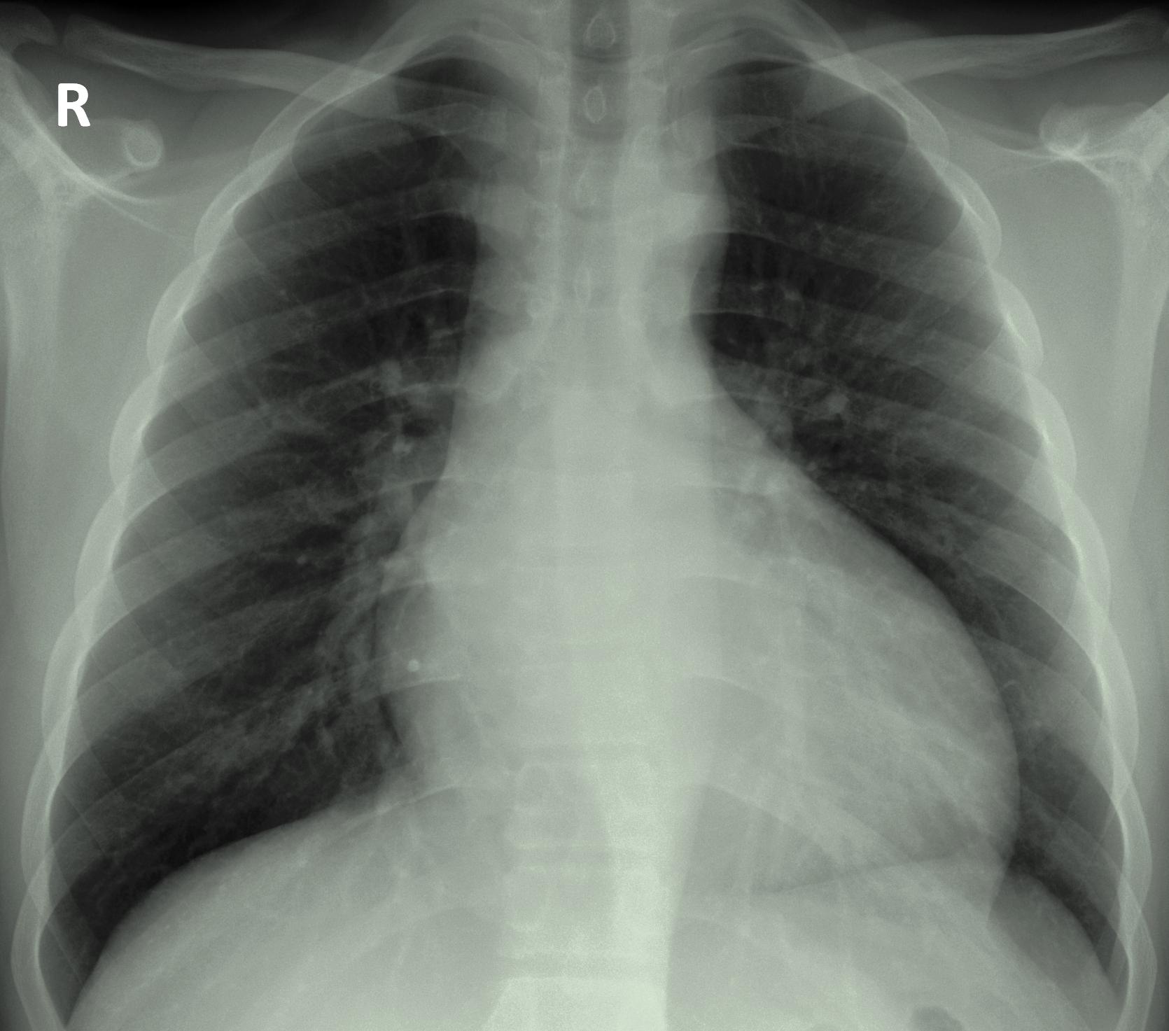
Shown below is a chest radiograph demonstrating [[left ventricular enlargement]] secondary to chronic aortic regurgitation as a result of increased [[left ventricular]] [[systolic]] pressure and volume overload. | |||
[[Image:Aortic regurgitation x-ray.jpg|400px]] | [[Image:Aortic regurgitation x-ray.jpg|left|400px]] | ||
<br clear="left"/> | |||
==References== | ==References== | ||
{{reflist|2}} | {{reflist|2}} | ||
{{WH}} | |||
{{WS}} | |||
[[CME Category::Cardiology]] | |||
[[Category:Disease]] | |||
[[Category:Cardiology]] | |||
[[Category:Valvular heart disease]] | [[Category:Valvular heart disease]] | ||
[[Category: | [[Category:Congenital heart disease]] | ||
[[Category: | [[Category:Surgery]] | ||
[[Category:Cardiac surgery]] | [[Category:Cardiac surgery]] | ||
[[Category: | [[Category:Emergency medicine]] | ||
[[Category: | [[Category:Intensive care medicine]] | ||
[[Category:Up-To-Date cardiology]] | |||
[[Category:Up-To-Date]] | |||
Latest revision as of 21:26, 20 February 2020
| Resident Survival Guide |
|
Aortic Regurgitation Microchapters |
|
Diagnosis |
|---|
|
Treatment |
|
Acute Aortic regurgitation |
|
Chronic Aortic regurgitation |
|
Special Scenarios |
|
Case Studies |
|
Aortic regurgitation chest x-ray On the Web |
|
American Roentgen Ray Society Images of Aortic regurgitation chest x-ray |
|
Risk calculators and risk factors for Aortic regurgitation chest x-ray |
Editor-In-Chief: C. Michael Gibson, M.S., M.D. [1]; Associate Editor(s)-in-Chief: Cafer Zorkun, M.D., Ph.D. [2]; Varun Kumar, M.B.B.S.; Lakshmi Gopalakrishnan, M.B.B.S; Usama Talib, BSc, MD [3]
Overview
Chest X-ray findings associated with aortic regurgitation may include left ventricular enlargement, cardiomegaly, prominent aortic root with valvular calcification, prosthetic valve dislodgement, or aortic dilation. If aortic regurgitation is severe, signs of pulmonary edema may also be present.[1]
Chest X-Ray
In patients with aortic regurgitation, chest radiograph may demonstrate any of the following findings:[1][2][3]
- Cardiomegaly
- Aortic dilation
- Increased cardiac silhouette (suggestive of aortic dissection)
- Widened mediastinum (suggestive of aortic root dilation)
- Pulmonary congestion (suggestive of pulmonary edema or pulmonary hypertension in severe AR)
- Prominent aortic root
- Aortic valve calcification
- Prosthetic valve dislodgement
Shown below is a chest radiograph demonstrating left ventricular enlargement secondary to chronic aortic regurgitation as a result of increased left ventricular systolic pressure and volume overload.

References
- ↑ 1.0 1.1 Bonow RO, Carabello B, de Leon AC, Edmunds LH, Fedderly BJ, Freed MD; et al. (1998). "ACC/AHA Guidelines for the Management of Patients With Valvular Heart Disease. Executive Summary. A report of the American College of Cardiology/American Heart Association Task Force on Practice Guidelines (Committee on Management of Patients With Valvular Heart Disease)". J Heart Valve Dis. 7 (6): 672–707. PMID 9870202.
- ↑ McWilliams E, Zehr K, Alshehri A (2016). "An unusual shadow above the aortic valve". Heart. 102 (24): 1942. doi:10.1136/heartjnl-2016-309932. PMID 27515955.
- ↑ Kuan PX, Tan PW, Jobli AT, Norsila AR (2016). "Discrepancy in blood pressure between the left and right arms - importance of clinical diagnosis and role of radiological imaging". Med J Malaysia. 71 (4): 206–208. PMID 27770122.