Abdominal pain, fever and jaundice: Difference between revisions
(Created page with "_NOTOC_") |
Iqra Qamar (talk | contribs) No edit summary |
||
| Line 1: | Line 1: | ||
_NOTOC_ | _NOTOC_ | ||
==Differential diagnosis== | |||
<span style="font-size:85%">'''Abbreviations:''' | |||
'''[[RUQ]]'''= Right upper quadrant of the abdomen, '''LUQ'''= Left upper quadrant, '''LLQ'''= Left lower quadrant, '''RLQ'''= Right lower quadrant, '''LFT'''= Liver function test, SIRS= [[Systemic inflammatory response syndrome]], '''[[ERCP]]'''= [[Endoscopic retrograde cholangiopancreatography]], '''IV'''= Intravenous, '''N'''= Normal, '''AMA'''= Anti mitochondrial antibodies, '''[[LDH]]'''= [[Lactate dehydrogenase]], '''GI'''= Gastrointestinal, '''CXR'''= Chest X ray, '''IgA'''= [[Immunoglobulin A]], '''IgG'''= [[Immunoglobulin G]], '''IgM'''= [[Immunoglobulin M]], '''CT'''= [[Computed tomography]], '''[[PMN]]'''= Polymorphonuclear cells, '''[[ESR]]'''= [[Erythrocyte sedimentation rate]], '''[[CRP]]'''= [[C-reactive protein]], TS= [[Transferrin saturation]], SF= Serum [[Ferritin]], SMA= [[Superior mesenteric artery]], SMV= [[Superior mesenteric vein]], ECG= [[Electrocardiogram]]</span> | |||
{| align="center" | |||
|- | |||
| | |||
{| style="border: 0px; font-size: 90%; margin: 3px;" align="center" | |||
! colspan="3" rowspan="3" style="background:#4479BA; color: #FFFFFF;" align="center" |Classification of pain in the abdomen based on etiology | |||
! rowspan="3" style="background:#4479BA; color: #FFFFFF;" align="center" |Disease | |||
| colspan="10" rowspan="1" style="background:#4479BA; color: #FFFFFF;" align="center" |'''Clinical manifestations''' | |||
! colspan="2" rowspan="2" style="background:#4479BA; color: #FFFFFF;" align="center" |Diagnosis | |||
! rowspan="3" style="background:#4479BA; color: #FFFFFF;" align="center" |Comments | |||
|- | |||
| colspan="6" rowspan="1" style="background:#4479BA; color: #FFFFFF;" align="center" |'''Symptoms''' | |||
! colspan="4" rowspan="1" style="background:#4479BA; color: #FFFFFF;" align="center" | Signs | |||
|- | |||
! style="background:#4479BA; color: #FFFFFF;" align="center" |Abdominal Pain | |||
! colspan="1" rowspan="1" style="background:#4479BA; color: #FFFFFF;" align="center" | Fever | |||
! style="background:#4479BA; color: #FFFFFF;" align="center" |Rigors and chills | |||
! style="background:#4479BA; color: #FFFFFF;" align="center" |Jaundice | |||
! style="background:#4479BA; color: #FFFFFF;" align="center" |Diarrhea | |||
! style="background:#4479BA; color: #FFFFFF;" align="center" |GI Bleed | |||
! style="background:#4479BA; color: #FFFFFF;" align="center" |Hypo- | |||
tension | |||
! colspan="1" rowspan="1" style="background:#4479BA; color: #FFFFFF;" align="center" | Guarding | |||
! style="background:#4479BA; color: #FFFFFF;" align="center" |Rebound Tenderness | |||
! style="background:#4479BA; color: #FFFFFF;" align="center" |Bowel sounds | |||
! colspan="1" rowspan="1" style="background:#4479BA; color: #FFFFFF;" align="center" | Lab Findings | |||
! style="background:#4479BA; color: #FFFFFF;" align="center" |Imaging | |||
|- | |||
! rowspan="13" style="background:#4479BA; color: #FFFFFF;" align="center" |Abdominal causes | |||
! colspan="1" rowspan="13" style="padding: 5px 5px; background: #DCDCDC;" align="center" | Inflammatory causes | |||
! rowspan="6" style="padding: 5px 5px; background: #DCDCDC;" align="center" |Pancreato-biliary disorders | |||
| colspan="1" rowspan="1" style="padding: 5px 5px; background: #DCDCDC;" align="center" | Acute suppurative cholangitis | |||
| style="padding: 5px 5px; background: #F5F5F5;" align="center" |[[RUQ]] | |||
| style="padding: 5px 5px; background: #F5F5F5;" align="center" | + | |||
| style="padding: 5px 5px; background: #F5F5F5;" align="center" | + | |||
| style="padding: 5px 5px; background: #F5F5F5;" align="center" | + | |||
| style="padding: 5px 5px; background: #F5F5F5;" align="center" | − | |||
| style="padding: 5px 5px; background: #F5F5F5;" align="center" | − | |||
| style="padding: 5px 5px; background: #F5F5F5;" align="center" | + | |||
| style="padding: 5px 5px; background: #F5F5F5;" align="center" | + | |||
| style="padding: 5px 5px; background: #F5F5F5;" align="center" | + | |||
| style="padding: 5px 5px; background: #F5F5F5;" align="left" |N | |||
| style="padding: 5px 5px; background: #F5F5F5;" align="left" | | |||
* Abnormal [[LFT]] | |||
* WBC >10,000 | |||
| style="padding: 5px 5px; background: #F5F5F5;" align="left" |Ultrasound shows [[biliary]] dilatation/stents/tumor | |||
| style="padding: 5px 5px; background: #F5F5F5;" align="left" |Septic shock occurs with features of [[SIRS]] | |||
|- | |||
| colspan="1" rowspan="1" style="padding: 5px 5px; background: #DCDCDC;" align="center" | [[Cholangitis|Acute cholangitis]] | |||
| style="padding: 5px 5px; background: #F5F5F5;" align="center" | [[RUQ]] | |||
| style="padding: 5px 5px; background: #F5F5F5;" align="center" | + | |||
| style="padding: 5px 5px; background: #F5F5F5;" align="center" | − | |||
| style="padding: 5px 5px; background: #F5F5F5;" align="center" | + | |||
| style="padding: 5px 5px; background: #F5F5F5;" align="center" | − | |||
| style="padding: 5px 5px; background: #F5F5F5;" align="center" | − | |||
| style="padding: 5px 5px; background: #F5F5F5;" align="center" | − | |||
| style="padding: 5px 5px; background: #F5F5F5;" align="center" | − | |||
| style="padding: 5px 5px; background: #F5F5F5;" align="center" | − | |||
| style="padding: 5px 5px; background: #F5F5F5;" align="left" |N | |||
| style="padding: 5px 5px; background: #F5F5F5;" align="left" | | |||
* Abnormal [[LFT]] | |||
| style="padding: 5px 5px; background: #F5F5F5;" align="left" |Ultrasound shows [[biliary]] dilatation/stents/tumor | |||
| style="padding: 5px 5px; background: #F5F5F5;" align="left" |Biliary drainage ([[Endoscopic retrograde cholangiopancreatography|ERCP]]) + IV antibiotics | |||
|- | |||
| colspan="1" rowspan="1" style="padding: 5px 5px; background: #DCDCDC;" align="center" | [[Acute cholecystitis|Acute cholecystitis]] | |||
| style="padding: 5px 5px; background: #F5F5F5;" align="center" | [[RUQ]] | |||
| style="padding: 5px 5px; background: #F5F5F5;" align="center" | + | |||
| style="padding: 5px 5px; background: #F5F5F5;" align="center" |− | |||
| style="padding: 5px 5px; background: #F5F5F5;" align="center" | + | |||
| style="padding: 5px 5px; background: #F5F5F5;" align="center" | − | |||
| style="padding: 5px 5px; background: #F5F5F5;" align="center" | − | |||
| style="padding: 5px 5px; background: #F5F5F5;" align="center" | − | |||
| style="padding: 5px 5px; background: #F5F5F5;" align="center" | − | |||
| style="padding: 5px 5px; background: #F5F5F5;" align="center" | − | |||
| style="padding: 5px 5px; background: #F5F5F5;" align="left" |Hypoactive | |||
| style="padding: 5px 5px; background: #F5F5F5;" align="left" | | |||
* [[Hyperbilirubinemia]] | |||
* [[Leukocytosis]] | |||
| style="padding: 5px 5px; background: #F5F5F5;" align="left" |Ultrasound shows gallstone and evidence of inflammation | |||
| style="padding: 5px 5px; background: #F5F5F5;" align="left" |[[Murphy's sign|Murphy’s sign]] | |||
|- | |||
| colspan="1" rowspan="1" style="padding: 5px 5px; background: #DCDCDC;" align="center" | [[Acute pancreatitis]] | |||
| style="padding: 5px 5px; background: #F5F5F5;" align="center" | [[Epigastric]] | |||
| style="padding: 5px 5px; background: #F5F5F5;" align="center" | + | |||
| style="padding: 5px 5px; background: #F5F5F5;" align="center" | − | |||
| style="padding: 5px 5px; background: #F5F5F5;" align="center" | ± | |||
| style="padding: 5px 5px; background: #F5F5F5;" align="center" | − | |||
| style="padding: 5px 5px; background: #F5F5F5;" align="center" | − | |||
| style="padding: 5px 5px; background: #F5F5F5;" align="center" | ± | |||
| style="padding: 5px 5px; background: #F5F5F5;" align="center" | + | |||
| style="padding: 5px 5px; background: #F5F5F5;" align="center" | + | |||
| style="padding: 5px 5px; background: #F5F5F5;" align="left" |N | |||
| style="padding: 5px 5px; background: #F5F5F5;" align="left" | | |||
* Increased [[amylase]] / [[lipase]] | |||
| style="padding: 5px 5px; background: #F5F5F5;" align="left" | | |||
* Ultrasound shows evidence of [[inflammation]] | |||
* CT scan shows severity of pancreatitis | |||
| style="padding: 5px 5px; background: #F5F5F5;" align="left" |Pain radiation to back | |||
|- | |||
| colspan="1" rowspan="1" style="padding: 5px 5px; background: #DCDCDC;" align="center" |[[Primary sclerosing cholangitis]] | |||
| style="padding: 5px 5px; background: #F5F5F5;" align="center" |[[RUQ]] | |||
| style="padding: 5px 5px; background: #F5F5F5;" align="center" | + | |||
| style="padding: 5px 5px; background: #F5F5F5;" align="center" | − | |||
| style="padding: 5px 5px; background: #F5F5F5;" align="center" | + | |||
| style="padding: 5px 5px; background: #F5F5F5;" align="center" | − | |||
| style="padding: 5px 5px; background: #F5F5F5;" align="center" | − | |||
| style="padding: 5px 5px; background: #F5F5F5;" align="center" | − | |||
| style="padding: 5px 5px; background: #F5F5F5;" align="center" | − | |||
| style="padding: 5px 5px; background: #F5F5F5;" align="center" | − | |||
| style="padding: 5px 5px; background: #F5F5F5;" align="left" |N | |||
| style="padding: 5px 5px; background: #F5F5F5;" align="left" | | |||
* Increased liver enzymes | |||
* Increased [[IgM]], [[IgG]]4 | |||
* [[Anti-neutrophil cytoplasmic antibody]] ([[p-ANCA]]) | |||
* [[Anti-nuclear antibody]] ([[ANA]]) | |||
* [[Anti-smooth muscle antibody]] (Anti-Sm) | |||
* Anti-endothelial antibody | |||
* Anti-cardiolipin antibody | |||
| style="padding: 5px 5px; background: #F5F5F5;" align="left" |ERCP and MRCP shows | |||
* Multiple segmental [[strictures]] | |||
* Mural irregularities | |||
* [[Biliary]] dilatation and diverticula | |||
* Distortion of biliary tree | |||
| style="padding: 5px 5px; background: #F5F5F5;" align="left" |The risk of [[cholangiocarcinoma]] in patients with primary sclerosing cholangitis is 400 times higher than the risk in the general population. | |||
|- | |||
| colspan="1" rowspan="1" style="padding: 5px 5px; background: #DCDCDC;" align="center" |[[Cholelithiasis]] | |||
| style="padding: 5px 5px; background: #F5F5F5;" align="center" |[[RUQ]]/[[Epigastric]] | |||
| style="padding: 5px 5px; background: #F5F5F5;" align="center" | ± | |||
| style="padding: 5px 5px; background: #F5F5F5;" align="center" | − | |||
| style="padding: 5px 5px; background: #F5F5F5;" align="center" | ± | |||
| style="padding: 5px 5px; background: #F5F5F5;" align="center" | − | |||
| style="padding: 5px 5px; background: #F5F5F5;" align="center" | − | |||
| style="padding: 5px 5px; background: #F5F5F5;" align="center" | − | |||
| style="padding: 5px 5px; background: #F5F5F5;" align="center" | − | |||
| style="padding: 5px 5px; background: #F5F5F5;" align="center" | − | |||
| style="padding: 5px 5px; background: #F5F5F5;" align="left" |N to hyperactive for dislodged stone | |||
| style="padding: 5px 5px; background: #F5F5F5;" align="left" | | |||
* [[Leukocytosis]] | |||
| style="padding: 5px 5px; background: #F5F5F5;" align="left" |Ultrasound shows [[gallstone]] | |||
| style="padding: 5px 5px; background: #F5F5F5;" align="left" |Fatty food intolerance | |||
|- | |||
!Gastric causes | |||
| style="padding: 5px 5px; background: #DCDCDC;" align="center" |[[Gastrointestinal perforation]] | |||
| style="padding: 5px 5px; background: #F5F5F5;" align="center" |Diffuse | |||
| style="padding: 5px 5px; background: #F5F5F5;" align="center" | + | |||
| style="padding: 5px 5px; background: #F5F5F5;" align="center" | ± | |||
| style="padding: 5px 5px; background: #F5F5F5;" align="center" | ± | |||
| style="padding: 5px 5px; background: #F5F5F5;" align="center" | − | |||
| style="padding: 5px 5px; background: #F5F5F5;" align="center" | +, depends on site | |||
| style="padding: 5px 5px; background: #F5F5F5;" align="center" | + | |||
| style="padding: 5px 5px; background: #F5F5F5;" align="center" | + | |||
| style="padding: 5px 5px; background: #F5F5F5;" align="center" | ± | |||
| style="padding: 5px 5px; background: #F5F5F5;" align="left" |Hyperactive/hypoactive | |||
| style="padding: 5px 5px; background: #F5F5F5;" align="left" | | |||
* WBC> 10,000 | |||
| style="padding: 5px 5px; background: #F5F5F5;" align="left" |Air under [[diaphragm]] in upright [[CXR]] | |||
| style="padding: 5px 5px; background: #F5F5F5;" align="left" |[[Hamman's sign]] | |||
|- | |||
! rowspan="2" |Intestinal causes | |||
| style="padding: 5px 5px; background: #DCDCDC;" align="center" |[[Inflammatory bowel disease]] | |||
| style="padding: 5px 5px; background: #F5F5F5;" align="center" |Diffuse | |||
| style="padding: 5px 5px; background: #F5F5F5;" align="center" | ± | |||
| style="padding: 5px 5px; background: #F5F5F5;" align="center" | − | |||
| style="padding: 5px 5px; background: #F5F5F5;" align="center" | ± | |||
| style="padding: 5px 5px; background: #F5F5F5;" align="center" | ± | |||
| style="padding: 5px 5px; background: #F5F5F5;" align="center" | [[Hematochezia]] | |||
| style="padding: 5px 5px; background: #F5F5F5;" align="center" |− | |||
| style="padding: 5px 5px; background: #F5F5F5;" align="center" |− | |||
| style="padding: 5px 5px; background: #F5F5F5;" align="center" |− | |||
| style="padding: 5px 5px; background: #F5F5F5;" align="left" |N/ Hyperactive | |||
| style="padding: 5px 5px; background: #F5F5F5;" align="left" | | |||
* [[Anti-neutrophil cytoplasmic antibody]] ([[P-ANCA]]) in [[Ulcerative colitis]] | |||
* [[Anti saccharomyces cerevisiae antibodies]] (ASCA) in [[Crohn's disease]] | |||
| style="padding: 5px 5px; background: #F5F5F5;" align="left" |[[String sign]] on [[abdominal x-ray]] in [[Crohn's disease]] | |||
| style="padding: 5px 5px; background: #F5F5F5;" align="left" | | |||
Extra intestinal findings: | |||
* [[Uveitis]] | |||
* [[Arthritis]] | |||
|- | |||
| style="padding: 5px 5px; background: #DCDCDC;" align="center" |[[Whipple's disease]] | |||
| style="padding: 5px 5px; background: #F5F5F5;" align="center" |Diffuse | |||
| style="padding: 5px 5px; background: #F5F5F5;" align="center" | ± | |||
| style="padding: 5px 5px; background: #F5F5F5;" align="center" | − | |||
| style="padding: 5px 5px; background: #F5F5F5;" align="center" | ± | |||
| style="padding: 5px 5px; background: #F5F5F5;" align="center" | + | |||
| style="padding: 5px 5px; background: #F5F5F5;" align="center" | − | |||
| style="padding: 5px 5px; background: #F5F5F5;" align="center" | ± | |||
| style="padding: 5px 5px; background: #F5F5F5;" align="center" | − | |||
| style="padding: 5px 5px; background: #F5F5F5;" align="center" | − | |||
| style="padding: 5px 5px; background: #F5F5F5;" align="left" |N | |||
| style="padding: 5px 5px; background: #F5F5F5;" align="left" | | |||
* [[Thrombocytopenia]] | |||
* [[Hypoalbuminemia]] | |||
* [[Small intestinal]] [[biopsy]] for [[Tropheryma whipplei]] | |||
| style="padding: 5px 5px; background: #F5F5F5;" align="left" |[[Whipple's disease other diagnostic studies|Endoscopy]] is used to confirm diagnosis. | |||
Images used to find complications | |||
*[[Whipple's disease x ray|Chest and joint x-ray]] | |||
*[[Whipple's disease CT|CT]] | |||
*[[Whipple's disease MRI|MRI]] | |||
*[[Whipple's disease ultrasound|Echocardiography]] | |||
| style="padding: 5px 5px; background: #F5F5F5;" align="left" |Extra intestinal findings: | |||
* [[Uveitis]] | |||
* [[Endocarditis]] | |||
* [[Encephalitis]] | |||
* [[Dementia]] | |||
* [[Hepatosplenomegaly]] | |||
* [[Arthritis]] | |||
* [[Ascites]] | |||
|- | |||
! rowspan="3" style="padding: 5px 5px; background: #DCDCDC;" align="center" |Hepatic causes | |||
| style="padding: 5px 5px; background: #DCDCDC;" align="center" |[[Hepatitis|Viral hepatitis]] | |||
| style="padding: 5px 5px; background: #F5F5F5;" align="center" |[[RUQ]] | |||
| style="padding: 5px 5px; background: #F5F5F5;" align="center" | + | |||
| style="padding: 5px 5px; background: #F5F5F5;" align="center" |− | |||
| style="padding: 5px 5px; background: #F5F5F5;" align="center" | + | |||
| style="padding: 5px 5px; background: #F5F5F5;" align="center" | + in Hep A and E | |||
| style="padding: 5px 5px; background: #F5F5F5;" align="center" | − | |||
| style="padding: 5px 5px; background: #F5F5F5;" align="center" | + in fulminant hepatitis | |||
| style="padding: 5px 5px; background: #F5F5F5;" align="center" | +in acute | |||
| style="padding: 5px 5px; background: #F5F5F5;" align="center" | + | |||
| style="padding: 5px 5px; background: #F5F5F5;" align="left" |N | |||
| style="padding: 5px 5px; background: #F5F5F5;" align="left" | | |||
* Abnormal LFTs | |||
* Viral serology | |||
| style="padding: 5px 5px; background: #F5F5F5;" align="left" |USG | |||
| style="padding: 5px 5px; background: #F5F5F5;" align="left" |Hep A and E have fecoral route of transmission and Hep B and C transmits via blood transfusion and sexual contact. | |||
|- | |||
| style="padding: 5px 5px; background: #DCDCDC;" align="center" |[[Liver mass|Liver masses]] | |||
| style="padding: 5px 5px; background: #F5F5F5;" align="center" |[[RUQ]] | |||
| style="padding: 5px 5px; background: #F5F5F5;" align="center" | + | |||
| style="padding: 5px 5px; background: #F5F5F5;" align="center" | + in [[Liver abscess]] | |||
| style="padding: 5px 5px; background: #F5F5F5;" align="center" | ± | |||
| style="padding: 5px 5px; background: #F5F5F5;" align="center" | − | |||
| style="padding: 5px 5px; background: #F5F5F5;" align="center" | + in [[Hepatocellular carcinoma]] | |||
| style="padding: 5px 5px; background: #F5F5F5;" align="center" | + in sepsis | |||
| style="padding: 5px 5px; background: #F5F5F5;" align="center" | + in [[Liver abscess]] | |||
| style="padding: 5px 5px; background: #F5F5F5;" align="center" | + in [[Liver abscess]] | |||
| style="padding: 5px 5px; background: #F5F5F5;" align="left" |N | |||
| style="padding: 5px 5px; background: #F5F5F5;" align="left" | | |||
* CBC | |||
* LFTs | |||
| style="padding: 5px 5px; background: #F5F5F5;" align="left" |USG | |||
| style="padding: 5px 5px; background: #F5F5F5;" align="left" | | |||
|- | |||
| style="padding: 5px 5px; background: #DCDCDC;" align="center" |[[Budd-Chiari syndrome|Budd-Chiari syndrome]] | |||
| style="padding: 5px 5px; background: #F5F5F5;" align="center" |[[RUQ]] | |||
| style="padding: 5px 5px; background: #F5F5F5;" align="center" | ± | |||
| style="padding: 5px 5px; background: #F5F5F5;" align="center" |− | |||
| style="padding: 5px 5px; background: #F5F5F5;" align="center" | ± | |||
| style="padding: 5px 5px; background: #F5F5F5;" align="center" | − | |||
| style="padding: 5px 5px; background: #F5F5F5;" align="center" | + in liver failure leading to varices | |||
| style="padding: 5px 5px; background: #F5F5F5;" align="center" |− | |||
| style="padding: 5px 5px; background: #F5F5F5;" align="center" |− | |||
| style="padding: 5px 5px; background: #F5F5F5;" align="center" |− | |||
| style="padding: 5px 5px; background: #F5F5F5;" align="left" |N | |||
| style="padding: 5px 5px; background: #F5F5F5;" align="left" | | |||
*Elevated [[Aspartate aminotransferase|serum aspartate aminotransferase]] and [[alanine aminotransferase]] levels may be more than five times the upper limit of the normal range. | |||
*Elevated serum [[alkaline phosphatase]] and [[Bilirubin|bilirubin levels]], decreased [[Albumin|serum albumin level]]. | |||
| style="padding: 5px 5px; background: #F5F5F5;" align="left" | | |||
{| style="border: 0px; font-size: 90%; margin: 3px;" align="center" | |||
|- | |||
| style="padding: 5px 5px; background: #F5F5F5;" align="left" |Findings on [[CT scan]] suggestive of Budd-Chiari syndrome include: | |||
*Early enhancement of the [[caudate lobe]] and [[central liver]] around the [[Inferior vena cavae|inferior vena cava]] | |||
*Delayed enhancement of the peripheral [[liver]] with accompanying central low density (flip-flop appearance) | |||
*Peripheral zones of the [[liver]] show reversed [[portal]] [[venous]] [[blood flow]] | |||
*In the [[chronic]] phase, there is [[caudate lobe]] enlargement and [[atrophy]] of the [[Liver|peripheral liver]] in affected areas | |||
|} | |||
| style="padding: 5px 5px; background: #F5F5F5;" align="left" |[[Ascitic tap|Ascitic fluid examination]] shows: | |||
*[[Total protein]] more than 2.5 g per deciliter | |||
*[[White blood cells]] are usually less than 500/μL. | |||
|- | |||
! style="padding: 5px 5px; background: #DCDCDC;" align="center" | Peritoneal causes | |||
| colspan="1" rowspan="1" style="padding: 5px 5px; background: #DCDCDC;" align="center" |[[Spontaneous bacterial peritonitis]] | |||
| style="padding: 5px 5px; background: #F5F5F5;" align="center" |Diffuse | |||
| style="padding: 5px 5px; background: #F5F5F5;" align="center" | + | |||
| style="padding: 5px 5px; background: #F5F5F5;" align="center" | + | |||
| style="padding: 5px 5px; background: #F5F5F5;" align="center" | + in cirrhotic patients | |||
| style="padding: 5px 5px; background: #F5F5F5;" align="center" | + | |||
| style="padding: 5px 5px; background: #F5F5F5;" align="center" | − | |||
| style="padding: 5px 5px; background: #F5F5F5;" align="center" | ± | |||
| style="padding: 5px 5px; background: #F5F5F5;" align="center" | + | |||
| style="padding: 5px 5px; background: #F5F5F5;" align="center" | + | |||
| style="padding: 5px 5px; background: #F5F5F5;" align="left" |Hypoactive | |||
| style="padding: 5px 5px; background: #F5F5F5;" align="left" | | |||
* Ascitic fluid [[PMN]]>250 cells/mm<small>³</small> | |||
* Culture: Positive for single organism | |||
| style="padding: 5px 5px; background: #F5F5F5;" align="left" |Ultrasound for evaluation of liver cirrhosis | |||
! style="padding: 5px 5px; background: #F5F5F5;" align="left" | | |||
|- | |||
|} | |||
|} | |||
{| | |||
|- | |||
| <figure-inline class="mw-default-size"><figure-inline>[[Image:Right_upper_quadrant.PNG|link=Right upper quadrant abdominal pain resident survival guide|339x339px]]</figure-inline></figure-inline>||<figure-inline class="mw-default-size"><figure-inline>[[Image:Epigastric_quadrant_pain.PNG|link=Epigastric pain resident survival guide|179x179px]]</figure-inline></figure-inline>||<figure-inline class="mw-default-size"><figure-inline>[[Image:Left_upper_quadrant.PNG|link=Left upper quadrant abdominal pain resident survival guide|329x329px]]</figure-inline></figure-inline> | |||
|- | |||
| <figure-inline class="mw-default-size"><figure-inline>[[Image:Right_flank_quadrant.PNG|link=Right flank pain resident survival guide|338x338px]]</figure-inline></figure-inline>||<figure-inline class="mw-default-size"><figure-inline>[[Image:Umbilical_pain.PNG|link=Umbilical region pain resident survival guide|165x165px]]</figure-inline></figure-inline>||<figure-inline class="mw-default-size"><figure-inline>[[Image:Left_flank_quadrant.PNG|link=Left flank quadrant abdominal pain resident survival guide|335x335px]]</figure-inline></figure-inline> | |||
|- | |||
| <figure-inline class="mw-default-size"><figure-inline>[[Image:Right_lower_quadrant.PNG|link=Right lower quadrant abdominal pain resident survival guide|338x338px]]</figure-inline></figure-inline>||<figure-inline class="mw-default-size"><figure-inline>[[Image:Hypogastric.PNG|link=Hypogastric pain resident survival guide|199x199px]]</figure-inline></figure-inline>||<figure-inline class="mw-default-size"><figure-inline>[[Image:Left_lower_quadrant.PNG|link=Left lower quadrant abdominal pain resident survival guide|335x335px]]</figure-inline></figure-inline> | |||
|} | |||
Revision as of 22:23, 27 November 2017
_NOTOC_
Differential diagnosis
Abbreviations: RUQ= Right upper quadrant of the abdomen, LUQ= Left upper quadrant, LLQ= Left lower quadrant, RLQ= Right lower quadrant, LFT= Liver function test, SIRS= Systemic inflammatory response syndrome, ERCP= Endoscopic retrograde cholangiopancreatography, IV= Intravenous, N= Normal, AMA= Anti mitochondrial antibodies, LDH= Lactate dehydrogenase, GI= Gastrointestinal, CXR= Chest X ray, IgA= Immunoglobulin A, IgG= Immunoglobulin G, IgM= Immunoglobulin M, CT= Computed tomography, PMN= Polymorphonuclear cells, ESR= Erythrocyte sedimentation rate, CRP= C-reactive protein, TS= Transferrin saturation, SF= Serum Ferritin, SMA= Superior mesenteric artery, SMV= Superior mesenteric vein, ECG= Electrocardiogram
| |||||||||||||||||||||||||||||||||||||||||||||||||||||||||||||||||||||||||||||||||||||||||||||||||||||||||||||||||||||||||||||||||||||||||||||||||||||||||||||||||||||||||||||||||||||||||||||||||||||||||||||||||||||||||||||||||||||
<figure-inline class="mw-default-size"><figure-inline> </figure-inline></figure-inline> </figure-inline></figure-inline> |
<figure-inline class="mw-default-size"><figure-inline>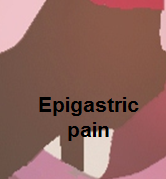 </figure-inline></figure-inline> </figure-inline></figure-inline> |
<figure-inline class="mw-default-size"><figure-inline>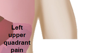 </figure-inline></figure-inline> </figure-inline></figure-inline>
|
<figure-inline class="mw-default-size"><figure-inline> </figure-inline></figure-inline> </figure-inline></figure-inline> |
<figure-inline class="mw-default-size"><figure-inline> </figure-inline></figure-inline> </figure-inline></figure-inline> |
<figure-inline class="mw-default-size"><figure-inline> </figure-inline></figure-inline> </figure-inline></figure-inline>
|
<figure-inline class="mw-default-size"><figure-inline>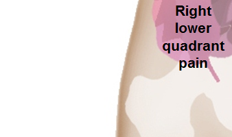 </figure-inline></figure-inline> </figure-inline></figure-inline> |
<figure-inline class="mw-default-size"><figure-inline>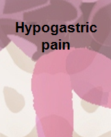 </figure-inline></figure-inline> </figure-inline></figure-inline> |
<figure-inline class="mw-default-size"><figure-inline>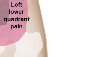 </figure-inline></figure-inline> </figure-inline></figure-inline>
|