ST elevation myocardial infarction EKG examples: Difference between revisions
Rim Halaby (talk | contribs) |
m Bot: Removing from Primary care |
||
| (13 intermediate revisions by one other user not shown) | |||
| Line 2: | Line 2: | ||
{{ST elevation myocardial infarction}} | {{ST elevation myocardial infarction}} | ||
{{CMG}} | {{CMG}} | ||
==EKG Examples== | ==EKG Examples== | ||
| Line 52: | Line 50: | ||
Shown below is an EKG demonstrating a 2 weeks old anterior infarction with [[Q wave]]s in [[Electrocardiogram#Precordial|V2]]-[[Electrocardiogram#Precordial|V4]] and persisting ST elevation, a sign of left [[ventricular aneurysm]] formation. | Shown below is an EKG demonstrating a 2 weeks old anterior infarction with [[Q wave]]s in [[Electrocardiogram#Precordial|V2]]-[[Electrocardiogram#Precordial|V4]] and persisting ST elevation, a sign of left [[ventricular aneurysm]] formation. | ||
[[Image:STEMI 18.jpg|center|800px]] | [[Image:STEMI 18.jpg|center|800px]] | ||
Copyleft image obtained courtesy of, http://en.ecgpedia.org/wiki/Main_Page | Copyleft image obtained courtesy of, http://en.ecgpedia.org/wiki/Main_Page | ||
---- | ---- | ||
| Line 78: | Line 72: | ||
---- | ---- | ||
Shown below is an EKG demonstrating | Shown below is an EKG demonstrating acute MI in a patient with [[LBBB]] | ||
[[Image:STEMI 26.jpg|center|800px]] | |||
Copyleft image obtained courtesy of, http://en.ecgpedia.org/wiki/Main_Page | |||
---- | |||
Shown below is an EKG demonstrating [[sinus rhythm]] with [[left bundle branch block]], comparison with an old EKG is mandatory to evaluate whether the [[LBBB]] is new (a sign of myocardial infarction) or old. | |||
[[Image:STEMI 12.jpg|center|800px]] | |||
Copyleft image obtained courtesy of, http://en.ecgpedia.org/wiki/Main_Page | Copyleft image obtained courtesy of, http://en.ecgpedia.org/wiki/Main_Page | ||
---- | ---- | ||
Shown below is an EKG | Shown below is an EKG showing Q wave in leads I and aVL consistent with an anterior myocardial infarction of undetermined age. | ||
[[ | [[File:Q wave anterior distribution 2.PNG|center|800px]] | ||
---- | ---- | ||
Shown below are two EKGs of the same subject: while the first EKG demonstrates no Q waves, the second EKG performed after a year shows Q wave in leads I and aVL consistent with an anterior myocardial infarction of undetermined age. | |||
Shown below is the first EKG. | |||
[[File:Q wave absent in anterior distribution.PNG|center|800px]] | |||
Shown below is the second EKG of the same subject. | |||
[[File:Q wave anterior distribution.PNG|center|800px]] | |||
===Lateral Myocardial Infarction=== | |||
Shown below is an EKG demonstrating [[sinus rhythm]] and a [[QRS]] with a rightward axis, as well as [[wide Q waves]] in leads [[Electrocardiogram#Limb|I]] and [[Electrocardiogram#Augmented lead|aVL]] as well as a poor R wave progression across the anterior chest leads. There is also slight [[ST elevation]] in leads [[Electrocardiogram#Limb|I]],[[Electrocardiogram#Augmented lead|aVL]] , and [[T wave inversion]] in the lateral leads. The EKG is consistent with a lateral wall myocardial infarction. | |||
[[Image:STEMI 35.jpg |center|800px]] | |||
Copyleft image obtained courtesy of, http://en.ecgpedia.org/wiki/Main_Page | |||
---- | |||
===Anterolateral Myocardial Infarction=== | ===Anterolateral Myocardial Infarction=== | ||
| Line 224: | Line 236: | ||
[[Image:STEMI_34.jpg|center|800px]] | [[Image:STEMI_34.jpg|center|800px]] | ||
Copyleft image obtained courtesy of, http://en.ecgpedia.org/wiki/File:De-Ami0010.jpg | Copyleft image obtained courtesy of, http://en.ecgpedia.org/wiki/File:De-Ami0010.jpg | ||
---- | ---- | ||
Shown below is an EKG demonstrating ST elevation in leads II, III and aVF and ST depression in leads V1, V2 and V3 depicting a [[posterior MI]]. | |||
[[Image:Posterior MI patient.jpg|center|800px]] | |||
---- | |||
===Right Ventricular Myocardial Infarction=== | ===Right Ventricular Myocardial Infarction=== | ||
| Line 243: | Line 259: | ||
---- | ---- | ||
==References== | ==References== | ||
{{Reflist|2}} | {{Reflist|2}} | ||
{{WH}} | |||
{{WS}} | |||
[[Category:Disease]] | [[Category:Disease]] | ||
| Line 261: | Line 270: | ||
[[Category:Intensive care medicine]] | [[Category:Intensive care medicine]] | ||
[[Category:Emergency medicine]] | [[Category:Emergency medicine]] | ||
[[Category:Up-To-Date]] | [[Category:Up-To-Date]] | ||
[[Category:Up-To-Date cardiology]] | [[Category:Up-To-Date cardiology]] | ||
[[Category:Mature chapter]] | [[Category:Mature chapter]] | ||
Latest revision as of 00:17, 30 July 2020
|
ST Elevation Myocardial Infarction Microchapters |
|
Differentiating ST elevation myocardial infarction from other Diseases |
|
Diagnosis |
|
Treatment |
|
|
Case Studies |
|
ST elevation myocardial infarction EKG examples On the Web |
|
Directions to Hospitals Treating ST elevation myocardial infarction |
|
Risk calculators and risk factors for ST elevation myocardial infarction EKG examples |
Editor-In-Chief: C. Michael Gibson, M.S., M.D. [1]
EKG Examples
Shown below is an EKG demonstrating the evolution of an infarct on the EKG. ST elevation, Q wave formation, T wave inversion, normalization with a persistent Q wave suggest STEMI.

Copyleft image obtained courtesy of ECGpedia, http://en.ecgpedia.org/wiki/File:AMI_evolutie.png
Anterior Myocardial Infarction
Shown below is an EKG demonstrating loss of R waves throughout the anterior wall (V1-V6). QS complexes in V3-V5. ST elevation in V1-V5 with terminal negative T waves.

Copyleft image obtained courtesy of, http://en.ecgpedia.org/wiki/Main_Page
Shown below is an EKG demonstrating acute anterior MI. LAD artery occlusion.
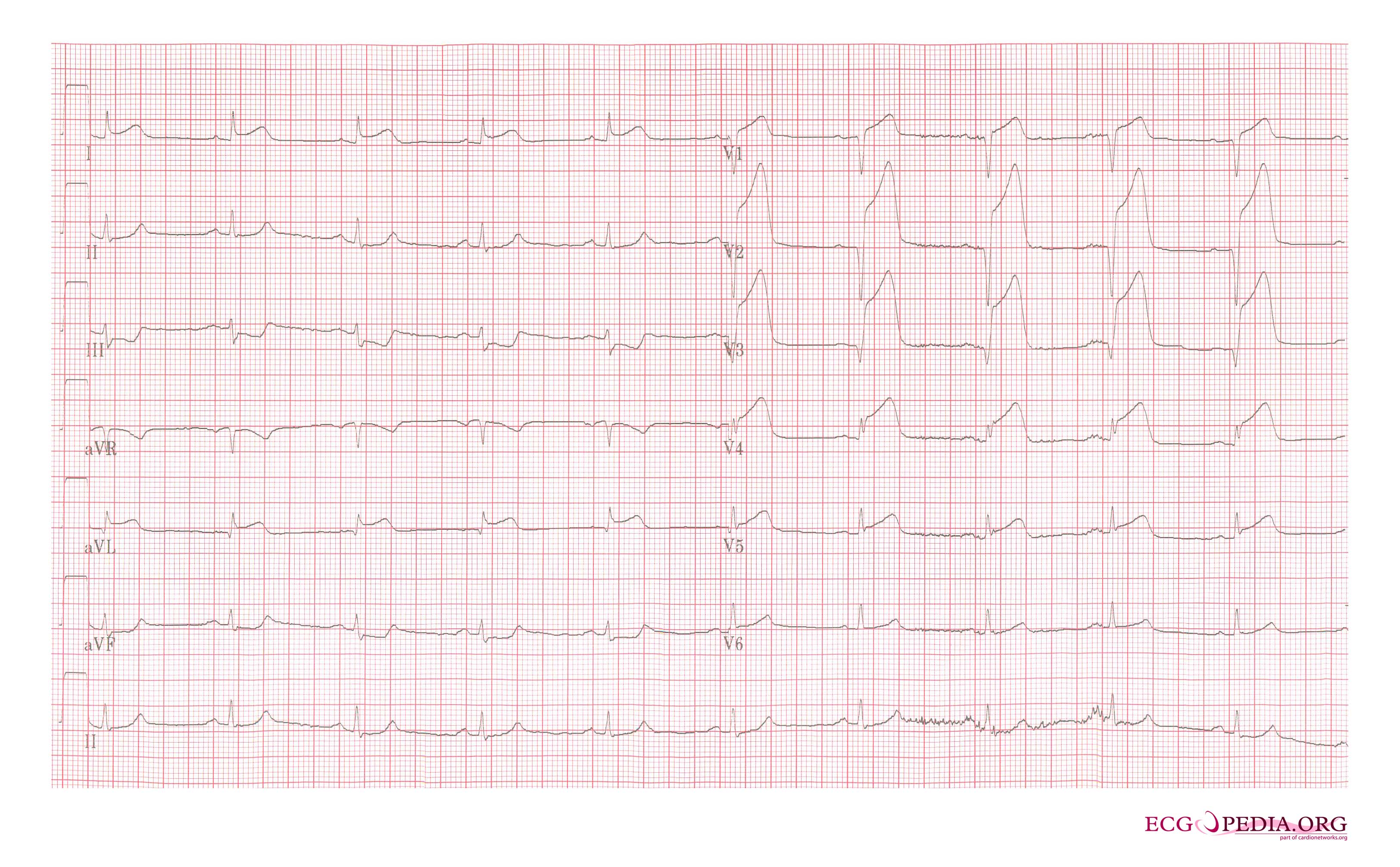
Copyleft image obtained courtesy of, http://en.ecgpedia.org/wiki/Main_Page
Shown below is an EKG showing sinus rhythm with anteroseptal myocardial infarction depicting ST elevation in V1-V6 and in lead I.
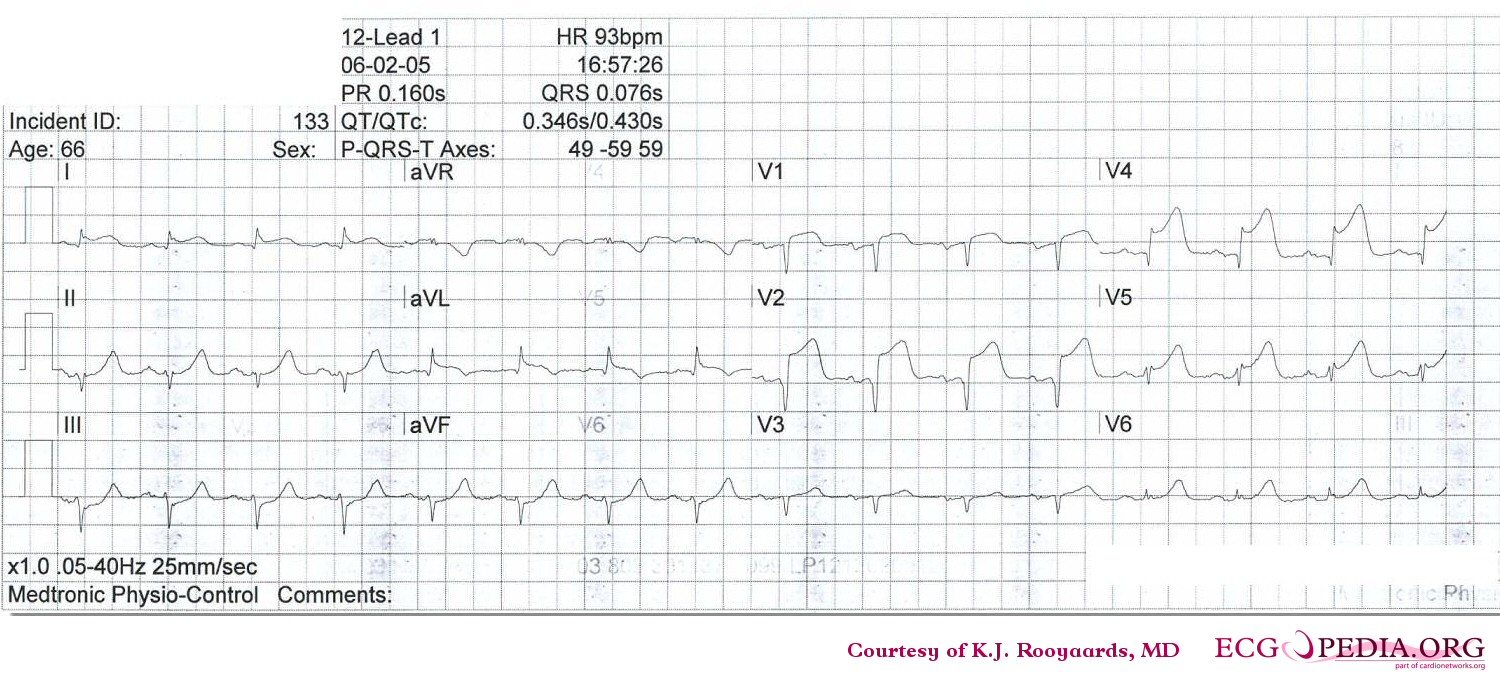
Copyleft image obtained courtesy of, http://en.ecgpedia.org/wiki/Main_Page
Shown below is an EKG demonstrating acute anterior myocardial infarction and left anterior hemiblock depicting ST elevation in precordial leads.

Copyleft image obtained courtesy of, http://en.ecgpedia.org/wiki/Main_Page
Shown below is an EKG demonstrating old anterior myocardial infarction and bifascicular block (RBBB and LAHB) as indicated in the anterior chest leads.
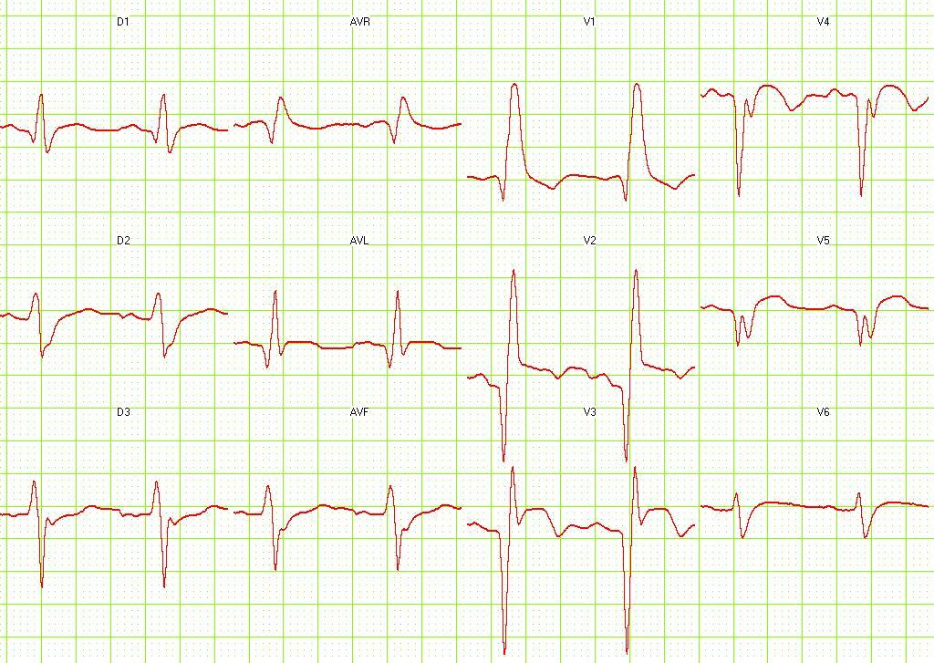
Copyleft image obtained courtesy of, http://en.ecgpedia.org/wiki/Main_Page
Shown below is an EKG illustrating acute MI with proximal LAD occlusion depicting ST elevation in anterior precordial leads.

Copyleft image obtained courtesy of, http://en.ecgpedia.org/wiki/Main_Page
Shown below is an EKG demonstrating a 2 days old anterior infarction with Q waves in V1-V4 with persisting ST elevation, a sign of left ventricular aneurysm formation.
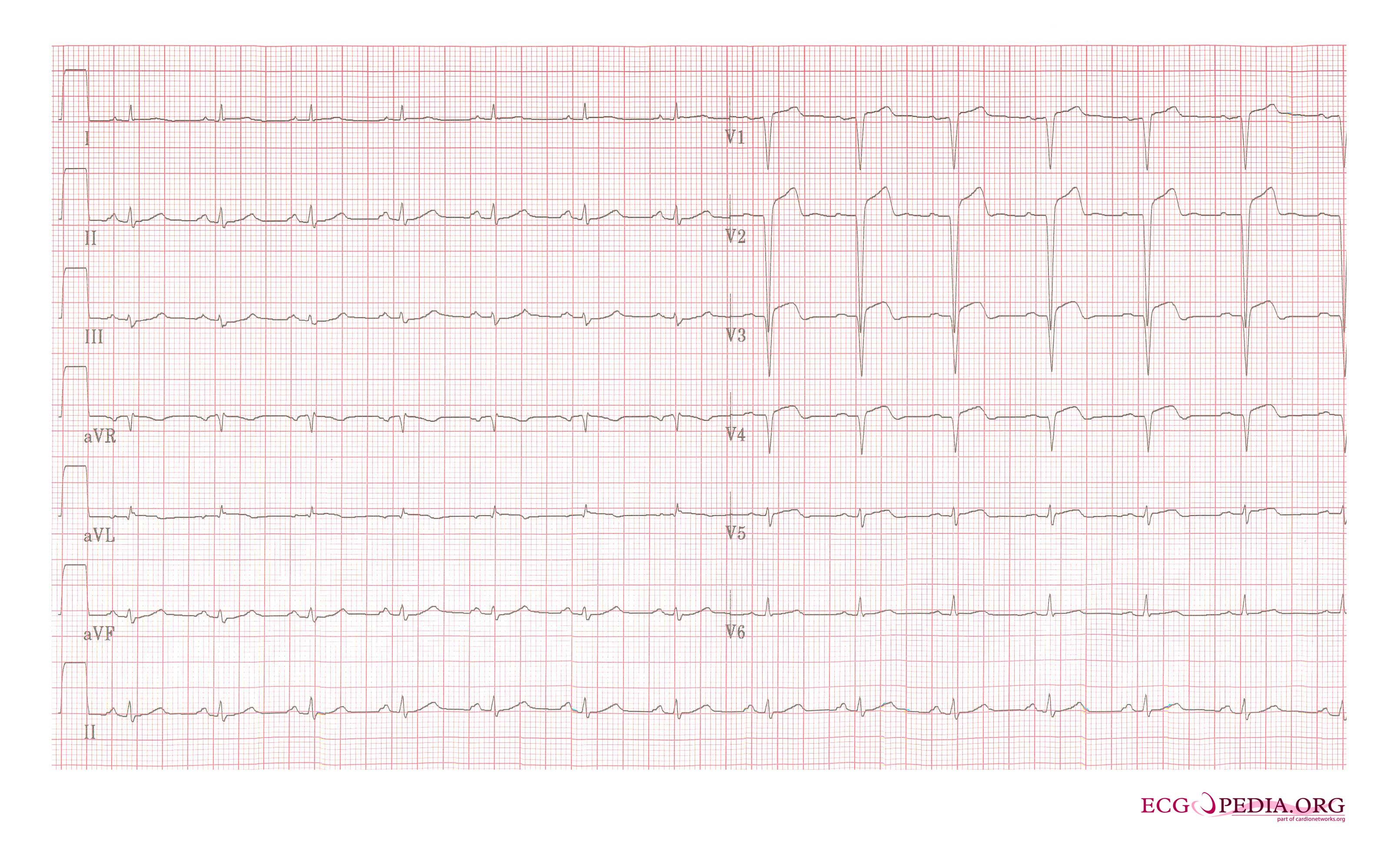
Copyleft image obtained courtesy of, http://en.ecgpedia.org/wiki/Main_Page
Shown below is an EKG demonstrating a 2 weeks old anterior infarction with Q waves in V2-V4 and persisting ST elevation, a sign of left ventricular aneurysm formation.
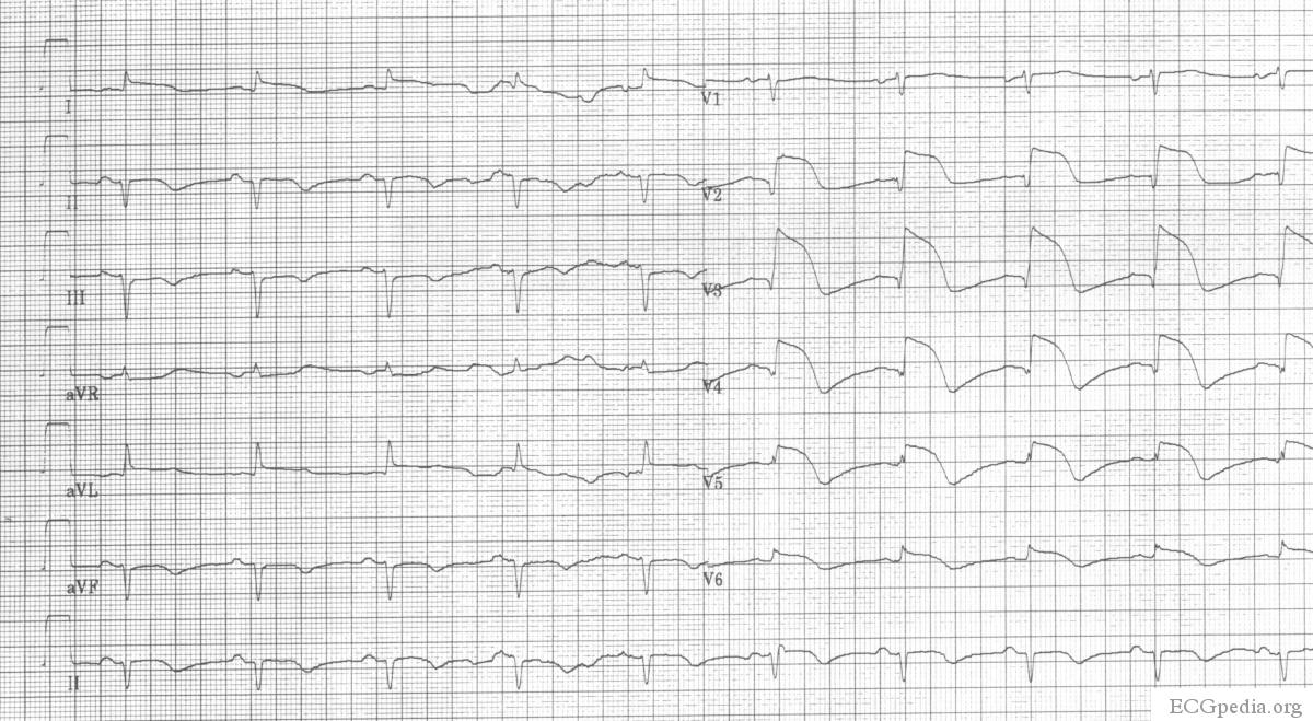
Copyleft image obtained courtesy of, http://en.ecgpedia.org/wiki/Main_Page
Shown below is an EKG showing ST elevation in the anterior precordial leads, low voltages in all the leads, poor R wave progression in the precordial leads.
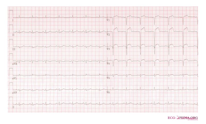
Copyleft image obtained courtesy of, http://en.ecgpedia.org/wiki/File:De-AMI_anterior_LAD_2days.jpg
Shown below is an EKG demonstrating ST segment elevation in precordial leads signifying anterior myocardial infarction.

Copyleft image obtained courtesy of, http://en.ecgpedia.org/wiki/File:De-AMI_anterior.png
Shown below is an EKG showing sinus rhythm with abnormal QRS and a Q wave in lead V2 which is suggestive of a previous anterior wall myocardial infarction.

Copyleft image obtained courtesy of, http://en.ecgpedia.org/wiki/File:E289.jpg
Shown below is an EKG demonstrating acute MI in a patient with LBBB

Copyleft image obtained courtesy of, http://en.ecgpedia.org/wiki/Main_Page
Shown below is an EKG demonstrating sinus rhythm with left bundle branch block, comparison with an old EKG is mandatory to evaluate whether the LBBB is new (a sign of myocardial infarction) or old.

Copyleft image obtained courtesy of, http://en.ecgpedia.org/wiki/Main_Page
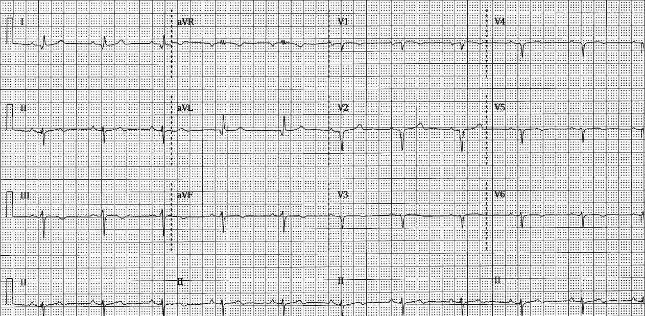
Shown below is an EKG showing Q wave in leads I and aVL consistent with an anterior myocardial infarction of undetermined age.
Shown below are two EKGs of the same subject: while the first EKG demonstrates no Q waves, the second EKG performed after a year shows Q wave in leads I and aVL consistent with an anterior myocardial infarction of undetermined age.
Shown below is the first EKG.

Shown below is the second EKG of the same subject.

Lateral Myocardial Infarction
Shown below is an EKG demonstrating sinus rhythm and a QRS with a rightward axis, as well as wide Q waves in leads I and aVL as well as a poor R wave progression across the anterior chest leads. There is also slight ST elevation in leads I,aVL , and T wave inversion in the lateral leads. The EKG is consistent with a lateral wall myocardial infarction.
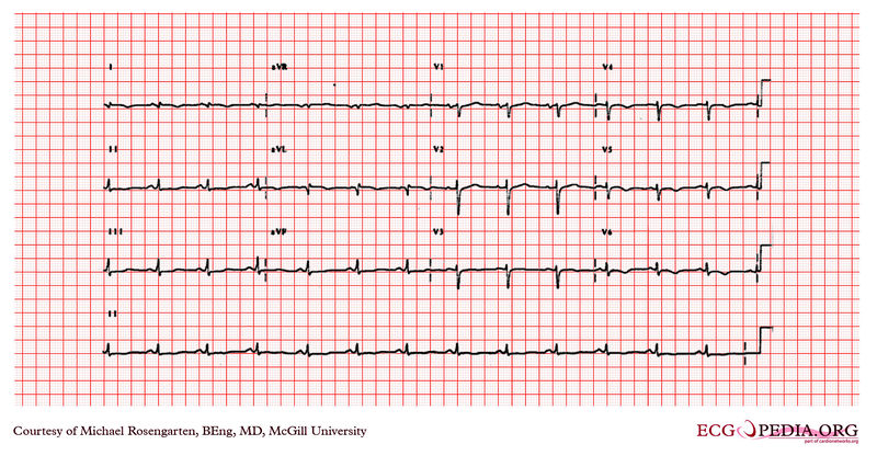
Copyleft image obtained courtesy of, http://en.ecgpedia.org/wiki/Main_Page
Anterolateral Myocardial Infarction
Shown below is an EKG demonstrating sinus rhythm. The remarkable feature is the poor R wave progression in the V1 and V2 leads and the ST elevation and T wave changes in leads V1 to V4 and I and aVL. The cardiogram suggests an anterior/ lateral MI possibly acute. There is also terminal P wave negativity in V1 suggesting a left atrial abnormality.
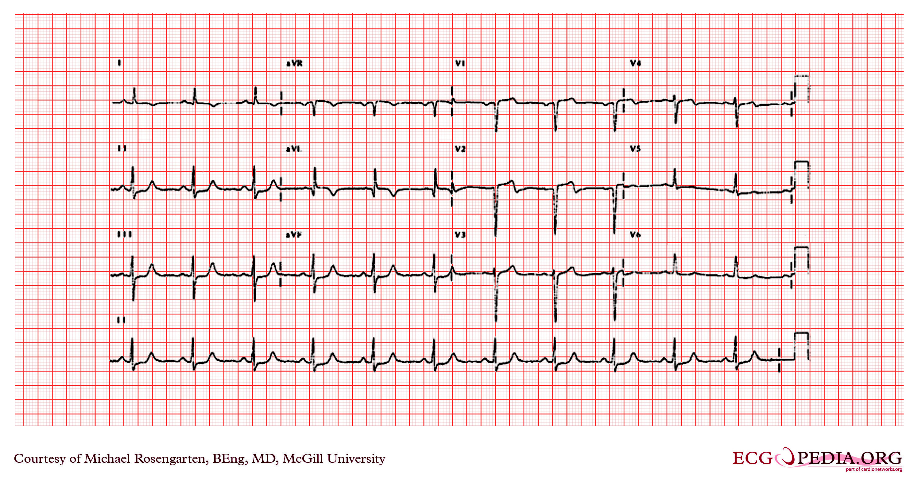
Copyleft image obtained courtesy of, http://en.ecgpedia.org/wiki/File:E209.jpg
Shown below is an EKG demonstrating acute myocardial infarction in in a patient with a pacemaker and LBBB. Concordant ST elevation in V5-V6 are clearly visible. There is discordant ST segment elevation > 5 mm in lead V3.
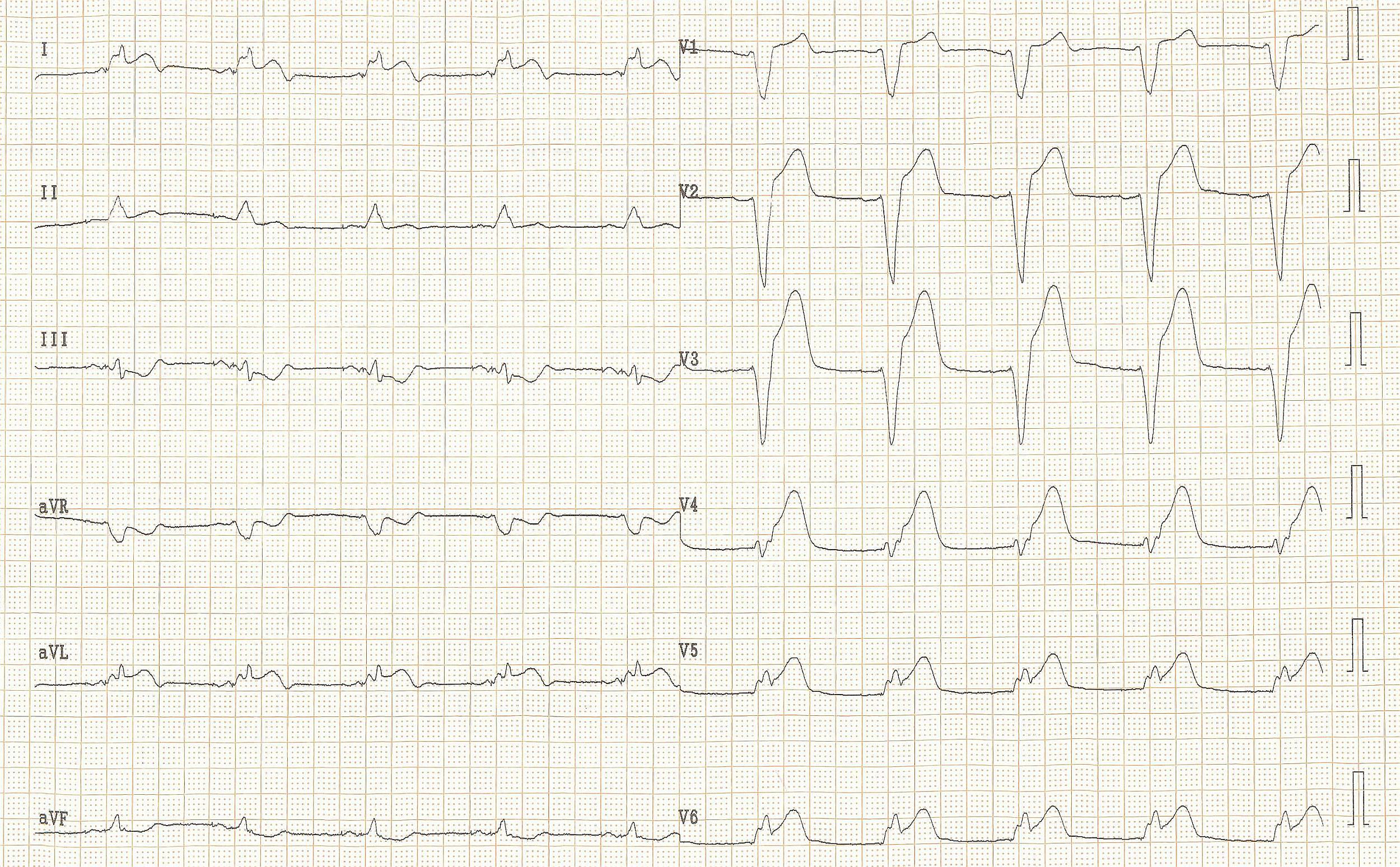
Copyleft image obtained courtesy of,http://en.ecgpedia.org/wiki/Main_Page
Shown below is an EKG demonstrating findings in the same patient as in the first example 2 months before the myocardial infarction. Normal LBBB pattern.
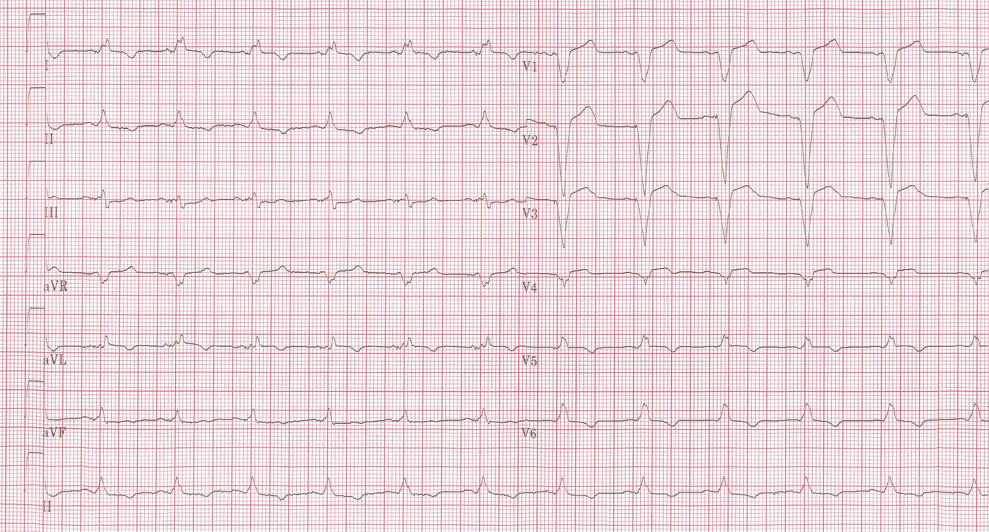
Copyleft image obtained courtesy of,http://en.ecgpedia.org/wiki/Main_Page
Shown below is an EKG showing ST elevation MI.
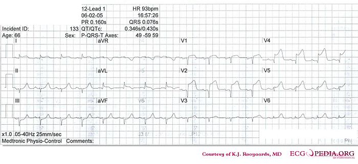
Copyleft image obtained courtesy of, http://en.ecgpedia.org/wiki/File:De-KJcasus12.jpg
Inferior Myocardial Infarction
Shown below is an EKG demonstrating ST elevation in the precordial and limb leads depicting acute inferior MI.

Copyleft image obtained courtesy of, http://en.ecgpedia.org/wiki/Main_Page
Shown below is an EKG demonstrating changes during acute inferior MI depicting ST elevation in leads II, III and aVF.
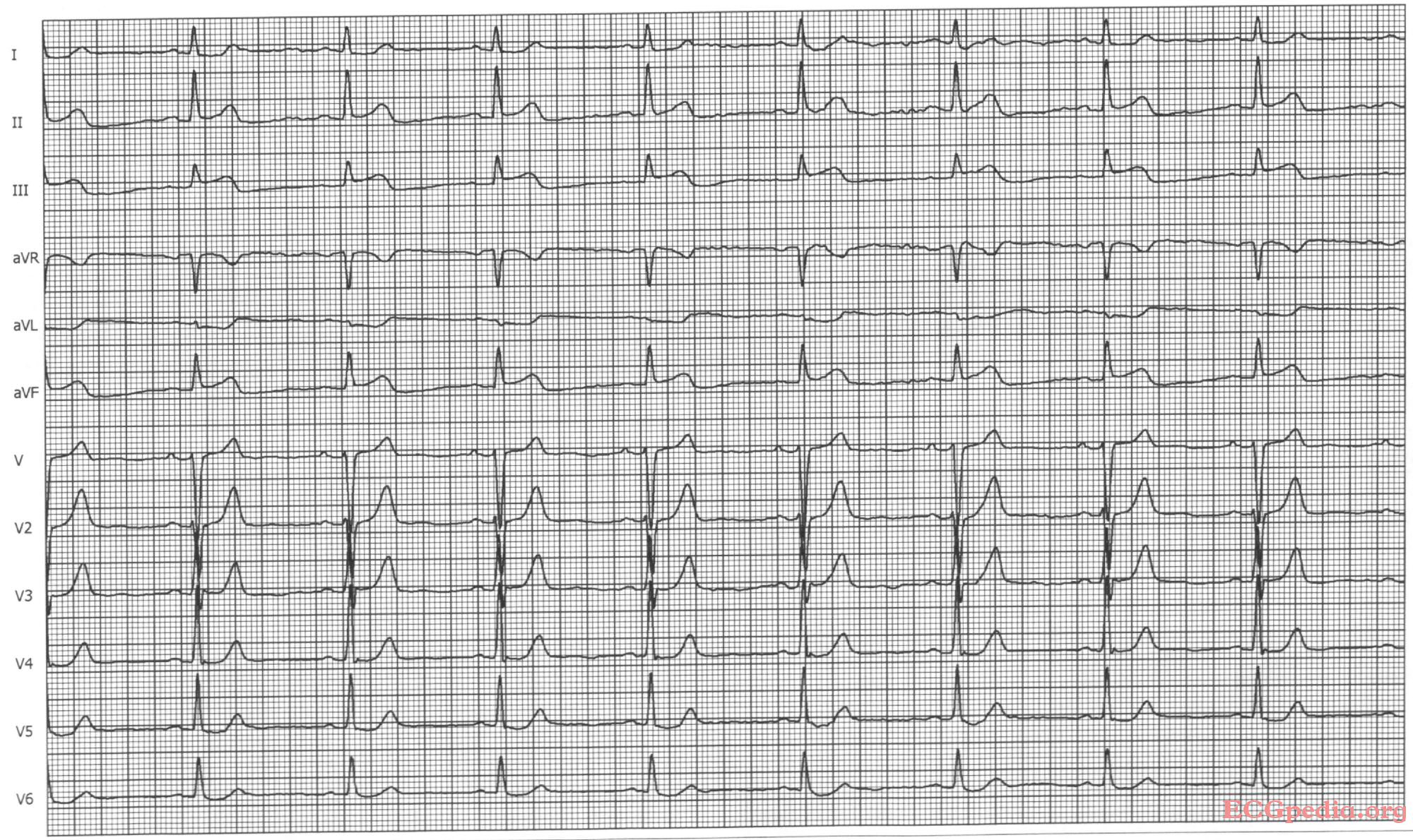
Copyleft image obtained courtesy of, http://en.ecgpedia.org/wiki/Main_Page
Shown below is an EKG demonstrating RBBB and inferior MI. Note to left axis deviation.
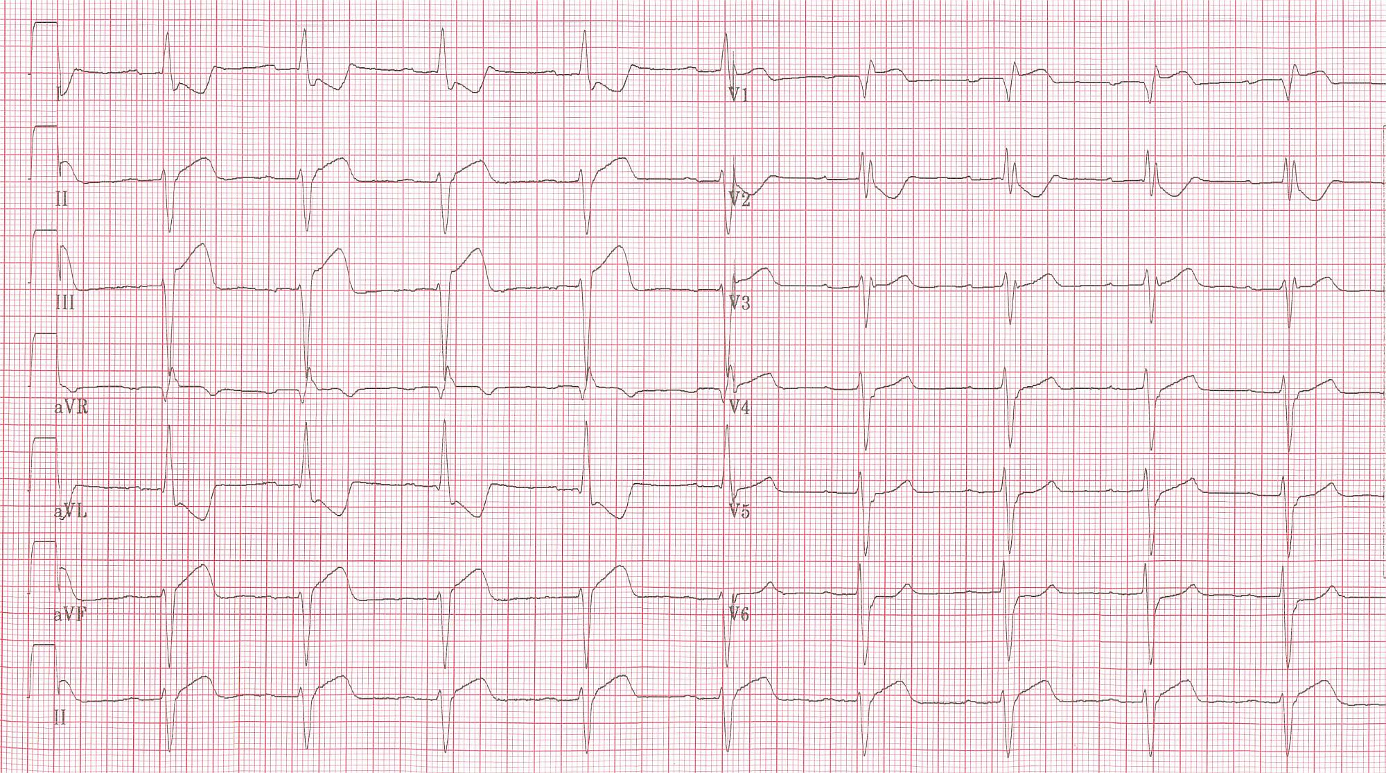
Copyleft image obtained courtesy of, http://en.ecgpedia.org/wiki/Main_Page
Shown below is an EKG demonstrating lead V4R in a patient with RBBB and inferior MI, which clearly shows ST elevation.
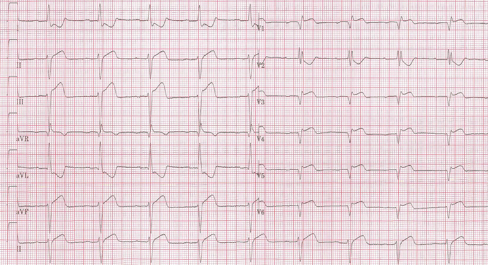
Copyleft image obtained courtesy of,http://en.ecgpedia.org/wiki/Main_Page
Shown below is an EKG demonstrating atrial fibrillation with inferior-posterior-lateral myocardial infarction and incomplete right bundle branch block. Lead I shows ST depression, suggestive of right coronary artery involvement.

Copyleft image obtained courtesy of, http://en.ecgpedia.org/wiki/Main_Page
Shown below is an EKG showing ST elevation in inferior leads.

Copyleft image obtained courtesy of, http://en.ecgpedia.org/wiki/File:De-Ami0007.jpg
Shown below is an EKG showing ST elevation MI.
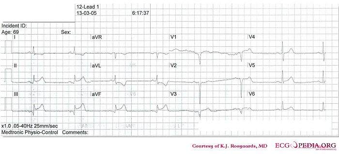
Copyleft image obtained courtesy of, http://en.ecgpedia.org/wiki/File:De-KJcasus16.jpg
Shown below is an EKG showing ST elevation in inferior leads.
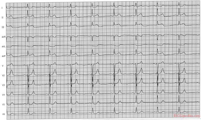
Copyleft image obtained courtesy of, http://en.ecgpedia.org/wiki/File:De-Ami0011.jpg
Shown below is an EKG demonstrating sinus rhythm. The QRS shows Q waves in the inferior leads which are wide (>30ms) and about 25% of the QRS height in aVF. There is also slight ST elevation in the inferior leads and T wave inversion. The EKG suggests an inferior wall infarction, probably old. (the best way to determine "old" is to see a previous cardiogram).
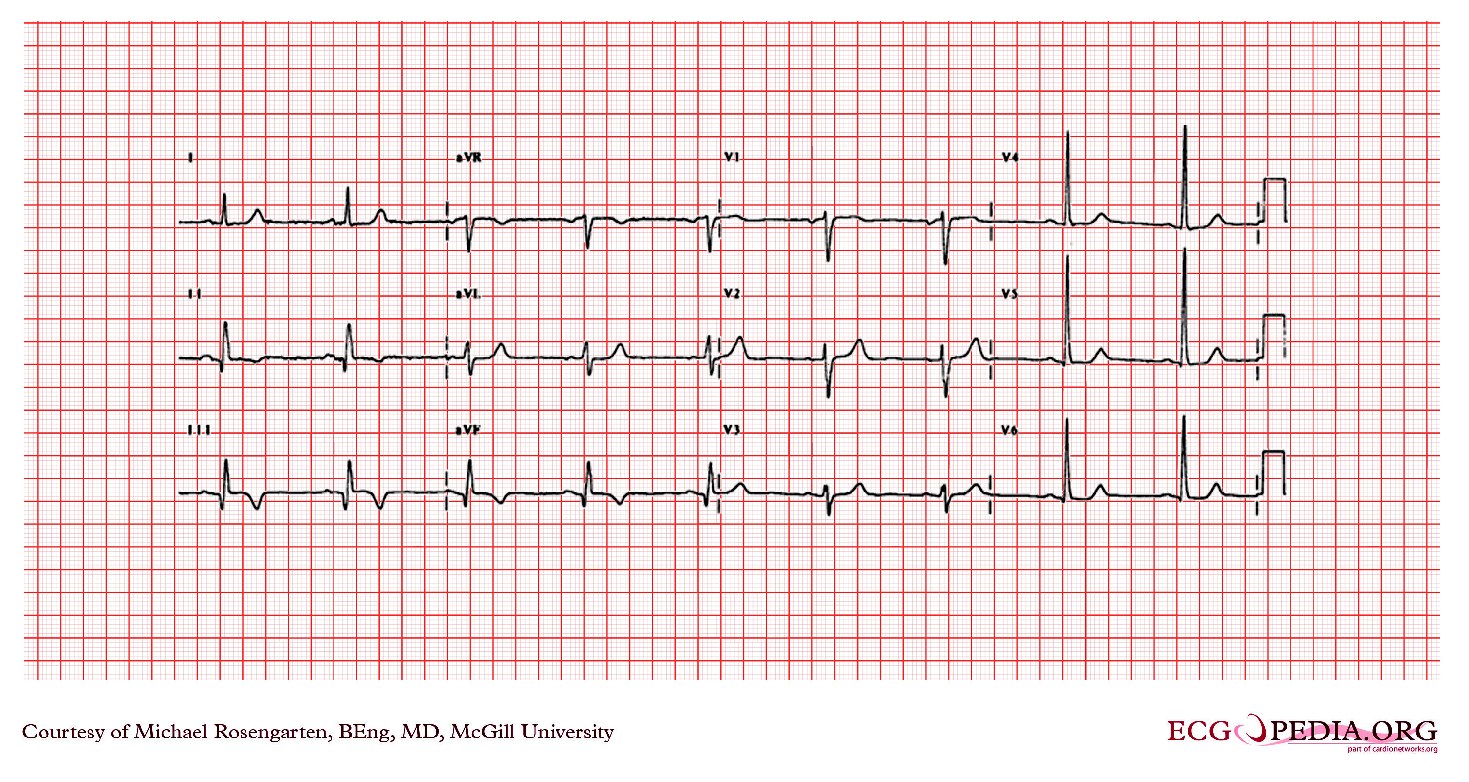
Copyleft image obtained courtesy of, http://en.ecgpedia.org/wiki/Main_Page
Posterior Myocardial Infarction
Shown below is an EKG with ST elevation in II, III, aVF (in III > II), ST depression in I, aVL, V2. Tall R in V2, otherwise normal QRS morphology. The findings are suggestive of acute posteroinferior MI.
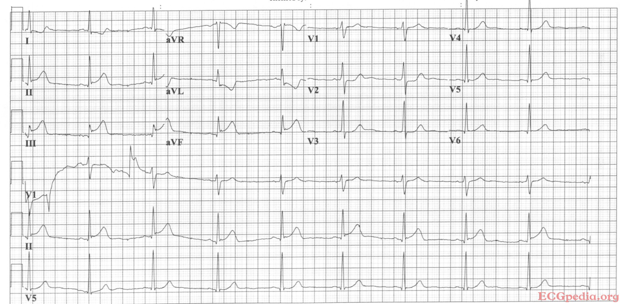
Copyleft image obtained courtesy of, http://en.ecgpedia.org/wiki/Main_Page
Shown below is an EKG demonstrating changes during acute posterolateral MI depicting ST depression in precordial leads V2-V6.
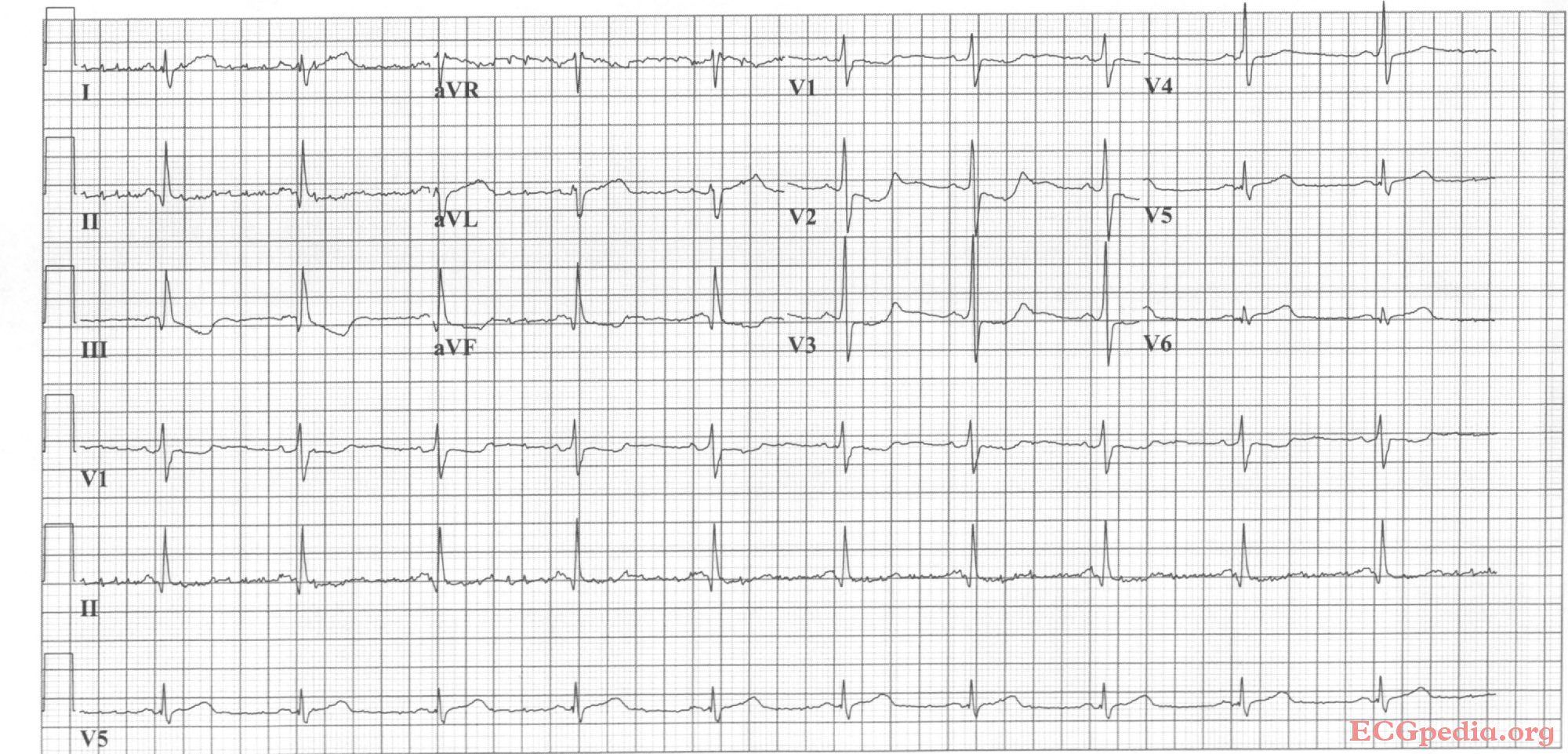
Copyleft image obtained courtesy of, http://en.ecgpedia.org/wiki/Main_Page
Infero-Posterior Myocardial Infarction
Shown below is an EKG with ST depression in V1, V4, tall R in V2. ST elevation in II, III, aVF, V5 and V6.
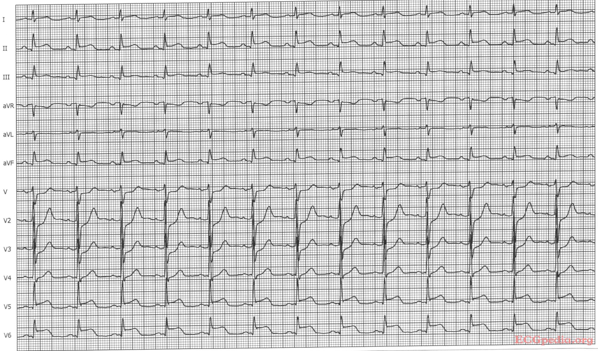
Copyleft image obtained courtesy of, http://en.ecgpedia.org/wiki/Main_Page
Shown below is an EKG with sinus bradycardia with first degree AV block and inferior-posterior-lateral myocardial infarction.
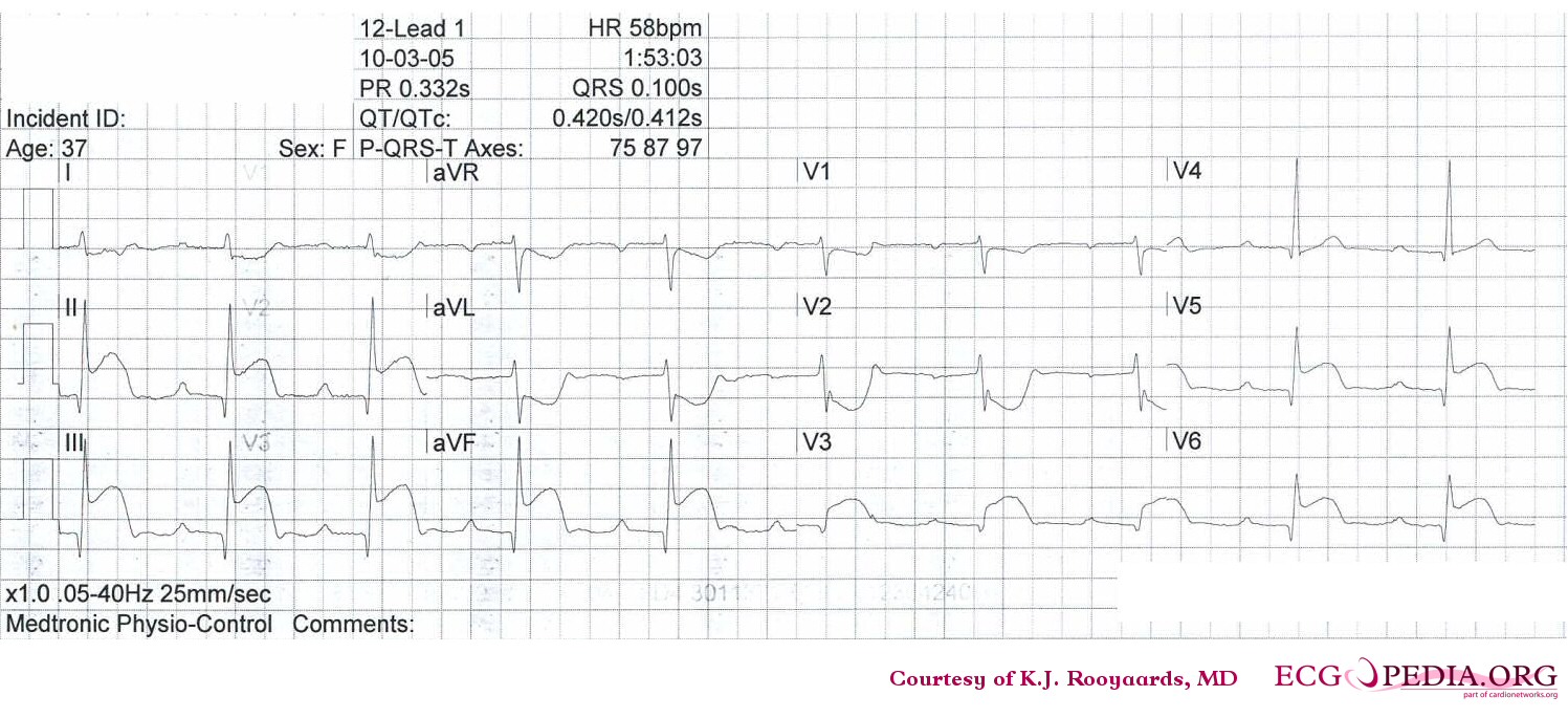
Copyleft image obtained courtesy of, http://en.ecgpedia.org/wiki/Main_Page
Shown below is an EKG depicting sinus bradycardia with inferior-lateral myocardial infarction.

Copyleft image obtained courtesy of, http://en.ecgpedia.org/wiki/Main_Page
Shown below is an EKG illustrating inferior-posterior myocardial infarction with complete AV block and ventricular escape rhythm with RBBB pattern and left axis, followed by sinus rhythm.
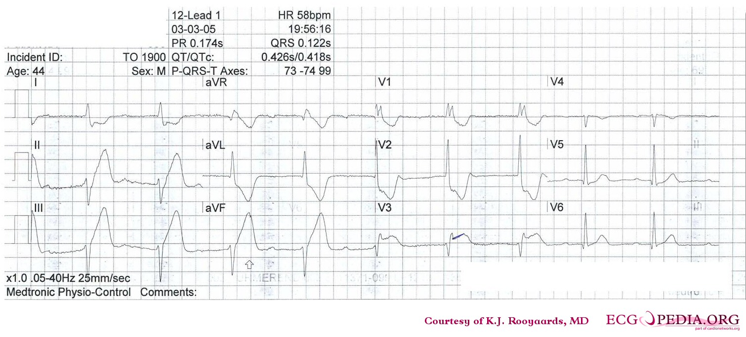
Copyleft image obtained courtesy of, http://en.ecgpedia.org/wiki/Main_Page
Shown below is an EKG demonstrating atrial fibrillation and inferior-posterior myocardial infarction.
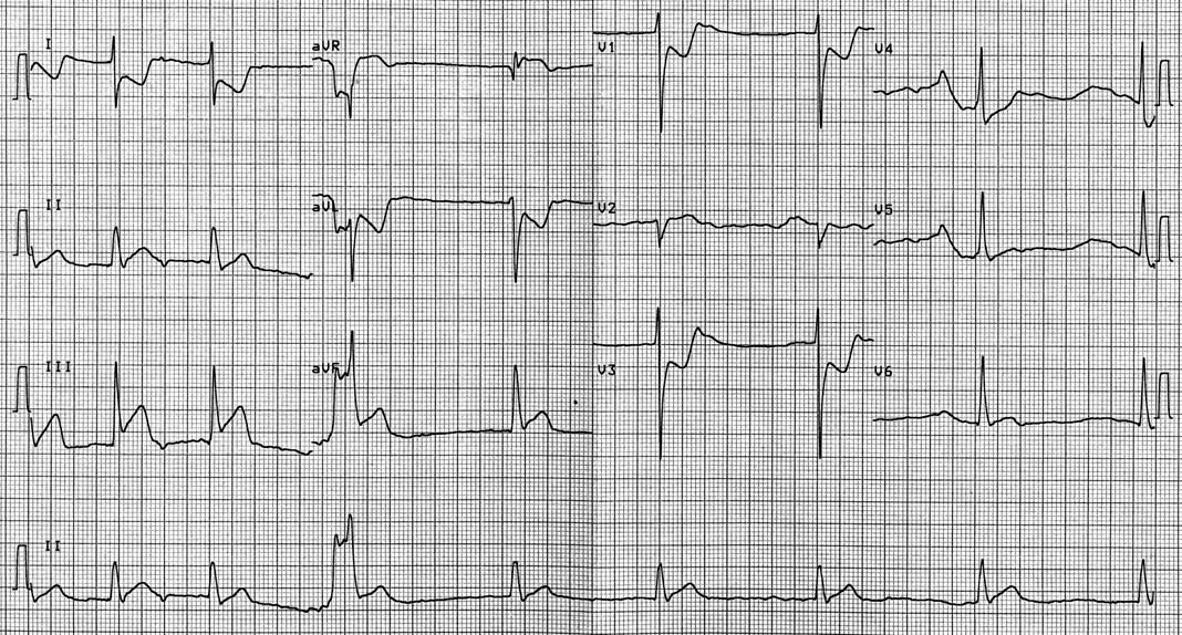
Copyleft image obtained courtesy of, http://en.ecgpedia.org/wiki/Main_Page
Shown below is an EKG demonstrating inferior-posterior-lateral myocardial infarction with a nodal escape rhythm

Copyleft image obtained courtesy of, http://en.ecgpedia.org/wiki/Main_Page
Shown below is an EKG showing ST elevation MI.
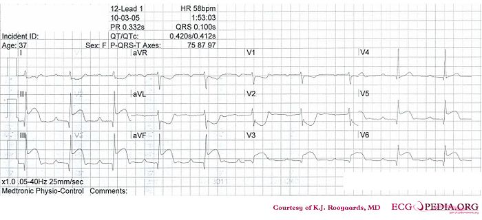
Copyleft image obtained courtesy of, http://en.ecgpedia.org/wiki/File:De-KJcasus13.jpg
Shown below is an EKG showing ST elevation MI.
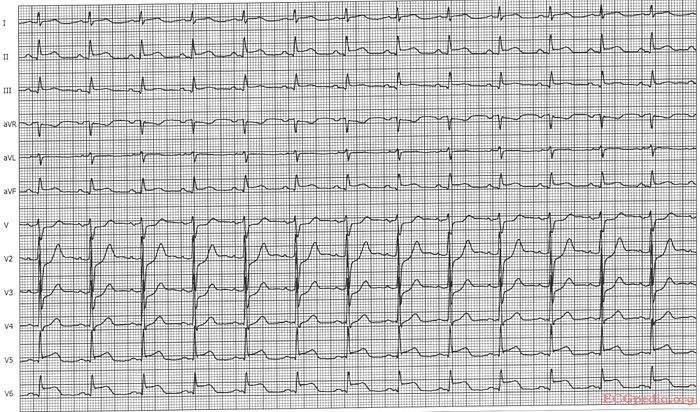
Copyleft image obtained courtesy of, http://en.ecgpedia.org/wiki/File:De-Ami0010.jpg
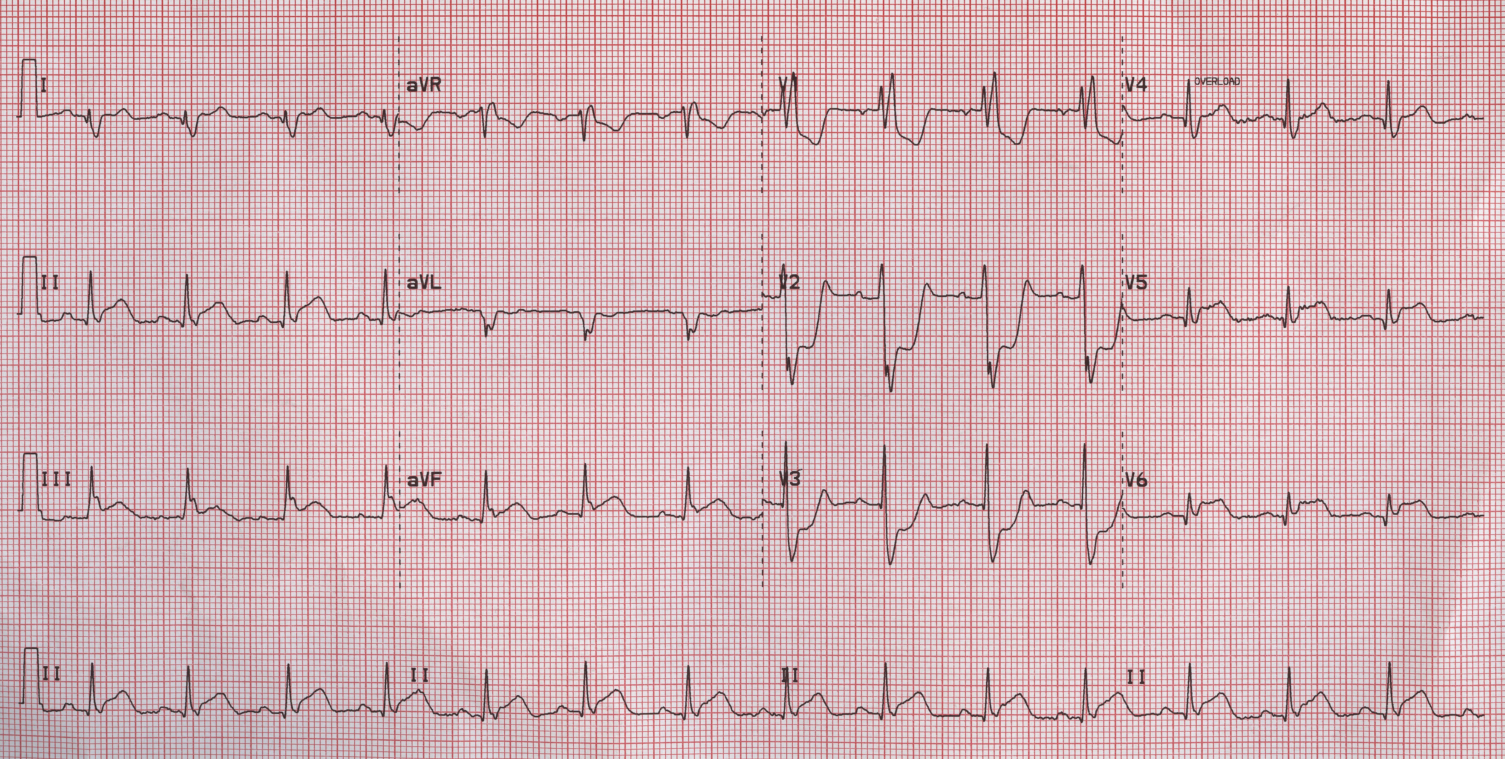
Shown below is an EKG demonstrating ST elevation in leads II, III and aVF and ST depression in leads V1, V2 and V3 depicting a posterior MI.
Right Ventricular Myocardial Infarction
Shown below is an EKG demonstrating ST elevation in lead V1 and aVR; reversal of V6.
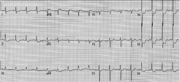
Copyleft image obtained courtesy of, http://en.ecgpedia.org/wiki/Main_Page
Shown below is an EKG demonstrating STEMI changes in the right precordial leads.
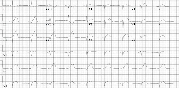
Copyleft image obtained courtesy of, http://en.ecgpedia.org/wiki/Main_Page
Shown below is an EKG demonstrating clear ST elevation in the right precordial leads. A coronary angiography revealed a proximal right coronary artery occlusion.
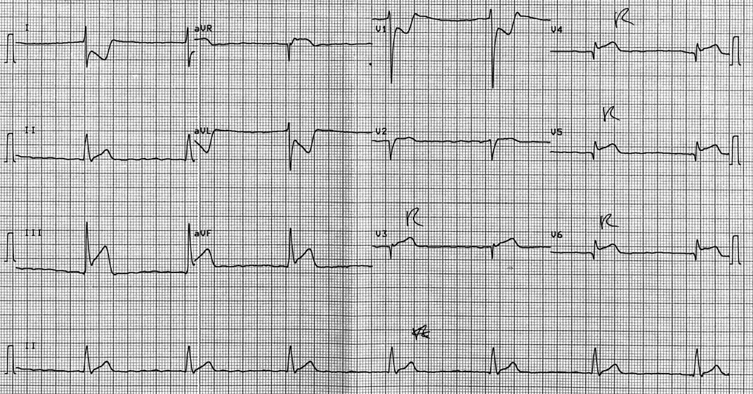
Copyleft image obtained courtesy of, http://en.ecgpedia.org/wiki/Main_Page