Umbilical cord
|
WikiDoc Resources for Umbilical cord |
|
Articles |
|---|
|
Most recent articles on Umbilical cord Most cited articles on Umbilical cord |
|
Media |
|
Powerpoint slides on Umbilical cord |
|
Evidence Based Medicine |
|
Clinical Trials |
|
Ongoing Trials on Umbilical cord at Clinical Trials.gov Trial results on Umbilical cord Clinical Trials on Umbilical cord at Google
|
|
Guidelines / Policies / Govt |
|
US National Guidelines Clearinghouse on Umbilical cord NICE Guidance on Umbilical cord
|
|
Books |
|
News |
|
Commentary |
|
Definitions |
|
Patient Resources / Community |
|
Patient resources on Umbilical cord Discussion groups on Umbilical cord Patient Handouts on Umbilical cord Directions to Hospitals Treating Umbilical cord Risk calculators and risk factors for Umbilical cord
|
|
Healthcare Provider Resources |
|
Causes & Risk Factors for Umbilical cord |
|
Continuing Medical Education (CME) |
|
International |
|
|
|
Business |
|
Experimental / Informatics |
Editor-In-Chief: C. Michael Gibson, M.S., M.D. [1]
Overview
In placental mammals, the umbilical cord is a tube that connects a developing embryo or fetus to the placenta. It normally contains three vessels, two arteries (Umbilical artery) and one vein (Umbilical vein), buried within Wharton's jelly, for the exchange of nutrient- and oxygen-rich blood between the embryo and placenta. The presence of only two vessels in the cord is sometimes related to abnormalities in the fetus, but may occur without accompanying abnormalities.
Explanation
The umbilical cord develops from the same sperm and egg from which the placenta and fetus develop, and contains remnants of the yolk sac and allantois. In humans, the umbilical cord in a full term neonate is usually about 50 centimetres (19.7 in) long and about 2 centimetres (0.75 in) diameter, shrinking rapidly in diameter in the after birth.

In the third stage of labour, after the child is born, the uterus spontaneously expels the neonate's placenta along with the cord from the mother's body, 10–45 minutes after the birth. However, the umbilical cord is generally clamped during or within minutes of birth and severed shortly after, a practice of "active management of labor" which has become increasingly controversial due to the lower transfer of placental blood to the neonate and associated stressors. The health benefits of non-clamping of the cord and delayed umbilical severance are receiving attention in medical journals.[1][2][3]
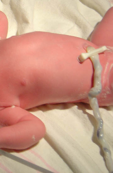
Today there are umbilical cord clamps which combine the cord clamps with the knife. These clamps are safer and faster, allowing one to first apply the cord clamp and then cut the umbilical cord. After the cord is clamped and cut (Western obstetrical protocol) the newborn wears a plastic clip on the navel area until the compressed region of the cord has dried and sealed sufficiently. The remaining umbilical stub remains for up to 2–3 weeks as it dries and then falls off. In nonseverance scenarios, also called lotus birth, the umbilical cord is wrapped up to within an inch of the newborn's belly, and the entire intact cord is allowed to dry like a sinew, which then falls off.[4]
Makeup and composition
The umbilical cord is made of Wharton's jelly, not ordinary skin and connective tissue. There are no nerves, so cutting it is not painful. There is ordinarily no significant loss of either infant or maternal blood while cutting the cord. In many Western cultures the umbilical cord is traditionally cut by the father of the baby. The cord contains two arteries which carry deoxygenated blood (from the fetus back to the mother) and one vein that carries oxygenated blood (from the mother to the fetus).
Cord blood
Recently, it has been discovered that the blood within the umbilical cord, known as cord blood, is a rich and readily available source of primitive, undifferentiated stem cells (i.e. CD34-positive and CD38-negative). These cord blood cells can be used for bone marrow transplant.
Others have chosen to have this blood diverted from the baby's umbilical blood transfer through early cord clamping and cutting, to freeze for long-term (and costly) storage at a cord blood bank should the child ever require the cord blood stem cells (for example, to replace bone marrow destroyed when treating leukemia). This practice is somewhat controversial, [citation needed] with critics asserting that early cord blood withdrawal actually increases the likelihood of childhood disease. The Royal College of Obstetricians and Gynaecologists 2006 opinion states, "There is still insufficient evidence to recommend directed commercial cord blood collection and stem-cell storage in low-risk families."
In the future, cord blood-derived embryonic-like stem cells (CBEs) may also be banked and matched with other patients, much like blood and transplanted tissues. The use of CBEs would eliminate the ethical difficulties associated with embryonic stem cells (ESCs).[5]
Problems
A number of abnormalities can affect the umbilical cord, which can cause problems that affect both mother and child:
- Nuchal cord
- Single umbilical artery
- Umbilical cord prolapse
- Umbilical cord knot
- Umbilical cord entanglement
- Vasa previa
- Velamentous cord insertion

Animals
In other mammals, the mother animal generally will gnaw the cord off separating the placenta from the baby. It is often consumed by the mother which nourishes her, and reduces tissue that would attract scavengers or predators. In chimpanzees, the mother focuses no attention on umbilical severance, instead staying still and nursing and holding her baby (with cord, placenta, and all) until the cord dries and separates within a day of birth, at which time she leaves the cord and placenta on the forest floor where it is recycled by scavengers. This was first documented by zoologists in the wild in 1974.[citation needed]
Other uses for the term "umbilical cord"
The term "umbilical cord" or just "umbilical" has also come to be used for other cords with similar functions, such as the hose connecting a surface-supplied diver to his surface supply of air and/or heating, or a space-suited astronaut to his spacecraft.
The phrase "cutting the umbilical cord" is used symbolically to describe a child's breaking away from the parental home.
Additional images
-
Diagram illustrating a later stage in the development of the umbilical cord.
-
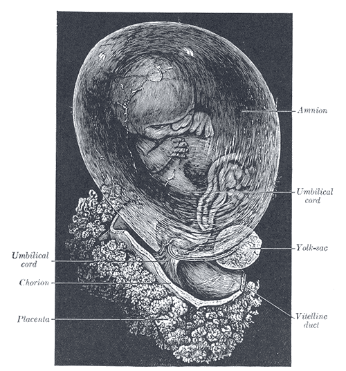
Fetus of about eight weeks, enclosed in the amnion. Magnified a little over two diameters.
-
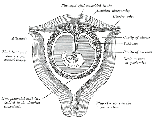
Sectional plan of the gravid uterus in the third and fourth month.
-
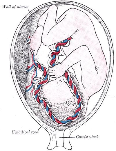
Fetus in utero, between fifth and sixth months.
-
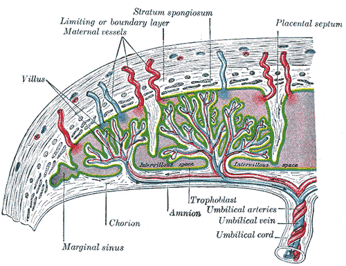
Scheme of placental circulation.
-
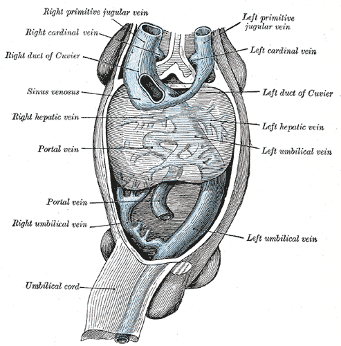
Human embryo with heart and anterior body-wall removed to show the sinus venosus and its tributaries.
References
- ↑ Hohmann M. (1985). "Early or late cord clamping? A question of optimal time" (Article in German). Wiener Klinische Wochenschrift, 97(11):497-500. PMID 4013344.
- ↑ Mercer J.S., B.R. Vohr, M.M. McGrath, J.F. Padbury, M. Wallach, W. Oh (2006). "Delayed cord clamping in very preterm infants reduces the incidence of intraventricular hemorrhage and late-onset sepsis: a randomized, controlled trial." Pediatrics, 117(4):1235-42. PMID 16585320.
- ↑ Hutton E.K., E.S. Hassan (2007). "Late vs early clamping of the umbilical cord in full-term neonates: systematic review and meta-analysis of controlled trials." Journal of the American Medical Association, 297(11):1257-58. PMID 17374818.
- ↑ Crowther S (2006). "Lotus birth: leaving the cord alone." The Practising Midwife, 9(6):12-14. PMID 16830839.
- ↑ "Cord blood yields 'ethical' embryonic stem cells.", Coghlin A. New Scientist, August 18, 2005. Accessed June 25, 2007.
ar:حبل سري da:Navlestreng de:Nabelschnur fi:Napanuora it:Funicolo ombelicale he:חבל הטבור la:Funiculus umbilicalis lt:Virkštelė nl:Navelstreng simple:Umbilical cord su:Ari-ari sv:Navelsträng
