Thromboembolism physical examination
|
Thromboembolism Microchapters |
|
Diagnosis |
|---|
|
Treatment |
|
Case Studies |
|
Thromboembolism On the Web |
|
American Roentgen Ray Society Images of Thromboembolism |
Please help WikiDoc by adding more content here. It's easy! Click here to learn about editing.
Editor-In-Chief: C. Michael Gibson, M.S., M.D. [1]
Physical Examination
There are several techniques during physical examination to increase the detection of DVT, such as measuring the circumference of the affected and the contralateral limb at a fixed point (to objectivate edema), and palpating the venous tract, which is often tender. Physical examination alone is unreliable for excluding the diagnosis of deep vein thrombosis.
Extremities
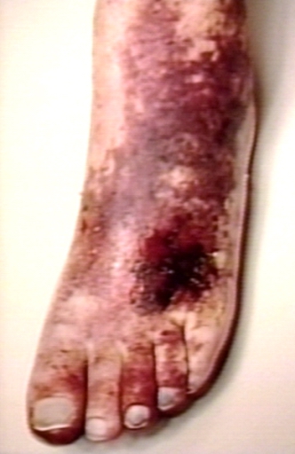
In phlegmasia alba dolens, the leg is pale and cool with a diminished arterial pulse due to spasm. It usually results from acute occlusion of the iliac and femoral veins due to DVT.
In phlegmasia cerulea dolens, there is an acute and nearly total venous occlusion of the entire extremity outflow, including the iliac and femoral veins. The leg is usually painful, cyanosed and oedematous. Venous gangrene may supervene.
- Homans' sign
- Pratt's sign: Squeezing of posterior calf elicits pain.
However, these medical signs do not perform well and are not included in clinical prediction rules that combine best findings in order to diagnose DVT.[1]
(Images courtesy of Charlie Goldberg, M.D., UCSD School of Medicine and VA Medical Center, San Diego, California)
-
Deep venous thrombosis: Right Lower Extremity DVT
-
Deep venous thrombosis: Left Lower Extremity DVT
-
Deep venous thrombosis: Left Lower Extremity DVT
Image:upper dvt.jpg|Deep venous thrombosis: Diffusely swollen RUE resulting from a PICC line induced thrombosis. Image:extremities_dvt4.jpg|Left Lower Extremity DVT:Note diffusely swollen left leg. skin changes on left are due to chronic venous insufficiency. </gallery>
-
 Thromboembolic event of popliteal artery
 -
 Fundoscopy: Central retinal vein thrombosis


References
- ↑ Wells PS, Owen C, Doucette S, Fergusson D, Tran H (2006). "Does this patient have deep vein thrombosis?". JAMA. 295 (2): 199–207. doi:10.1001/jama.295.2.199. PMID 16403932.