Thromboembolism MRI
Jump to navigation
Jump to search
|
Thromboembolism Microchapters |
|
Diagnosis |
|---|
|
Treatment |
|
Case Studies |
|
Thromboembolism On the Web |
|
American Roentgen Ray Society Images of Thromboembolism |
Editor-In-Chief: C. Michael Gibson, M.S., M.D. [1]
MRI
- Gadolinium-enhanced MRI is a non-invasive diagnostic modality and has the advantage of no contrast exposure.
- A potential benefit of MR, is that is incredibly sensitive, perhaps even better than contrast venography, in imaging clot in the inferior vena cava (IVC) and pelvic veins, and these images can be obtained at the same time as the lung scan.
- It needs to be pointed out, that although the criticism of using CT and MR angio lacks sensitivity when examining the subsegmental arteries, inter-reader agreement was only 66% with pulmonary angiography in PIOPED.
(Images shown below are courtesy of RadsWiki)
-
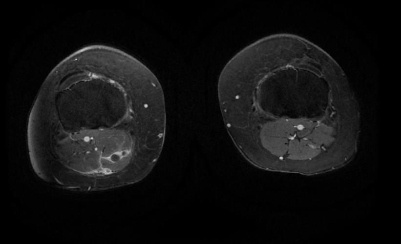
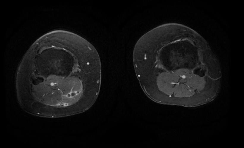
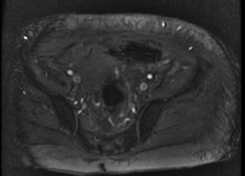 2D TOF GRE MRV images: Bilateral deep vein thrombosis
 -
 2D TOF GRE MRV images: Bilateral deep vein thrombosis
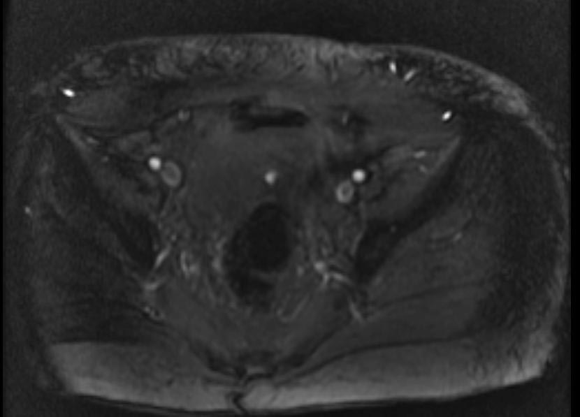
 -
 2D TOF GRE MRV images: Bilateral deep vein thrombosis


-
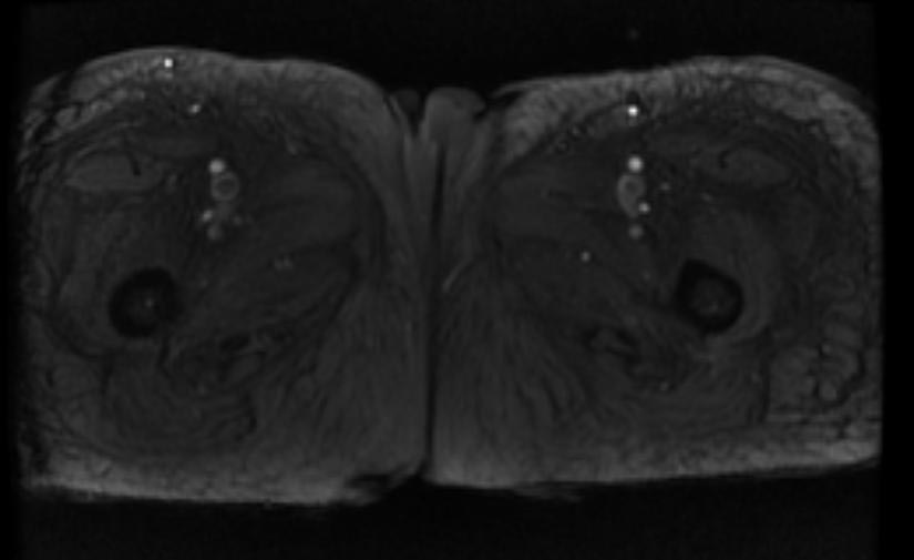
 MRI: Superficial vein thrombosis
 -
 MRI: Superficial vein thrombosis
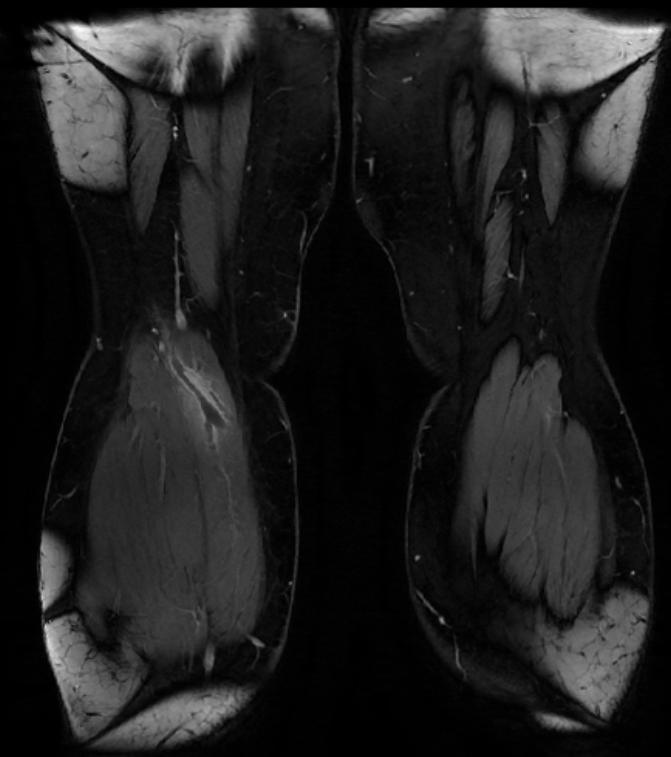
 -
 MRI: Superficial vein thrombosis


