Chest

|
WikiDoc Resources for Chest |
|
Articles |
|---|
|
Media |
|
Evidence Based Medicine |
|
Clinical Trials |
|
Ongoing Trials on Chest at Clinical Trials.gov Clinical Trials on Chest at Google
|
|
Guidelines / Policies / Govt |
|
US National Guidelines Clearinghouse on Chest
|
|
Books |
|
News |
|
Commentary |
|
Definitions |
|
Patient Resources / Community |
|
Directions to Hospitals Treating Chest Risk calculators and risk factors for Chest
|
|
Healthcare Provider Resources |
|
Continuing Medical Education (CME) |
|
International |
|
|
|
Business |
|
Experimental / Informatics |
Editor-In-Chief: C. Michael Gibson, M.S., M.D. [1]
Overview
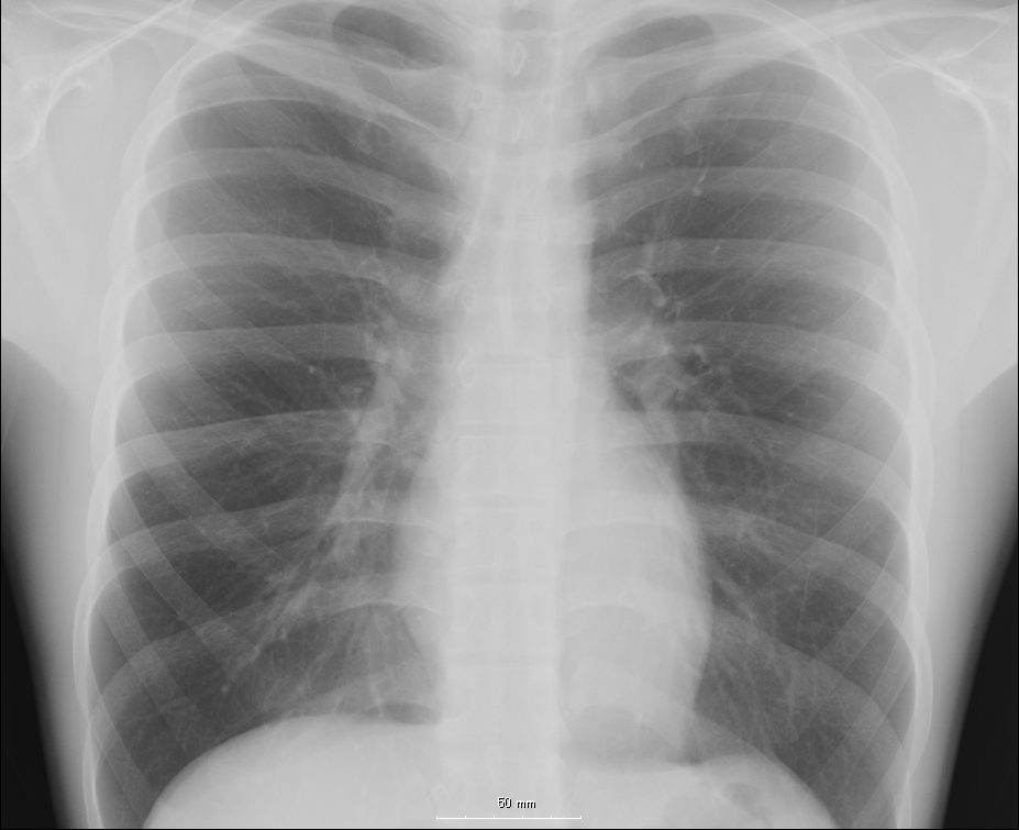
The chest is a part of the anatomy of humans and various other animals.
Human chest anatomy
In human, the chest is the region of the body between the neck and the abdomen, along with its internal organs and other contents. It is mostly protected and supported by the ribcage, spine, and shoulder girdle. Contents of the chest include the following:
- organs
- muscles
- internal structures
- arteries and veins

- bones
- the shoulder socket containing the upper part of the humerus
- scapula
- sternum
- thoracic portion of the spine
- collarbone
- ribcage
- Floating ribs
- external structures
- thoracic abdomen (stomach, kidney/adrenal, pancreas, spleen, and lower oesophagus)
In humans, the portion of the chest protected by the ribcage is also called the thorax.
Chest injury
Injury to the chest (also referred to as chest trauma, thoracic injury, or thoracic trauma) results in up to ¼ of all deaths due to trauma in the United States.[1]
In the human body, the chest is the body region between the neck and diaphragm in the front of the body. The corresponding area in an animal can be referred to as the chest. The chest holds many important internal, and is protected by the ribcage.
It is important to realize that the shape of the chest does not correspond to that of the bony thorax which encloses the heart and lungs; all the breadth of the shoulders is due to the shoulder girdle, and contains the axilla and the head of the humerus. In the middle line the suprasternal notch is seen above, while about three fingers' breadth below it a transverse ridge can be felt, which is known as (Ludovic's angle) and marks the junction between the manubrium and gladiolus of the sternum. Level with this line the second ribs join the sternum, and when these are found the lower ribs may be easily counted in a moderately thin subject. At the lower part of the sternum, where the seventh or last true ribs join it, the ensiform cartilage begins, and over this there is often a depression popularly known as the pit of the stomach.
The nipple in the male is situated in front of the fourth rib or a little below; vertically it lies a little external to a line drawn down from the middle of the clavicle; in the female it is not so constant. A little below it the lower limit of the great pectoral muscle is seen running upward and outward to the axilla; in the female this is obscured by the breast, which extends from the second to the sixth rib vertically and from the edge of the sternum to the mid-axillary line laterally. The female nipple is surrounded for half an inch by a more or less pigmented disc, the areola. The apex of a normal heart is in the fifth left intercostal space, three and a half inches from the mid-line.
References
- ↑ Shahani, Rohit, MD. (2005). Penetrating Chest Trauma. eMedicine. Retrieved 2005-02-05.
See also
- Thoracic cavity
- Pectus excavatum
- Pectus carinatum
- Breast
- Chest hair
- Barechested
Template:Human anatomical features Template:Torso general
ar:صدر
ay:Ñuñu
de:Brust
ln:Bontólo
qu:Qhasqu
simple:Chest
sk:Hrudník
fi:Rinta
sv:Bröst (könsneutralt)
tl:Dibdib
yi:ברוסט קאסטען
[[zh:胸]