Pilon fracture

| Pilon fracture | |
| ICD-10 | S42.2-S42.4 |
|---|---|
| ICD-9 | 812 |
| eMedicine | emerg/199 orthoped/271 orthoped/199 |
|
WikiDoc Resources for Pilon fracture |
|
Articles |
|---|
|
Most recent articles on Pilon fracture Most cited articles on Pilon fracture |
|
Media |
|
Powerpoint slides on Pilon fracture |
|
Evidence Based Medicine |
|
Clinical Trials |
|
Ongoing Trials on Pilon fracture at Clinical Trials.gov Trial results on Pilon fracture Clinical Trials on Pilon fracture at Google
|
|
Guidelines / Policies / Govt |
|
US National Guidelines Clearinghouse on Pilon fracture NICE Guidance on Pilon fracture
|
|
Books |
|
News |
|
Commentary |
|
Definitions |
|
Patient Resources / Community |
|
Patient resources on Pilon fracture Discussion groups on Pilon fracture Patient Handouts on Pilon fracture Directions to Hospitals Treating Pilon fracture Risk calculators and risk factors for Pilon fracture
|
|
Healthcare Provider Resources |
|
Causes & Risk Factors for Pilon fracture |
|
Continuing Medical Education (CME) |
|
International |
|
|
|
Business |
|
Experimental / Informatics |
Editor-In-Chief: C. Michael Gibson, M.S., M.D. [1]; Associate Editor(s)-in-Chief: Mohammadmain Rezazadehsaatlou[2].
Synonyms and Keywords: Pilon fracture, Plafond fracture
Overview
Pilon fracture is a fracture of the distal part of the tibia bone (also called the shinbone). Meanwhile, the injured tibia may break into one or multiple pieces.
Historical Perspective [1]
There are no reliable information regarding the historical perspective of the Pilon fracture but:
The word "Pilon" is the French word for "pestle" which was first introduced by Étienne Destot into orthopedic literature in 1911.
Causes [2]
The Pilon fractures most often result from high-energy trauma like: a car or motorcycle accident, fall from height, or skiing accident due to the rotational or axial-loading forces to the tibia bone. Meanwhile, there are other causes responsible for this type of fracture such as:
- Pressure during car accidents
- Twisted ankle side to side
- Rotated ankle side to side
- Rolling ankle in or out
- Hyper-flextion
- Hyper-Extention
- Tripping
- Falling from a height
- Jumping from a height
As a person age, two factors cause higher risk of fractures:
- Weaker bones
- Greater risk of falling
Stress fractures as a common causes of fractures can be found due to the repeated stresses and strains. Importantly children having more physically active lifestyles than adults, are also prone to fractures. People with any underlying diseases such as osteoporosis, infection, or a tumor affecting their bones having a higher risk of fractures. As mentioned in previous chapters, this type of fracture is known as a pathological fracture. Stress fractures, which result from repeated stresses and strains, commonly found among professional sports people, are also common causes of fractures.
Life-threatening Causes
- There are no life-threatening causes of Pilon fracture, however complications resulting from Pilon fracture is common.
Common Causes
Common causes of Pilon fracture may include:
Less Common Causes
Less common causes of Pilon fracture include conditions that predisposes to fracture:
Causes by Organ System
| Cardiovascular | No underlying causes |
| Chemical/Poisoning | No underlying causes |
| Dental | No underlying causes |
| Dermatologic | No underlying causes |
| Drug Side Effect | No underlying causes |
| Ear Nose Throat | No underlying causes |
| Endocrine | No underlying causes |
| Environmental | No underlying causes |
| Gastroenterologic | No underlying causes |
| Genetic | No underlying causes |
| Hematologic | No underlying causes |
| Iatrogenic | No underlying causes |
| Infectious Disease | No underlying causes |
| Musculoskeletal/Orthopedic | Osteoporosis and osteopenia. |
| Neurologic | No underlying causes |
| Nutritional/Metabolic | Osteoporosis and osteopenia. |
| Obstetric/Gynecologic | No underlying causes |
| Oncologic | No underlying causes |
| Ophthalmologic | No underlying causes |
| Overdose/Toxicity | No underlying causes |
| Psychiatric | No underlying causes |
| Pulmonary | No underlying causes |
| Renal/Electrolyte | No underlying causes |
| Rheumatology/Immunology/Allergy | No underlying causes |
| Sexual | No underlying causes |
| Trauma | Falling of car accident to on side of Ankle . |
| Urologic | No underlying causes |
| Miscellaneous | No underlying causes |
Causes in Alphabetical Order
List the causes of the disease in alphabetical order:
Pathophysiology [3]
The main etiology of the Pilon fracture is thought to excessive inversion stress to the ankle joint.
Mechanism [4]
In 80% of ankle fractures the foot is in supination position while, in 20% of fractures the foot is in pronation position. Meanwhli,e the Pilon fracture is caused by a falling or direct trauma to the ankle joint. The form and severity of this fracture depends on the position of the ankle joint at the moment of the trauma. The ankle joint is flexible but the medial side of the ankle joint is rigid because the medial malleolus is attached to the tibia and also the medial collateral ligaments are very strong. Also, lateral wall of the ankle include: the fibula, syndesmosis and lateral collateral ligaments play important rolls in this flexibility. This lateral wall of the ankle allows the talus to move in lateral and dorsal sides easily. The fibula has no weight-bearing roll but it provide a flexible lateral support. The syndesmosis is formed by the anterior and posterior tibiofibular ligaments which is the fibrous connection between the fibula and tibia.
If we imagine the ankle as a ring in which bones and ligaments play an important role in the maintaining the stability of this joint. Meanwhile, if this ring is broken in one place it remains stable but when it is broken in two places, the ring is unstable and it is at the higher risk of fracture. Consequently the ankle is unstable when both the medial and the lateral malleoli are fractured.
Pathophysiology[3]
Its known that the Pilon fracture in normal healthy adults can be caused due to the high-energy trauma (e.g., motor vehicle accidents), sport related injuries, falling from height. But it should be noted that the most important Risk factors for insufficiency fractures is chronic metabolic disease such as steoporosis, osteopenia, eating-disordered behavior, higher age, prolonged corticosteroid usage, female gender, lower BMI, history of a recent falling, and prior fracture.
- The pattern of bone fracture and severity of injury depends on variety of factors such as:
- Patients age
- Patients Weight
- Patients past medical history specifically any bone diseases affecting the quality of bone (such as osteoporosis, malignancies)
- Energy of trauma
- Bone quality
- Position of the specific organ during the trauma
- The below-mentioned processes cause decreased bone mass density:
- Autophagy is the mechanism through which osteocytes evade oxidative stress.
- The capability of autophagy in cells decreases as they age, a major factor of aging.
- As osteocytes grow, viability of cells decrease thereby decreasing the bone mass density.
Differentiating Pilon fracture from other Diseases [5][6]
In the orthopedic medicine its important to know that the Pilon fracture should be evaluated using radiography for both confirming diagnosis and also for evaluating the surrounding tissues.
- Acute compartment syndrome of ankle joint
- Ankle Dislocation
- Soft tissue Injury around the ankle ,
- Deep Venous thrombosis
- Thrombophlebitis
- Foot Fracture
- Gout
- Pseudogout
- Rheumatoid Arthritis
- Tibia Fracture
- Fibula Fracture
- bimalleolar fracture
- trimalleolar fracture
- triplane fracture
- Tillaux fracture
- Bosworth fracture
- Wagstaffe-Le Forte fracture
- Charcot-Marie-Tooth disease: in cases with repeated Pilon fractures
Epidemiology and Demographics[3]
The Pilon fracture accounts for around 1-10% of all fractures and 5-10 % of tibial fractures. It is more common in adults aged 30s to 40s due to the fall from height or a motor vehicle crash which is more common in this age group.
There are different risk factors that predispose patient for the Pilon fracture that include:
- High-risk contact sports
- Higher age (elderly adults are higher prone to such fractures)
- Reduced bone density (osteoporosis)
- Direct blow
- Road / traffic accidents
- Falling
- Direct trauma to the ankle
- Taking part in any rough or high-impact sport
- Street fights, gunshot wounds, and domestic violence, may also cause the Pilon fracture
- Road traffic accidents.
The Pilon fracture may be classified based on the exact location of fracture:
| Ruedi and Allgower Classification of Pilon Fractures | |
|---|---|
| Type I | Articular involvement with minimal or no displacement |
| Type II | Articular displacement but with minimal or no comminution |
| Type IIII | Marked comminution as well as the articular impaction |
AO/OTA classification for the distal tibial Fracture:
| AO/OTA classification for the distal tibial Fracture | |
|---|---|
| A: Distal tibial metaphyseal without intra-articular injury | 1: Simple Fracture |
| 2: Comminuted Fracture | |
| 3: Severely comminuted Fracture | |
| B: Distal tibial metaphyseal with partial articular fractures | 1: Pure split Fracture |
| 2: Split Fracture with depression | |
| 3: Split Fracture with depression with multiple fragments | |
| C: Distal tibial metaphyseal involves the entire joint area | 1: Simple split Fracture affecting the articular surface and the metaphysis |
| 2: Articular split Fracture with a multifragmentary metaphysis split | |
| 3: Fracture with multiple fragments of the articular surface and the metaphysis area | |
Osteoporosis is an important risk factor for human affecting human bone especially in men with the age of older than 50 years old and postmenopausal and women.
Based on the US Preventive Services Task Force (USPSTF) there are three groups of patients need to be screened for the osteoporosis:
- · Men with no history of osteoporosis
- · Women with the age of 65≤ year old, with no previous history of pathological fracture due to the osteoporosis
- · Women with the age of <65 years, with 10-year fracture risk of not less than a 65-year-old white woman (who has not any other risk factor)
Accordingly women older than age of 50 are the main target for the osteoporosis screening. There is no specific recommendation to screen men for the osteoporosis.
The USPSTF recommendations from 2002 included:
Meanwhile, there are two major modalities for the osteoporosis screening:
- · Dual energy x-ray absorptiometry (DXA) of the hip and lumbar spine bones
- · Quantitative ultrasonography of the calcaneus
*It should be noted of the two above mentioned modalities for screening the ultrasonograhy is preferred to the DXA due to its lower cost, lower ionizing radiation, more availability.
After the primary evaluation of the osteoporosis, the further evaluation are required in some cases such as:
· Women with normal bone density or mild osteopenia: T-score of greater than −1.50 – should have screening for 15 years.
· Women with moderate osteopenia: T-score of −1.50 to −1.99 – should have screening for 5 years.
· Women with advanced osteopenia: T-score of −2.00 to −2.49 - should have screening for 1 year.
Natural History [13]
In cases with untreated Pilon fracture the malunion and deformity of arm can be occurred.
The overall complication rate in the treatment of Pilon fracture were found in around 40% of cases:
- Neurovascular compromise: such as Ulna nerve damage
- Compartment syndrome
- Chronic disability of the DRUJ
- Physeal Injury
- Malunion of the radius
- Nonunion
- Infection
- Refracture following plate removal
- Neural injury
- Instability of the DRUJ
- Loss of Motion (Stiffness)
- Posttraumatic Arthritis
- Heterotopic Ossification
Successful treatment of Pilon fracture depends on the on-time interventions such as: accurate diagnosis and appropriate treatment and referral. Complex open fractures with soft-tissue injuries have a worse prognosis than isolated closed Pilon fractures.
Diagnosis
The diagnosis of a Pilon fracture should be confirmed using a radiographic examination.
History and Symptoms [3]
The related signs and symptoms include:
- Deformity
- Skin lacerations
- Open fractures
- Erythema
- Edema
- Stiffness
- Decreased range of motion
- Tenderness
- Loss of function of the ankle
- Difficulties in detection of pulses
- Nerve damage
In the physical exam the orthopedic surgeon should check the vascular status and amount of swelling in the ankle. In MULTI-trauma patients or in comatose or obtunded patients a tense compartment with neurological signs or stretch pain should be considered as the compartment syndrome, and the compartment pressures should be measured and monitored. Normally the pain and soft-tissue swelling are found at the injury site. This injury should be confirmed using a radiographic evaluations.
Physical Examination[3]
The related signs and symptoms include:
- Edema of the ankle
- Most of the time the edema will be a non-pitting edema
- Depends on the edema extent, it may even lead to compartment syndrome in the anterior and internal compartment of the ankle
- Bruising
- As a manifestation of internal injury to the local vessels by trauma or fractures bone
- Decrease in range of motion of the ankle
- Movement of the ankle will be painful if possible at all
- Tenderness
- Deformity
- Fractured bone deformity may be touchable in the internal side of the ankle if the fracture is displaced
In the physical exam the orthopedic surgeon should check the vascular status and amount of swelling in the ankle. In polytrauma patients or in comatose or obtunded patients a tense compartment with neurological signs or stretch pain should be considered as the compartment syndrome, and the compartment pressures should be measured and monitored.
Physical examination of patients with Pilon fracture is usually remarkable for swelling, tenderness, bruises, ecchymosis, deformity and restricted range of motion of the ankle.
Appearance of the Patient
- Patients with Pilon fracture usually appears normal unless the patients had a high energy trauma causing the open wound fracture.
Vital Signs
- Weak pulse may be seen when associated with polytrauma.
- Low blood pressure with normal pulse pressure may be present due to compound fracture with blood loss.
Skin
- Skin examination of patients with Pilon fracture includes:
HEENT
- HEENT examination of patients with Pilon fracture usually normal.
Neck
- Neck examination of patients with Pilon fracture is usually normal
Lungs
- Pulmonary examination of patients with Pilon fracture usually normal
Heart
- Cardiovascular examination of patients with Pilon fracture usually normal
Abdomen
- Abdominal examination of patients with Pilon fracture usually normal
Back
- Back examination of patients with Pilon fracture usually normal
Genitourinary
- Genitourinary examination of patients with Pilon fracture usually normal
Neuromuscular
- Neuromuscular examination of patients with Pilon fracture is usually normal
- However, some patients may develop neuropraxia of the branch of the Ulnar nerve resulting in decreased sensation of thumb, index and middle finger.
Laboratory Findings[15]
There is a limited laboratory tests useful in the diagnosis of bone fractures such as the Pilon fracture. Meanwhile, aged men and women may have some abnormalities in their laboratory findings suggestive of osteoporosis.
Laboratory tests for the diagnosis of osteoporosis are:
- Complete blood count (CBC)
- Serum total calcium level
- Serum Ionized calcium level
- Serum phosphate level
- Serum alkaline phosphatase level
- Serum 25-(OH)-vitamin D level
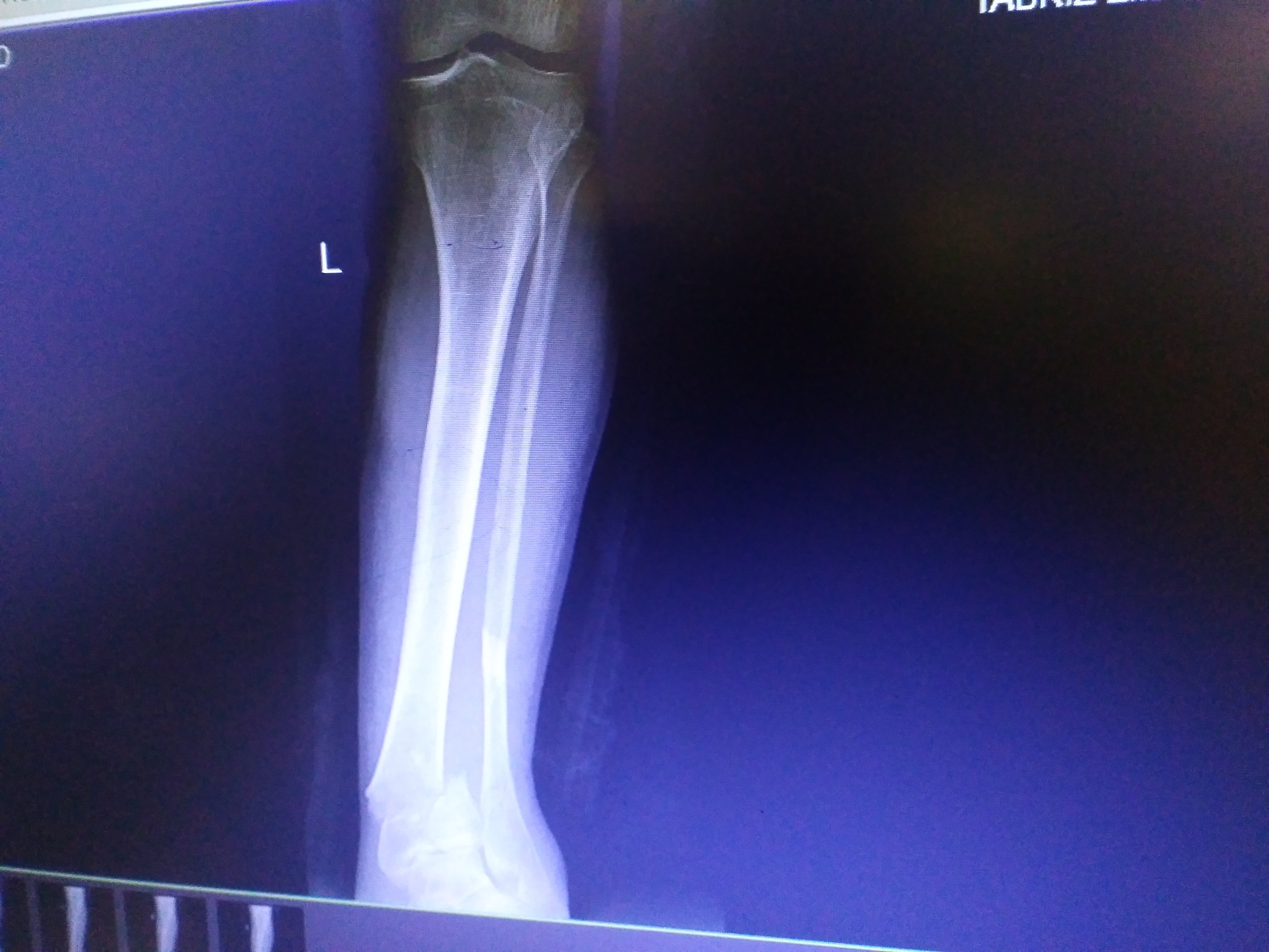
X Ray[16]
The orthopedic surgeon should consider to have at least two radiographic projections (ie, anteroposterior [AP] and lateral) of the ankle. These show the fracture, the extent of displacement, and the extent of comminution. The orthopedic surgeon should pay serious attention toward finding any foreign bodies in open fractures and gunshot injuries. Also imperative is to include the elbow and wrist joint in the radiographs of Ankle fracture to ensure that the distal radioulnar joint injuries are not missed.
-
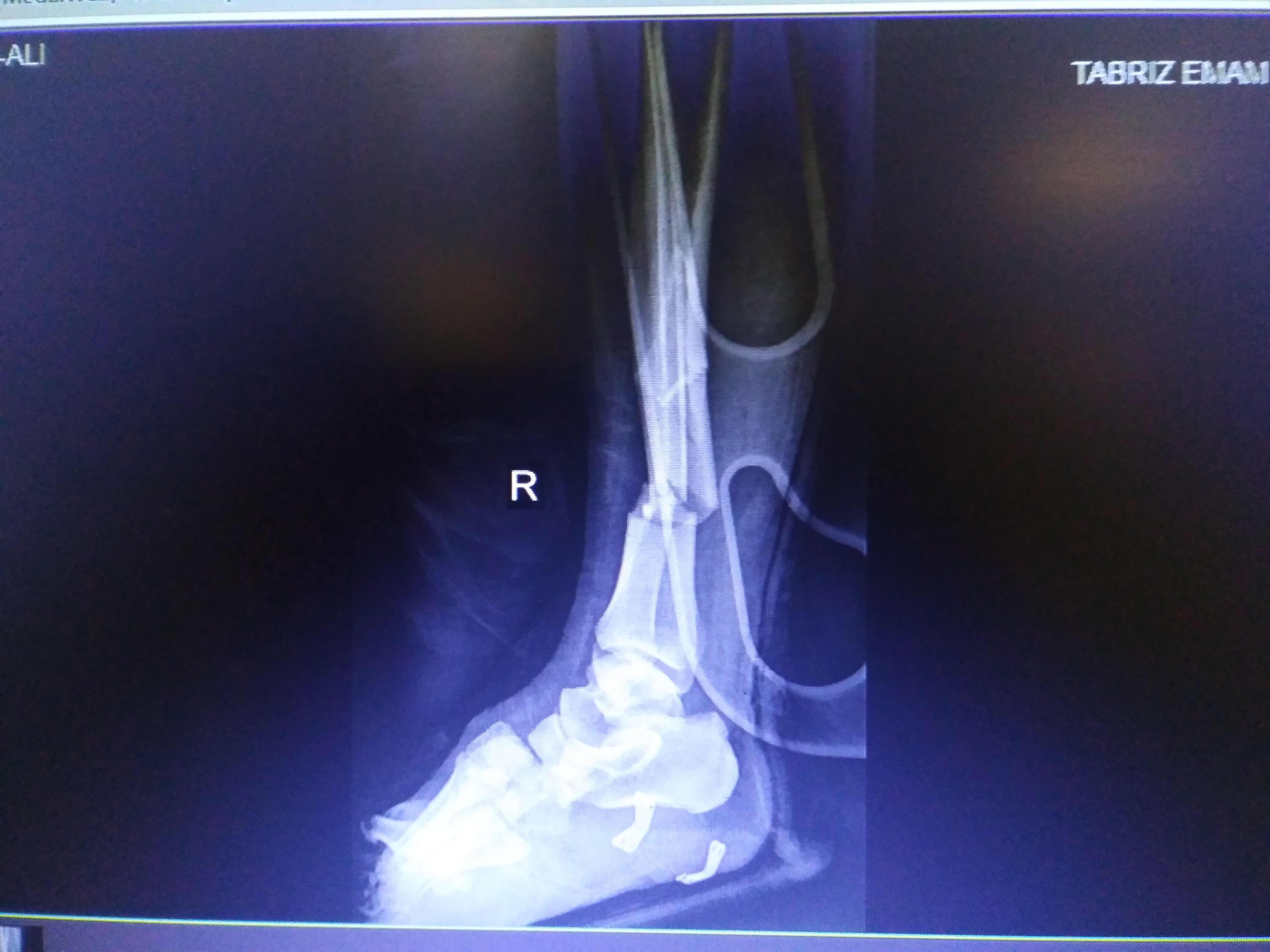
 There is a comminuted distal tibial fracture extending into the tibial plafond.
 -
 There is a comminuted distal tibial fracture extending into the tibial plafond.
 -
 There is a comminuted distal tibial fracture extending into the tibial plafond.


CT [16]
- CT-scan in the case of the Pilon fractureis the best modality if you can not have an exclusive diagnosis by X-ray itself can not be made.
-
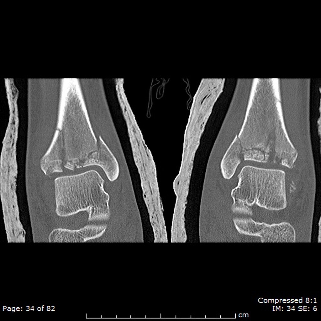 Severely comminuted tibial plafond fractures that extend into the metadiaphyseal regions which are bilateral and nearly symmetric. On the right, there is approximately 3mm of articular offest anteriorly, but otherwise the articular fragments are minimally displaced. There is 1mm medial offset of the medial malleolar fracture fragment on the left and 2mm on the right. There are several small sub-millimeter intraarticular fragments bilaterally.
 -
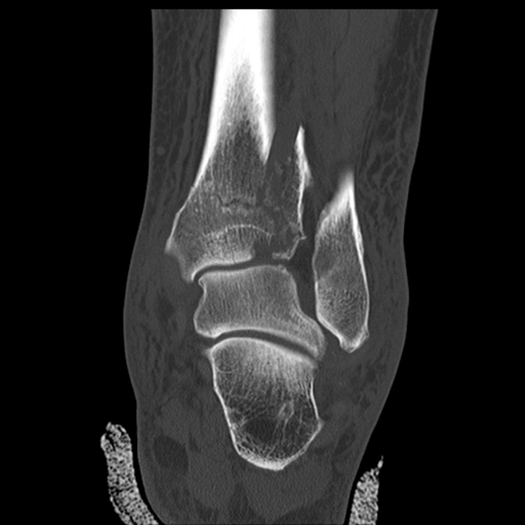 There is a comminuted distal tibial fracture extending into the tibial plafond, representing a Pilon fracture. There are also associated fractures of the talar dome and tip of the lateral malleolus.
.jpg)
.jpg)
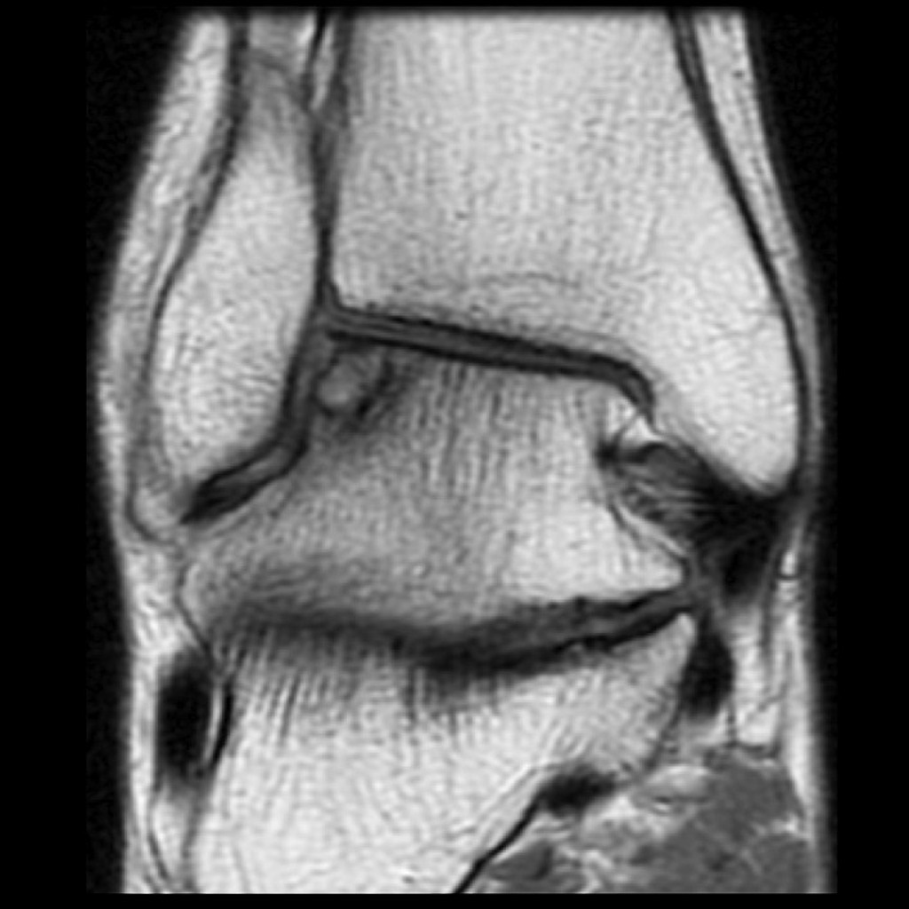
MRI [16]
- Magnetic resonance imaging (MRI) is an expensive technique that should not be used routinely.
- MRI is a powerful diagnostic tool to assess the abnormalities of the bone, ligaments and soft tissues associated with the Pilon fracture, but it is known as a limited utility in radioulnar injuries and is not indicated in uncomplicated foankle earm fractures.
- Meanwhile, the MRI can be useful in in following mentioned evaluations:
- Evaluation of occult fractures
- Evaluation of the post-traumatic or avascular necrosis of carpal bones
- Evaluation of tendons
- Evaluation of nerve
- Evaluation of carpal tunnel syndrome
-
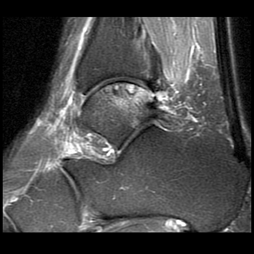 PD Subchondral fracture of the talar dome. Note the intact overlying cartilage.
 -
 T2 fat sat Subchondral fracture of the talar dome. Note the intact overlying cartilage.
.jpg)

Other Imaging Findings[16]
There are no other imaging findings associated with Pilon fracture
Other Diagnostic Studies[16]
There are no other Diagnostic studies associated with Pilon fracture
Immediate stabilization of patients is the first step. Then the radial fracture and the DRUJ stabilization is recommended in these cases. Open ankle fractures considered as a surgical emergency. Pilon fracture occurs in younger patients who are skeletally immature; the normally they treated using a closed reduction and casting. Since closed reduction and cast application have led to unsatisfactory results. Then, Almost always the open reduction are necessary for the Pilon fracture. There are controversies regarding the indications for intramedullary nailing of ankle fractures.
Non-Operative Treatment[19]
- The first step in managing a patient with a fracture is to stabilize the patient if he/she is unstable due to blood loss, etc by giving them intravenous fluids and giving them some painkillers if the pain is severe.
- In children, the usual plan is to attempt closed reduction followed by cast immobilization. In adults, treatment with immobilization in a molded long arm cast can be used in those rare occasions of a non-displaced fracture of the ankle joint. If the fracture shifts in position, it may require surgery to put the bones back together.
- Rigid immobilization is suggested in preference to removable splints in nonoperative treatment for the management of the Pilon fracture
- For all patients with Pilon fracture, a post-reduction true lateral radiograph is suggested.
- Operative fixation is suggested in preference to cast fixation for fractures with post-reduction radial shortening greater than 3 mm, dorsal tilt greater than 10º, or intra-articular displacement or step-off greater than 2 mm.
- Patients probably do not need to begin early wrist motion routinely after stable fracture fixation.
- Adjuvant treatment of Pilon fracture with vitamin C is suggested for the prevention of disproportionate pain
- Lateral epicondylar fractures should be immobilized for 7 days with patients elbow flexed at 90º, with the supinated ankle, and the extended wrist for relaxing the extensor muscles.
Complications of Non-surgical therapy
Failure of non-surgical therapy is common:
- Re-displacement to its original position even in a cast
- Stiffness
- Post traumatic osteoarthritis leading to wrist pain and loss of function
- Other risks specific to cast treatment include:
- Compression of the swollen arm causing compartment syndrome
- Reflex sympathetic dystrophy is a serious complication
- Stiffness is universal following a prolonged period of immobilization and swelling
Returning to the normal physical activity after Pilon fracturecan take weeks to months of therapy under supervision an orthopedist. Meanwhile, a physiotherapy can be helpful for patient to achieve the normal wrist and elbow function caused by the immobilization. All adult Pilon fracture should be considered to be treated with open reduction and internal fixation (ORIF).
External fixation: For severe open fractures Open reduction and internal fixation: For distal Pilon fracture which depending on each patients condition the following may be needed:
Nerve placement Bone grafting Osteotomy Arthrodesis
-
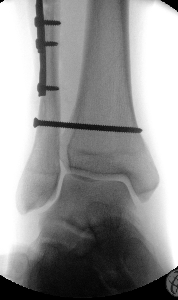 Screw-plate stabilization of the fibular fracture and trans-syndesmotic screw stabilizing the ankle mortise.

Operation [23]
- There are a variety of methods and implants useful to stabilize the Pilon fracturee, ranging from closed reduction and percutaneous pin fixation to the use of intra-medullary devices.
- However, the most common fixation methods to treat complex Pilon fracture include external fixation, and open reduction and internal fixation.
External Fixation With or Without Percutaneous Pin Fixation
- Ankle spanning external fixation employs ligamentotaxis to restore and maintain length, alignment, and rotation of bone.
- Reduction is typically obtained through closed or minimally open methods and preserves the fracture biology.
- The addition of percutaneous pins enhances the ability to reduce and stabilize fracture fragments.
Complications of External Fixation
- Pin tract infection
- Injury to the Superficial branch of the nerve
- Complex regional pain syndrome
Open reduction and internal fixation with plates and screws
- This is the most common type of surgical repair for Pilon fracture
- During this type of procedure, the bone fragments are first repositioned (reduced) into their normal alignment.
- The bones held together with special screws and metal plates attached to the outer surface of the bone.
Complications of open reduction and internal fixation with plates and screws =
- Infection
- Damage to nerves and blood vessels
- Synostosis
- Nonunion
Pain Management [24]
Pain after an injury or surgery is a natural part of the healing process.
Medications are often prescribed for short-term pain relief after surgery or an injury such as:
- opioids
- non-steroidal anti-inflammatory drugs (NSAIDs)
- local anesthetics
Be aware that although opioids help relieve pain after surgery or an injury, they are a narcotic and can be addictive. It is important to use opioids only as directed by doctor.
Interventions[24]
The following options can be helpful for patients to rehabilitate after their fracture :
- Joints mobilization
- compression bandage
- Soft tissue massage
- Exercises and Activity modification
- Complex Pilon fracture warrant individualized immobilization and rehabilitation strategies.
- Because most multifragmentary Pilon fracture are the result of high-energy injuries, a prolonged period of wrist immobilization and soft-tissue rest may be beneficial and has not been shown to affect clinical outcomes.
- The ankle is typically immobilized for 6 weeks post-operatively in a splint with Full weight bearing commences at approximately 3 months post-operatively after consolidation of the fracture is noted on radiographs.
- The presence of varying degrees of ankle stiffness is inevitable and may result from poor pain control, lack of effort in controlled mobilization, edema, concomitant ipsilateral lower extremity fractures, or peripheral nerve injuries. Early stretching and mobilization of the intrinsic and extrinsic tendons of the hand is important to prevent finger stiffness. Edema control can be initiated with compression gloves, digital massage, and active and passive ROM of the ankle. A home exercise program or outpatient occupational therapy is started immediately post-operatively to maintain full range of motion of the ankle and limit the development of intrinsic muscle tightnes
There are various preventive options to reduce the incidence of the Pilon fracture
- Using ankle guards during practicing sports (skating, biking)
- Using ankle guards during driving motorbikes
- Avoid falls in elderly individuals
- Prevention and/or treatment of osteoporosis
- Healthy diet
It should be noted that the Post-menopausal women specially older than the age of 65 are at the higher risk of osteoporosis consequently these type of patients at greater risk for the pathological fractures .
So the Calcium and vitamin D supplementation play important role in increasing the bone mineral density (BMD) consequently decrease the risk of fracture in these type of patients. Also, avoiding excessive alcohol and quitting smoking play important role in this regard.
Detecting osteoporosis
- DEXA(dual-energy x-ray absorptiometry) scan
- Serum calcium and vitamin D levels
- Ultrasonography of the calcaneus
Pharmacological therapy
- The primary goal for the treatment of osteoporosis is to reduce longtime fracture risk in patients. Increasing bone mineral density (BMD) in response to the treatment is far less important than improvement of clinical aspects of osteoporosis, i.e., osteoporoticfracture. Therefore, most of the drugs efficacy is measured by the extent they improve the fracture risk instead of increasing BMD.
- During the treatment, if a single fracture happens, it does not necessarily indicate treatment failure or the need to be started on an alternative treatment or patient referral to a specialist.
- Calcium and vitamin D supplementation have been found to be effective in reducing the long term fracture risk, significantly. In order to suggest the people to use vitamin D and calcium supplements, the physician needs to make sure that patient is not able to obtain the nutrients through the daily intake. The available supplemental ions of calcium include calcium carbonate, calcium citrate, and vitamin D3 in various dosage forms.
Life style modifications[14]
- Exercise: Exercise promotes the mineralization of bone and bone accumulation particularly during growth. High impact exercise, in particular, has been shown to prevent the development of osteoporosis. However, it can have a negative effect on bone mineralization in cases of poor nutrition, such as anorexia nervosa and celiac disease.
- Nutrition: A diet high in calcium and vitamin D prevents bone loss. Patients at risk for osteoporosis, such as persons with chronic steroid use are generally treated with vitamin D and calcium supplementation. In renal disease, more active forms of vitamin D, such as 1,25-dihydroxycholecalciferol or calcitriol are used; as the kidney cannot adequately generate calcitriol from calcidiol (25-hydroxycholecalciferol), which is the storage form of vitamin D.
- By quitting smoking, osteoporosis as well as other diseases can be prevented.
- Avoiding excessive alcohol intake or drinking only in moderation.
See also
- triplane fracture
- Tillaux fracture
- Bosworth fracture
- pilon fracture
- Wagstaffe-Le Forte fracture
- Maisonneuve Fracture
References
- ↑ 1.0 1.1 Krettek C, Bachmann S (February 2015). "[Pilon fractures. Part 2: Repositioning and stabilization technique and complication management]". Chirurg (in German). 86 (2): 187–201, quiz 202–3. doi:10.1007/s00104-014-2917-5. PMID 25673229.
- ↑ Busel GA, Watson JT, Israel H (June 2017). "Evaluation of Fibular Fracture Type vs Location of Tibial Fixation of Pilon Fractures". Foot Ankle Int. 38 (6): 650–655. doi:10.1177/1071100717695348. PMID 28288519.
- ↑ 3.0 3.1 3.2 3.3 3.4 Schweigkofler U, Benner S, Hoffmann R (June 2015). "[Pilon Fractures]". Z Orthop Unfall (in German). 153 (3): 335–54, quiz 355–6. doi:10.1055/s-0035-1545983. PMID 26114568.
- ↑ Sommer C, Nork SE, Graves M, Blauth M, Rudin M, Stoffel K (December 2017). "Quality of fracture reduction assessed by radiological parameters and its influence on functional results in patients with pilon fractures-A prospective multicentre study". Injury. 48 (12): 2853–2863. doi:10.1016/j.injury.2017.10.031. PMID 29079366.
- ↑ Gaulke R, Krettek C (August 2017). "[Tibial pilon fractures : Advoidance and therapy of complications]". Unfallchirurg (in German). 120 (8): 658–666. doi:10.1007/s00113-017-0366-6. PMID 28540568.
- ↑ Liu J, Smith CD, White E, Ebraheim NA (April 2016). "A Systematic Review of the Role of Surgical Approaches on the Outcomes of the Tibia Pilon Fracture". Foot Ankle Spec. 9 (2): 163–8. doi:10.1177/1938640015620637. PMID 26644032.
- ↑ Zelle BA, Gruen GS, McMillen RL, Dahl JD (June 2014). "Primary Arthrodesis of the Tibiotalar Joint in Severely Comminuted High-Energy Pilon Fractures". J Bone Joint Surg Am. 96 (11): e91. doi:10.2106/JBJS.M.00544. PMID 24897748.
- ↑ Liu Z, Xu H, Zhang Z, Teng Y, Wu M, Tao S (October 2013). "[Effectiveness observation of staged treatment of open Pilon fracture combined with soft tissue defect]". Zhongguo Xiu Fu Chong Jian Wai Ke Za Zhi (in Chinese). 27 (10): 1185–9. PMID 24397128.
- ↑ 9.0 9.1 9.2 Wiebking U (August 2017). "[Pilon fractures : Review of diagnostics and classification]". Unfallchirurg (in German). 120 (8): 632–639. doi:10.1007/s00113-017-0363-9. PMID 28567532.
- ↑ Leonetti D, Tigani D (October 2017). "Pilon fractures: A new classification system based on CT-scan". Injury. 48 (10): 2311–2317. doi:10.1016/j.injury.2017.07.026. PMID 28774706.
- ↑ Gauthé R, Desseaux A, Rony L, Tarissi N, Dujardin F (June 2016). "Ankle fractures in the elderly: Treatment and results in 477 patients". Orthop Traumatol Surg Res. 102 (4 Suppl): S241–4. doi:10.1016/j.otsr.2016.03.001. PMID 27033840.
- ↑ Krettek C, Bachmann S (January 2015). "[Pilon fractures. Part 1: Diagnostics, treatment strategies and approaches]". Chirurg (in German). 86 (1): 87–101, quiz 102–4. doi:10.1007/s00104-014-2895-7. PMID 25591416.
- ↑ 13.0 13.1 13.2 Kottmeier SA, Madison RD, Divaris N (September 2018). "Pilon Fracture: Preventing Complications". J Am Acad Orthop Surg. 26 (18): 640–651. doi:10.5435/JAAOS-D-17-00160. PMID 30134307.
- ↑ 14.0 14.1 14.2 De-Las-Heras-Romero J, Lledo-Alvarez AM, Lizaur-Utrilla A, Lopez-Prats FA (June 2017). "Quality of life and prognostic factors after intra-articular tibial pilon fracture". Injury. 48 (6): 1258–1263. doi:10.1016/j.injury.2017.03.023. PMID 28365069.
- ↑ 15.0 15.1 Wang Y, Wang J, Luo CF (August 2016). "Modified posteromedial approach for treatment of posterior pilon variant fracture". BMC Musculoskelet Disord. 17: 328. doi:10.1186/s12891-016-1182-9. PMC 4974710. PMID 27496169.
- ↑ 16.0 16.1 16.2 16.3 16.4 Klaue K, Cronier P (September 2015). "[Pilon fractures]". Unfallchirurg (in German). 118 (9): 795–801, quiz 802–3. doi:10.1007/s00113-015-0054-3. PMID 26293486.
- ↑ Klaue K (August 2017). "[Operative access for treatment of pilon fractures]". Unfallchirurg (in German). 120 (8): 648–651. doi:10.1007/s00113-017-0377-3. PMID 28674752.
- ↑ Jia SH, Huang CL, Xu HW, Gong SL (June 2016). "[Surgical treatment for posterior Pilon fracture through posterolateral approach]". Zhongguo Gu Shang (in Chinese). 29 (6): 557–60. PMID 27534089.
- ↑ Mittlmeier T, Wichelhaus A (August 2017). "[Treatment strategy and planning for pilon fractures]". Unfallchirurg (in German). 120 (8): 640–647. doi:10.1007/s00113-017-0383-5. PMID 28717978.
- ↑ Rotter R, Gierer P (August 2017). "[Operative techniques and results of tibial pilon fractures]". Unfallchirurg (in German). 120 (8): 652–657. doi:10.1007/s00113-017-0371-9. PMID 28638931.
- ↑ 21.0 21.1 Liu P, Guo Y, Wen Y, Wang W (2017). "Clinical application of arthroscopy-assisted minimally invasive therapy in Chinese elderly with type III Pilon fracture". Clin Interv Aging. 12: 2033–2038. doi:10.2147/CIA.S140272. PMC 5716396. PMID 29238178.
- ↑ Abou Elatta MM, Assal F, Basheer HM, El Morshidy AF, Elglaind SM, Abdalla MA (February 2017). "The use of dynamic external fixation in the treatment of dorsal fracture subluxations and pilon fractures of finger proximal interphalangeal joints". J Hand Surg Eur Vol. 42 (2): 182–187. doi:10.1177/1753193416674155. PMID 27756830.
- ↑ Zhang SB, Zhang YB, Wang SH, Zhang H, Liu P, Zhang W, Ma JL, Wang J (April 2017). "Clinical efficacy and safety of limited internal fixation combined with external fixation for Pilon fracture: A systematic review and meta-analysis". Chin. J. Traumatol. 20 (2): 94–98. doi:10.1016/j.cjtee.2016.06.012. PMC 5392718. PMID 28359592.
- ↑ 24.0 24.1 Ren T, Ding L, Xue F, He Z, Xiao H (June 2015). "Risk factors for surgical site infection of pilon fractures". Clinics (Sao Paulo). 70 (6): 419–22. doi:10.6061/clinics/2015(06)06. PMC 4462573. PMID 26106960.
- ↑ Huebner EJ, Iblher N, Kubosch DC, Suedkamp NP, Strohm PC (2014). "Distal tibial fractures and pilon fractures". Acta Chir Orthop Traumatol Cech. 81 (3): 167–76. PMID 24945386.
- ↑ Chen Z, Chen D, Yang H, Wu W, Dai Z (October 2015). "[360 degrees INTERNAL FIXATION BY DOUBLE APPROACHES FOR HIGH-ENERGY CLOSED Pilon FRACTURES]". Zhongguo Xiu Fu Chong Jian Wai Ke Za Zhi (in Chinese). 29 (10): 1226–9. PMID 26749728.
- ↑ Hutson JJ (March 2008). "Salvage of pilon fracture nonunion and infection with circular tensioned wire fixation". Foot Ankle Clin. 13 (1): 29–68, vi. doi:10.1016/j.fcl.2007.11.004. PMID 18328415.
- ↑ Kim GB, Shon OJ, Park CH (April 2018). "Treatment of AO/OTA Type C Pilon Fractures Through the Anterolateral Approach Combined With the Medial MIPO Technique". Foot Ankle Int. 39 (4): 426–432. doi:10.1177/1071100717746628. PMID 29359596.
- ↑ Sukur E, Akman YE, Gokcen HB, Ozyurek EC, Senel A, Ozturkmen Y (September 2017). "Open reduction in pilon variant posterior malleolar fractures: Radiological and clinical evaluation". Orthop Traumatol Surg Res. 103 (5): 703–707. doi:10.1016/j.otsr.2017.05.012. PMID 28606840.