Multiple myeloma MRI
|
Multiple myeloma Microchapters |
|
Diagnosis |
|---|
|
Treatment |
|
Case Studies |
|
Multiple myeloma MRI On the Web |
|
American Roentgen Ray Society Images of Multiple myeloma MRI |
Editor-In-Chief: C. Michael Gibson, M.S., M.D. [1] Associate Editor(s)-in-Chief: Hannan Javed, M.D.[2]; Haytham Allaham, M.D. [3]; Shyam Patel [4]
Overview
MRI may be diagnostic of multiple myeloma.[1]Findings on MRI suggestive of multiple myeloma include infiltration and replacement of the bone marrow.[2][3] MRI is a more expensive test than an X-ray.
MRI
Magnetic resonance imaging (MRI) is more sensitive than simple X-ray in the detection of lytic lesions of multiple myeloma.[2] The International Myeloma Working Group (IMWG) recently proposed revised imaging criteria for a diagnosis of multiple myeloma. The MRI criteria is the presence of greater than 1 focal lesion of at least 5mm in size. Of note, MRI is the most expensive of the imaging tests. X-rays may sometimes be a sufficient substitute for assessing for the presence of lytic lesions for patients who are symptomatic. However, X-rays carry a risk for radiation exposure, unlike MRI. Clinical judgment should be used when determining whether to order MRI or X-rays to assess for lytic lesions. MRI may supersede skeletal survey, especially when vertebral disease is suspected.[2]
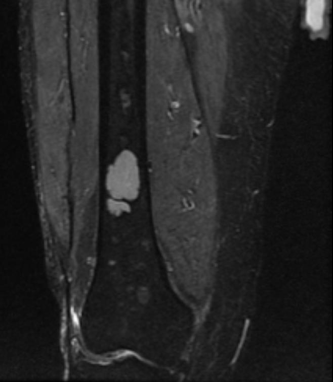
- Shown below is a series of MRI images of long bones involved in multiple myeloma. (Images courtesy of RadsWiki)
-
-
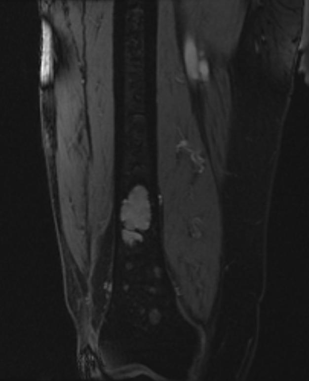 ([http://www.radswiki.net Images courtesy of RadsWiki


- Shown below is a series of MRI images in a multiple myeloma patient complaining of back pain.


References
- ↑ Gerecke C, Fuhrmann S, Strifler S, Schmidt-Hieber M, Einsele H, Knop S (July 2016). "The Diagnosis and Treatment of Multiple Myeloma". Dtsch Arztebl Int. 113 (27–28): 470–6. doi:10.3238/arztebl.2016.0470. PMC 4973001. PMID 27476706.
- ↑ 2.0 2.1 2.2 Multiple myeloma. Radiopaedia(2015) http://radiopaedia.org/articles/multiple-myeloma-1 Accessed on September, 20th 2015
- ↑ Reisenbuckler C (2014). "Multiple myeloma and diagnostic imaging". Radiol Technol. 85 (4): 391–410, quiz 411–3. PMID 24614435.