Mastoiditis CT
|
Mastoiditis Microchapters |
|
Diagnosis |
|---|
|
Treatment |
|
Case Studies |
Editor-In-Chief: C. Michael Gibson, M.S., M.D. [1]; Associate Editor(s)-in-Chief: Mehrian Jafarizade, M.D [2]
Overview
High Resolution CT scans of the temporal bone in mastoiditis patients are the preferred diagnostic tool and may reveal mastoiditis and its complications. CT findings in acute mastoiditis are: partial-to-complete opacification of mastoid air cells, erosion of mastoid air cell bony septum, mastoid cortex destruction and irregularity, periosteal thickening, periosteal disruption, and subperiosteal abscess. CT findings in subacute and chronic mastoiditis are: markers for inflammation, sclerosis, or opacification of mastoid process, tympanic membrane changes including thickening, retraction, tympanic membrane perforation, or calcification, ossicle erosion or other possible causes for hearing loss, determination of cholesteatoma, intratemporal complications such as petrositis, labyrinthitis, subperiosteal abscess, or labyrinthine fistula, intracranial complications such as brain abscess and meningitis, presence of fibrous tissue, tympanosclerosis, formation of new bone matter, ossicle erosion, and displacement and extension of cholesteatoma to sinuses.
CT
High Resolution CT scans of the temporal bone in mastoiditis patients are the preferred diagnostic tool and may reveal complications including the following: [1][2][3][4][5]
Acute mastoiditis
- Partial-to-complete opacification of mastoid air cells due to fluid accumulation and thickening of the mucosa that lines the middle ear
- Erosion of mastoid air cell bony septum
- Mastoid cortex destruction and irregularity
- Periosteal thickening or periosteal disruption
- Subperiosteal abscess
Sub acute and chronic mastoiditis
Primary imaging findings for chronic mastoiditis include the following:
- Markers of inflammation
- Sclerosis or opacification of mastoid process
- Tympanic membrane changes including thickening, retraction, tympanic membrane perforation, or calcification
- Ossicle erosion or other possible causes for hearing loss
- Determination of cholesteatoma
- Intratemporal complications including petrositis, labyrinthitis, subperiosteal abscess, or labyrinthine fistula
- Intracranial complications including brain abscess or meningitis
- Presence of fibrous tissue
- Tympanosclerosis
- Formation of new bone matter
- Ossicle erosion and displacement
- Extension of cholesteatoma to sinuses
High Resolution CT scanning (HRCT) is the best tool to evaluate ossicular chain, tympanic cavity walls, and the mastoid itself; however, HRCT is unable to differentiate between different types of effusions in the tympanic cavity, and evaluation of cholesteatoma can be challenging.
CT Examples of Otitis Media
-
 Selected CT images showing soft tissue density material occupying partially the left mastoid air cells with cortical destruction of the posterior mastoid wall in continuity with a retroauricular fluid collection containing air-bubbles with peripheral enhancement. Case courtesy of Dr Ammar Haouimi, Radiopaedia.org, rID: 69818 From Radiopaedia Image Library. https://radiopaedia.org/cases/acute-mastoiditis-with-retroauricular-abscess?lang=us
 -
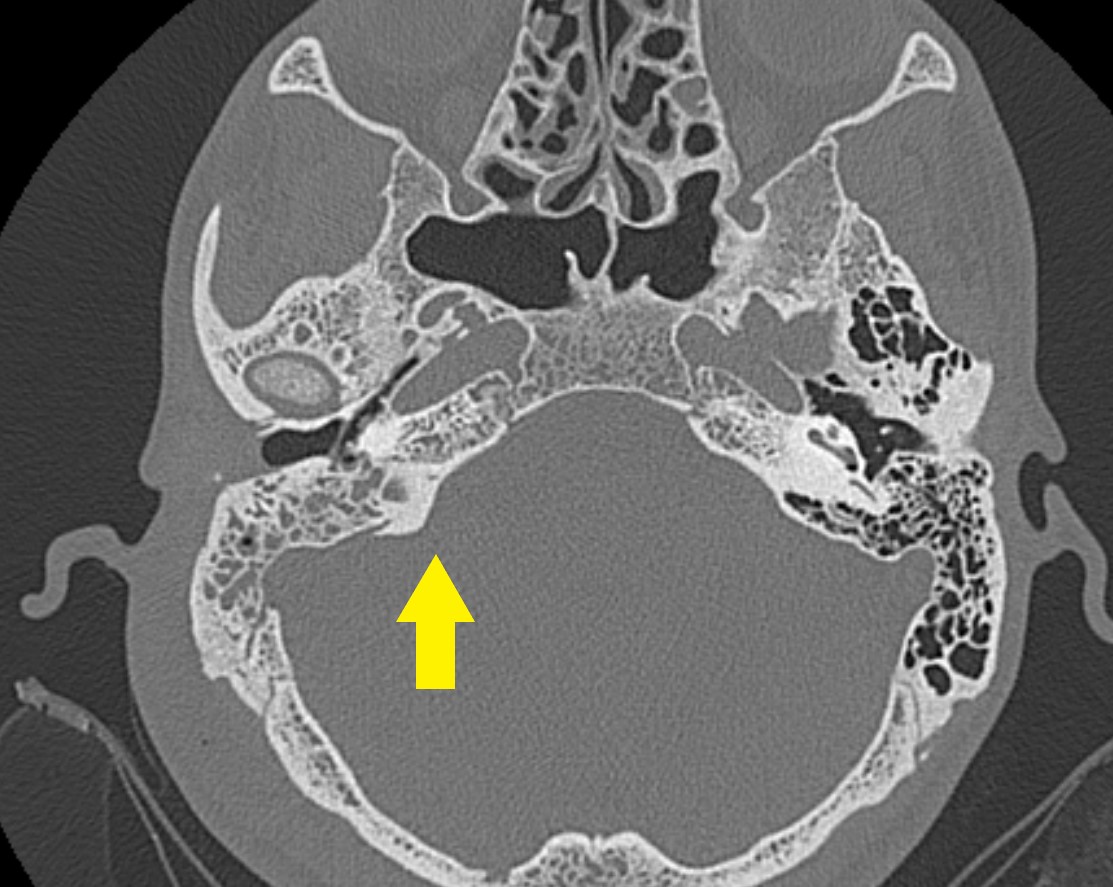
![The left middle ear is filled by soft tissue completely encasing the auditory ossicles. There is some residual ventilation near the tympanic ostium of the Eustachian tube. In this region, a linear calcification can also be seen. In the right middle ear, there are also some strands with soft tissue attenuation neighbouring the tympanic membrane, which is retracted on both sides. The right ossicles are not affected. Bilateral mastoid sclerosis is also present. There are no bony destructions hinting at a cholesteatoma. Case presented by Dr. Roberto Schubert.From Radiopaedia Image Library. [6]](/images/b/ba/Mastoiditis.jpg) The left middle ear is filled by soft tissue completely encasing the auditory ossicles. There is some residual ventilation near the tympanic ostium of the Eustachian tube. In this region, a linear calcification can also be seen. In the right middle ear, there are also some strands with soft tissue attenuation neighbouring the tympanic membrane, which is retracted on both sides. The right ossicles are not affected. Bilateral mastoid sclerosis is also present. There are no bony destructions hinting at a cholesteatoma. Case presented by Dr. Roberto Schubert.From Radiopaedia Image Library. [6]
The left middle ear is filled by soft tissue completely encasing the auditory ossicles. There is some residual ventilation near the tympanic ostium of the Eustachian tube. In this region, a linear calcification can also be seen. In the right middle ear, there are also some strands with soft tissue attenuation neighbouring the tympanic membrane, which is retracted on both sides. The right ossicles are not affected. Bilateral mastoid sclerosis is also present. There are no bony destructions hinting at a cholesteatoma. Case presented by Dr. Roberto Schubert.From Radiopaedia Image Library. [6]

![The left middle ear is filled by soft tissue completely encasing the auditory ossicles. There is some residual ventilation near the tympanic ostium of the Eustachian tube. In this region, a linear calcification can also be seen. In the right middle ear, there are also some strands with soft tissue attenuation neighbouring the tympanic membrane, which is retracted on both sides. The right ossicles are not affected. Bilateral mastoid sclerosis is also present. There are no bony destructions hinting at a cholesteatoma. Case presented by Dr. Roberto Schubert.From Radiopaedia Image Library. [6]](/index.php/File:Mastoiditis.jpg)
References
- ↑ Lin HW, Shargorodsky J, Gopen Q (2010). "Clinical strategies for the management of acute mastoiditis in the pediatric population". Clin Pediatr (Phila). 49 (2): 110–5. doi:10.1177/0009922809344349. PMID 19734439.
- ↑ Stähelin-Massik J, Podvinec M, Jakscha J, Rüst ON, Greisser J, Moschopulos M, Gnehm HE (2008). "Mastoiditis in children: a prospective, observational study comparing clinical presentation, microbiology, computed tomography, surgical findings and histology". Eur. J. Pediatr. 167 (5): 541–8. doi:10.1007/s00431-007-0549-1. PMID 17668240.
- ↑ Trojanowska A, Drop A, Trojanowski P, Rosińska-Bogusiewicz K, Klatka J, Bobek-Billewicz B (2012). "External and middle ear diseases: radiological diagnosis based on clinical signs and symptoms". Insights Imaging. 3 (1): 33–48. doi:10.1007/s13244-011-0126-z. PMC 3292638. PMID 22695997.
- ↑ Pellegrini S, Gonzalez Macchi ME, Sommerfleck PA, Bernáldez PC (2012). "Intratemporal complications from acute otitis media in children: 17 cases in two years". Acta Otorrinolaringol Esp. 63 (1): 21–5. doi:10.1016/j.otorri.2011.06.007. PMID 21982482.
- ↑ van den Aardweg MT, Rovers MM, de Ru JA, Albers FW, Schilder AG (2008). "A systematic review of diagnostic criteria for acute mastoiditis in children". Otol. Neurotol. 29 (6): 751–7. doi:10.1097/MAO.0b013e31817f736b. PMID 18617870.
- ↑ "Middle ear | Radiology Reference Article | Radiopaedia.org".