Knee examination
| Knee examination | |
 | |
|---|---|
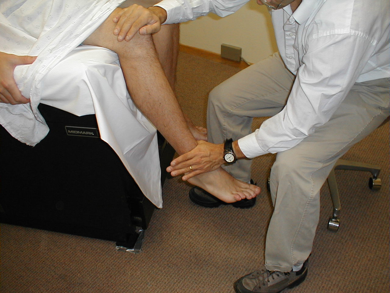
| Knee Extension |
|
WikiDoc Resources for Knee examination |
|
Articles |
|---|
|
Most recent articles on Knee examination Most cited articles on Knee examination |
|
Media |
|
Powerpoint slides on Knee examination |
|
Evidence Based Medicine |
|
Clinical Trials |
|
Ongoing Trials on Knee examination at Clinical Trials.gov Trial results on Knee examination Clinical Trials on Knee examination at Google
|
|
Guidelines / Policies / Govt |
|
US National Guidelines Clearinghouse on Knee examination NICE Guidance on Knee examination
|
|
Books |
|
News |
|
Commentary |
|
Definitions |
|
Patient Resources / Community |
|
Patient resources on Knee examination Discussion groups on Knee examination Patient Handouts on Knee examination Directions to Hospitals Treating Knee examination Risk calculators and risk factors for Knee examination
|
|
Healthcare Provider Resources |
|
Causes & Risk Factors for Knee examination |
|
Continuing Medical Education (CME) |
|
International |
|
|
|
Business |
|
Experimental / Informatics |
Editor-In-Chief: C. Michael Gibson, M.S., M.D. [1]
The knee examination, in medicine, is performed as part of a physical examination, or when a patient presents with knee pain or a history that suggests a pathology of the knee joint.
The exam includes several parts:
- position/lighting/draping
- inspection
- palpation
- motion
The latter three steps are often remembered with the saying look, feel, move.
Position/lighting/draping
Position - for most of the exam the patient should be supine and the bed or examination table should be flat. The patient's hands should remain at his or her sides with the head resting on a pillow. The knees and hips should be in the anatomical position (knee extend, hip neither flexed or extend).
Lighting - adjusted so that it is ideal.
Draping - both of the patient's knees should be exposed so that the quadriceps muscles can be assessed.
Video: Understanding Knee Pain
{{#ev:youtube|BVWApDz6zmQ}}
Inspection done while the patient is standing
The knee should be examined for:
- Baker's cyst
- genu recurvatum
- Valgus deformity (knock-kneed)
- Varus deformity (bowlegged)
- Gait - antalgic gait?
Inspection done while supine
The knee should be examined for:
- Masses
- Scars
- Lesions
- Signs of trauma/previous surgery
- Swelling (edema - particular in the medial fossa (the depression medial to the patella)
- erythema (redness)
- Muscle bulk and symmetry (in particular atrophy of the medial aspect of the quadriceps muscle - vastus medialis)
- Displacement of the patella (knee cap)
Palpation
An inflamed knee exhibits tumor (swelling), rubor (redness), calor (heat), dolor (pain). Swelling and redness should be evident by inspection. Pain is gained by history and heat by palpation.
- Temperature change - using the back of the hand one should feel the temperature of the knee below the patella, over the patella, and above the patella. Normally, the patella is cool relative to above and below the knee. A complete exam involves comparing the knees to one another.
- joint line tenderness - this is done by flexing the knee and palpating the joint line with the thumb.
- Effusions, test for
- Patellar tap - useful for large effusions
- Ballottement - defined as a palpatory technique for detecting or examining a floating object in the body
- Bulge sign - useful for smaller effusions
Motion
The patient should be asked to move their knee. Full range of motion is 0-135 degrees. If the patient has full range of motion and can move their knee on their own it is not necessary to move the knee passively.
- examination of crepitus - clicking of the joint with motion
Ligament tests
- Anterior drawer sign - tests the anterior cruciate ligament (ACL)
- Posterior drawer sign - tests the posterior cruciate ligament
- Lachman test (ACL)
- Medial collateral ligament
- Lateral collateral ligament
- McMurray test
- medial meniscus is tested by external rotation + lateral force (mnemonic Mel)
- lateral meniscus is tested by internal rotation + medial force
Knee Extension (L 2, 3, 4):
Have the seated patient steadily press their lower extremity into your hand against resistance. Test each leg separately. Extension is mediated by the quadriceps muscle group, which is innervated by the femoral nerve.
(Image courtesy of Charlie Goldberg, M.D., UCSD School of Medicine and VA Medical Center, San Diego, California)
-
Knee extension
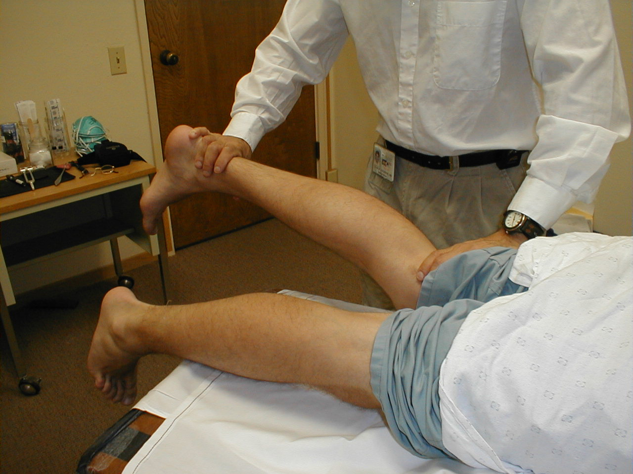
Knee flexion (L 5; S 1, 2):
Have the patient rest prone. Then have them pull their heel up and off the table against resistance. Each leg is tested separately. Flexion is mediated by the hamstring muscle group, via branches of the sciatic nerve.
(Image courtesy of Charlie Goldberg, M.D., UCSD School of Medicine and VA Medical Center, San Diego, California)
-
Knee flexion
Explanation of Knee Examination
Video 1
{{#ev:youtube|_StElDL5A64}}
Video 2
{{#ev:youtube|fNUGyNYVhqE}}
Video 3
{{#ev:youtube|NpSu6m9_mpA}}
See also
External Links
- For more information on Knee Replacement
