Horseshoe kidney CT
Jump to navigation
Jump to search
|
Horseshoe kidney Microchapters |
|
Diagnosis |
|---|
|
Treatment |
|
Case Studies |
|
Horseshoe kidney CT On the Web |
|
American Roentgen Ray Society Images of Horseshoe kidney CT |
Editor-In-Chief: C. Michael Gibson, M.S., M.D. [1]; Associate Editor(s)-in-Chief:
Overview
CT Scan with contrast is imaging technique of choice. CT Scan of abdomen and pelvis with and without contract is used to look for location of kidneys. It is accurate in determining the anatomical location and also help in differentiating the parenchymal from fibrous isthmus.
CT scan
Abdominopelvic CT scan may be helpful in the diagnosis of horseshoe kidney. CT scan accurately delineate:[1][2]
- Degree and anatomical location of the horseshoe kidney.
- Degree of malrotation.
- Help differentiating the parenchymal from fibrous isthmus.
- Showing the relation of the isthmus to surrounding structures.
- Evaluate associated renal parenchymal changes and collecting system abnormalities.
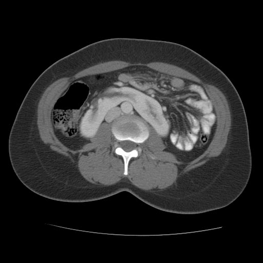
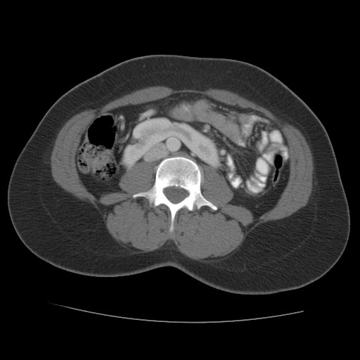
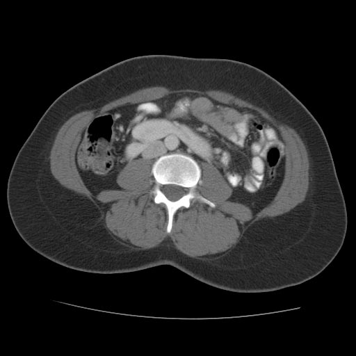
Patient #1: CT images demonstrate a horseshoe kidney
-
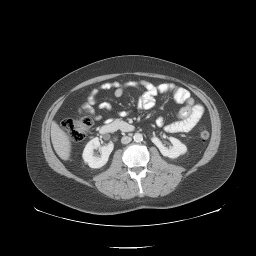 CT image demonstrates a horseshoe kidney
 -
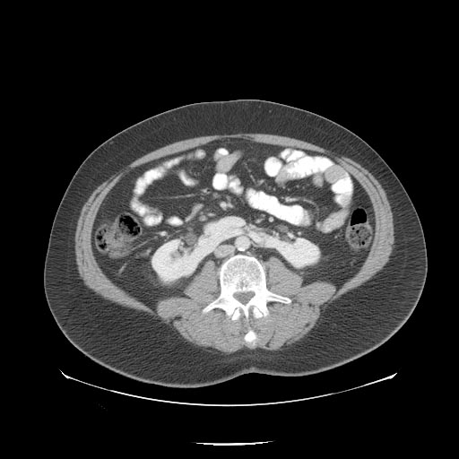 CT image demonstrates a horseshoe kidney


-
 CT image demonstrates a horseshoe kidney
 -
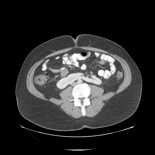 CT image demonstrates a horseshoe kidney


Patient #2: CT images demonstrate a horseshoe kidney
-
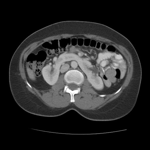 CT image demonstrates a horseshoe kidney
 -
 CT image demonstrates a horseshoe kidney


-
 CT image demonstrates a horseshoe kidney
 -
 CT image demonstrates a horseshoe kidney


References
- ↑ Shah HU, Ojili V (2017). "Multimodality imaging spectrum of complications of horseshoe kidney". Indian J Radiol Imaging. 27 (2): 133–140. doi:10.4103/ijri.IJRI_298_16. PMC 5510309. PMID 28744072.
- ↑ O’Brien, J; Buckley, O; Doody, O; Ward, E; Persaud, T; Torreggiani, W (2008). "Imaging of horseshoe kidneys and their complications". Journal of Medical Imaging and Radiation Oncology. 52 (3): 216–226. doi:10.1111/j.1440-1673.2008.01950.x. ISSN 1754-9477.