Pericardium
|
Pericarditis Microchapters |
|
Diagnosis |
|---|
|
Treatment |
|
Surgery |
|
Case Studies |
|
Pericardium On the Web |
|
American Roentgen Ray Society Images of Pericardium |
Editor-In-Chief: C. Michael Gibson, M.S., M.D. [1]
Overview
The pericardium is a double-walled sac that contains the heart and the roots of the great vessels.
Layers
There are two layers to the pericardial sac: the fibrous pericardium and the serous pericardium. The serous pericardium, in turn, is divided into two layers, the parietal pericardium, which is fused to and inseparable from the fibrous pericardium, and the visceral pericardium, which is in fact epicardium, or the outer surface of the heart.
In between the parietal and visceral pericardial layers there is a potential space called the pericardial cavity. It is normally lubricated by a film of pericardial fluid. Too much fluid in the cavity (such as in a pericardial effusion) can result in pericardial tamponade, compression of the heart within the pericardial sac.
Pericardial Sinuses
There are two small chambers or sinuses are located where the visceral and parietal pericardia are continuous with one another within the pericardial cavity.
The pericardial sinuses are:
- Oblique pericardial sinus
- Transverse pericardial sinus


Additional images
-
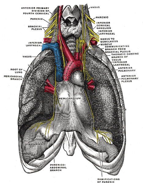
The phrenic nerve and its relations with the vagus nerve.
-
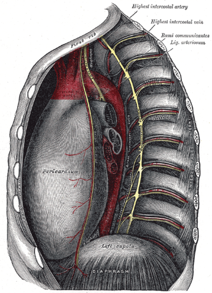
Thoracic portion of the sympathetic trunk.
-
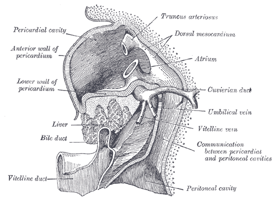
Liver with the septum transversum. Human embryo 3 mm. long.
-
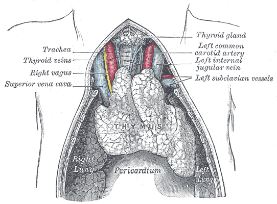
The thymus of a full-time fetus, exposed in situ.


Diseases of the Pericardium
- Pericarditis is an inflammatory condition of the pericardium.
- Pericardial effusion is fluid accumulation in the pericardial sac.
- Constrictive pericarditis occurs when there is a scar encasing the heart which constricts filling of the heart chronically.
- Cardiac tamponade is a medical emergency in which fluid in the pericardial sac restricts the filling of the heart acutely. This requires drainage surgically or by pericardiocentesis.
External links
- Template:SUNYAnatomyLabs - "Mediastinum: Pericardium (pericardial sac)"
- Template:NormanAnatomy (Template:NormanAnatomyFig)
de:Herzbeutel it:Pericardio la:Pericardium ms:Perikardium nl:Pericard nn:Hjartepose fi:Perikardium