Pericarditis x ray
|
Pericarditis Microchapters |
|
Diagnosis |
|---|
|
Treatment |
|
Surgery |
|
Case Studies |
|
Pericarditis x ray On the Web |
|
American Roentgen Ray Society Images of Pericarditis x ray |
Editor-In-Chief: C. Michael Gibson, M.S., M.D. [1]Associate Editor(s)-in-Chief: Homa Najafi, M.D.[2]
Overview
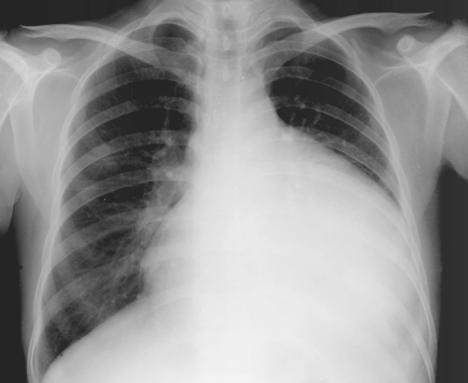
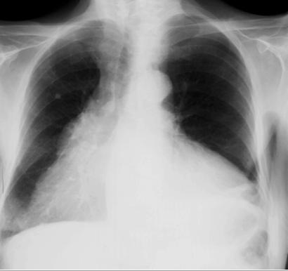
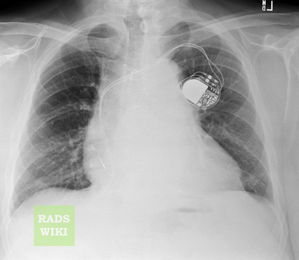
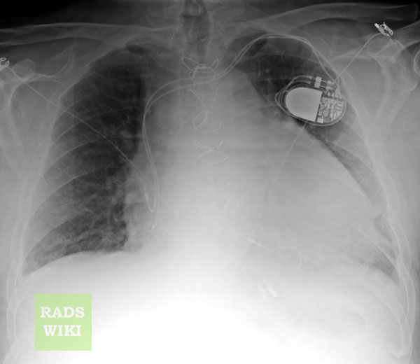
A flask-shaped, enlarged cardiac silhouette will be observed on chest x-ray in pericarditis complicated with pericardial effusion or tamponade. A mass may also be seen when malignancy is the cause. Calcification of pericardium may be noted in constrictive pericarditis.
Uncomplicated Pericarditis
In uncomplicated pericarditis, in the absence of a pericardial effusion or cardiac tamponade, the heart will not be enlarged on chest x ray.
Complicated Pericarditis
A flask-shaped, enlarged cardiac silhouette will be observed on chest X ray in the setting of pericardial effusion or cardiac tamponade.
Chest X Ray Examples


Images shown below are courtesy of RadsWiki


Pericardial Masses

Pericardial Calcification

2015 ESC Guidelines on the Diagnosis and Treatment of Pericarditis (DO NOT EDIT)[1]
Recommendations for the general diagnostic work-up of pericardial diseases
| Class I |
| 1. In all cases of suspected pericardial disease a first diagnostic evaluation is recommended with:
– ECG – transthoracic echocardiography – routine blood tests, including markers of inflammation (i.e., CRP and/or ESR), white blood cell count with differential count, renal function and liver tests and myocardial lesion tests (CK, troponins). 2. CT and/or CMR are recommended as second-level testing for diagnostic workup in pericarditis. 3. Pericardiocentesis or surgical drainage are indicated for cardiac tamponade or suspected bacterial and neoplastic pericarditis. 4. Further testing is indicated in high-risk patients (defined as above) according to the clinical conditions. (Level of Evidence: C)
|
Recommendations for diagnosis of acute pericarditis
| Class I |
| 1. ECG is recommended in all patients with suspected acute pericarditis.
2. Transthoracic echocardiography is recommended in all patients with suspected acute pericarditis. 3. Chest X-ray is recommended in all patients with suspected acute pericarditis. 4. Assessment of markers of inflammation (i.e. CRP) and myocardial injury (i.e. CK, troponin) is recommended in patients with suspected acute pericarditis. (Level of Evidence: C)
|
Recommendations for the diagnosis of constrictive pericarditis
| Class I |
| 1. Transthoracic echocardiography is recommended in all patients with suspected constrictive pericarditis.
2. Chest X-ray (frontal and lateral views)with adequate technical characteristics is recommended in all patients with suspected constrictive pericarditis. 3. CT and/or CMR are indicated as second-level imaging techniques to assess calcifications (CT), pericardial thickness, degree and extension of pericardial involvement. 4. Cardiac catheterization is indicated when non-invasive diagnostic methods do not provide a definite diagnosis of constriction. (Level of Evidence: C)
|
References
- ↑ Adler, Yehuda; Charron, Philippe; Imazio, Massimo; Badano, Luigi; Barón-Esquivias, Gonzalo; Bogaert, Jan; Brucato, Antonio; Gueret, Pascal; Klingel, Karin; Lionis, Christos; Maisch, Bernhard; Mayosi, Bongani; Pavie, Alain; Ristić, Arsen D.; Sabaté Tenas, Manel; Seferovic, Petar; Swedberg, Karl; Tomkowski, Witold (2015). "2015 ESC Guidelines for the diagnosis and management of pericardial diseases". European Heart Journal. 36 (42): 2921–2964. doi:10.1093/eurheartj/ehv318. ISSN 0195-668X.