Necrotizing fasciitis pathophysiology: Difference between revisions
m (Bot: Removing from Primary care) |
|||
| (9 intermediate revisions by 4 users not shown) | |||
| Line 4: | Line 4: | ||
==Overview== | ==Overview== | ||
The pathophysiology of necrotizing fasciitis is common to all types but the speed of development and associated clinical features differs depending on the causative organisms. Following transmission, the bacteria uses the entry site to invade the [[Fascia|fascial planes]] which causes the wide spread necrosis of [[Fascia|superficial fascia]], [[Fascia|deep fascia]], [[subcutaneous fat]], [[nerves]], [[arteries]], and [[veins]]. Necrotizing fasciitis can be a serious complication of [[omphalitis]] in the [[neonate]].The pathogenesis of necrotizing fasciitis is the result of bacterial and host factors. The exact pathogenesis of type 1 necrotizing fasciitis is not fully understood but [[polymicrobial]] species work | The pathophysiology of necrotizing fasciitis is common to all types, but the speed of development and associated clinical features differs depending on the causative organisms. Following transmission, the bacteria uses the entry site to invade the [[Fascia|fascial planes]] which causes the wide spread necrosis of [[Fascia|superficial fascia]], [[Fascia|deep fascia]], [[subcutaneous fat]], [[nerves]], [[arteries]], and [[veins]]. Necrotizing fasciitis can be a serious complication of [[omphalitis]] in the [[neonate]]. The pathogenesis of necrotizing fasciitis is the result of bacterial and host factors. The exact pathogenesis of type 1 necrotizing fasciitis is not fully understood but [[polymicrobial]] species work [[Synergistic|synergistically]] to enhance the spread of infection. [[Streptococcus|Group A streptococcus]] is the most common causative agent of type 2 necrotizing fasciitis. Bacterial [[virulence factors]], [[exotoxins]], [[superantigens]] and [[host]] [[immune system]] plays a major role in the pathogenesis of type II necrotizing fasciitis. Recurrent necrotizing fasciitis is caused by [[MRSA]]. On gross pathology, the characteristic findings of necrotizing fasciitis include [[Emphysema|subcutaneous emphysema]], skin sloughing, bulae and [[necrosis]]. [[Inflammation|Inflammatory changes]] are seen on microscopic histopathology.<ref name="pmid25593960">{{cite journal| author=Misiakos EP, Bagias G, Patapis P, Sotiropoulos D, Kanavidis P, Machairas A| title=Current concepts in the management of necrotizing fasciitis. | journal=Front Surg | year= 2014 | volume= 1 | issue= | pages= 36 | pmid=25593960 | doi=10.3389/fsurg.2014.00036 | pmc=4286984 | url=https://www.ncbi.nlm.nih.gov/entrez/eutils/elink.fcgi?dbfrom=pubmed&tool=sumsearch.org/cite&retmode=ref&cmd=prlinks&id=25593960 }} </ref> | ||
==Pathophysiology== | ==Pathophysiology== | ||
The pathophysiology of necrotizing fasciitis is common to all types of | The pathophysiology of necrotizing fasciitis is common to all types of necrotizing fasciitis, but the speed of development and associated clinical features differs depending on the causative organisms.<ref name="pmid25593960">{{cite journal| author=Misiakos EP, Bagias G, Patapis P, Sotiropoulos D, Kanavidis P, Machairas A| title=Current concepts in the management of necrotizing fasciitis. | journal=Front Surg | year= 2014 | volume= 1 | issue= | pages= 36 | pmid=25593960 | doi=10.3389/fsurg.2014.00036 | pmc=4286984 | url=https://www.ncbi.nlm.nih.gov/entrez/eutils/elink.fcgi?dbfrom=pubmed&tool=sumsearch.org/cite&retmode=ref&cmd=prlinks&id=25593960 }} </ref> | ||
*The transmission of pathogens occurs through the following routes:<ref name="pmid18476182">{{cite journal| author=Lynch CM, Pinelli DM, Cruse CW, Spellacy WN, Sinnott JT, Shashy RG| title=Maternal death from postpartum necrotizing fasciitis arising in an episiotomy: a case report. | journal=Infect Dis Obstet Gynecol | year= 1997 | volume= 5 | issue= 5 | pages= 341-4 | pmid=18476182 | doi=10.1155/S1064744997000598 | pmc=2364577 | url=https://www.ncbi.nlm.nih.gov/entrez/eutils/elink.fcgi?dbfrom=pubmed&tool=sumsearch.org/cite&retmode=ref&cmd=prlinks&id=18476182 }} </ref><ref name="pmid20542593">{{cite journal| author=Morgan MS| title=Diagnosis and management of necrotising fasciitis: a multiparametric approach. | journal=J Hosp Infect | year= 2010 | volume= 75 | issue= 4 | pages= 249-57 | pmid=20542593 | doi=10.1016/j.jhin.2010.01.028 | pmc= | url=https://www.ncbi.nlm.nih.gov/entrez/eutils/elink.fcgi?dbfrom=pubmed&tool=sumsearch.org/cite&retmode=ref&cmd=prlinks&id=20542593 }} </ref> | *The transmission of pathogens occurs through the following routes:<ref name="pmid18476182">{{cite journal| author=Lynch CM, Pinelli DM, Cruse CW, Spellacy WN, Sinnott JT, Shashy RG| title=Maternal death from postpartum necrotizing fasciitis arising in an episiotomy: a case report. | journal=Infect Dis Obstet Gynecol | year= 1997 | volume= 5 | issue= 5 | pages= 341-4 | pmid=18476182 | doi=10.1155/S1064744997000598 | pmc=2364577 | url=https://www.ncbi.nlm.nih.gov/entrez/eutils/elink.fcgi?dbfrom=pubmed&tool=sumsearch.org/cite&retmode=ref&cmd=prlinks&id=18476182 }} </ref><ref name="pmid20542593">{{cite journal| author=Morgan MS| title=Diagnosis and management of necrotising fasciitis: a multiparametric approach. | journal=J Hosp Infect | year= 2010 | volume= 75 | issue= 4 | pages= 249-57 | pmid=20542593 | doi=10.1016/j.jhin.2010.01.028 | pmc= | url=https://www.ncbi.nlm.nih.gov/entrez/eutils/elink.fcgi?dbfrom=pubmed&tool=sumsearch.org/cite&retmode=ref&cmd=prlinks&id=20542593 }} </ref> | ||
:*External trauma (e.g., [[laceration]], [[abrasion]], [[burn]], insect bite) | :*External trauma (e.g., [[laceration]], [[abrasion]], [[burn]], insect bite) | ||
| Line 14: | Line 14: | ||
:*[[Perirectal abscess]] | :*[[Perirectal abscess]] | ||
:*[[Decubitus ulcer]] | :*[[Decubitus ulcer]] | ||
*Following transmission, the bacteria uses the entry site to invade the [[Fascia|fascial planes]] which causes the wide spread necrosis of [[Fascia|superficial fascia]], [[Fascia|deep fascia]], | *Following transmission, the bacteria uses the entry site to invade the [[Fascia|fascial planes]] which causes the wide spread necrosis of [[Fascia|superficial fascia]], [[Fascia|deep fascia]], [[subcutaneous fat]], [[nerves]], [[arteries]], and [[veins]]. | ||
[[subcutaneous fat]], [[nerves]], [[arteries]], and [[veins]]. | |||
*Superficial skin and deeper muscles are typically spared. | *Superficial skin and deeper muscles are typically spared. | ||
*In late stages, lesions develop [[liquefactive necrosis]] at all tissue levels. | *In late stages, lesions develop [[liquefactive necrosis]] at all tissue levels. | ||
| Line 29: | Line 28: | ||
'''Type 1 NF''' | '''Type 1 NF''' | ||
*The exact pathogenesis of type 1 necrotizing fasciitis is not fully understood. | *The exact pathogenesis of type 1 necrotizing fasciitis is not fully understood. | ||
*It is thought that type 1 NF is caused by polymicrobial species that work | *It is thought that type 1 NF is caused by polymicrobial species that work [[Synergistic|synergistically]] to enhance the spread of infection. | ||
*Synergistic NF is comparatively slow process evolving over days. | *Synergistic NF is comparatively slow process evolving over days. | ||
*It usually develops in the area where [[gut flora]] breaches the [[mucosa]], entering the tissue planes. | *It usually develops in the area where [[gut flora]] breaches the [[mucosa]], entering the tissue planes. | ||
| Line 35: | Line 34: | ||
*[[Streptococcus|Group A streptococcus]] is the most common causative agent of type 2 NF.<ref name="pmid10925760">{{cite journal| author=Leitch HA, Palepu A, Fernandes CM| title=Necrotizing fasciitis secondary to group A streptococcus. Morbidity and mortality still high. | journal=Can Fam Physician | year= 2000 | volume= 46 | issue= | pages= 1460-6 | pmid=10925760 | doi= | pmc=2144855 | url=https://www.ncbi.nlm.nih.gov/entrez/eutils/elink.fcgi?dbfrom=pubmed&tool=sumsearch.org/cite&retmode=ref&cmd=prlinks&id=10925760 }} </ref><ref name="pmid23753218">{{cite journal| author=Shiroff AM, Herlitz GN, Gracias VH| title=Necrotizing soft tissue infections. | journal=J Intensive Care Med | year= 2014 | volume= 29 | issue= 3 | pages= 138-44 | pmid=23753218 | doi=10.1177/0885066612463680 | pmc= | url=https://www.ncbi.nlm.nih.gov/entrez/eutils/elink.fcgi?dbfrom=pubmed&tool=sumsearch.org/cite&retmode=ref&cmd=prlinks&id=23753218 }} </ref> | *[[Streptococcus|Group A streptococcus]] is the most common causative agent of type 2 NF.<ref name="pmid10925760">{{cite journal| author=Leitch HA, Palepu A, Fernandes CM| title=Necrotizing fasciitis secondary to group A streptococcus. Morbidity and mortality still high. | journal=Can Fam Physician | year= 2000 | volume= 46 | issue= | pages= 1460-6 | pmid=10925760 | doi= | pmc=2144855 | url=https://www.ncbi.nlm.nih.gov/entrez/eutils/elink.fcgi?dbfrom=pubmed&tool=sumsearch.org/cite&retmode=ref&cmd=prlinks&id=10925760 }} </ref><ref name="pmid23753218">{{cite journal| author=Shiroff AM, Herlitz GN, Gracias VH| title=Necrotizing soft tissue infections. | journal=J Intensive Care Med | year= 2014 | volume= 29 | issue= 3 | pages= 138-44 | pmid=23753218 | doi=10.1177/0885066612463680 | pmc= | url=https://www.ncbi.nlm.nih.gov/entrez/eutils/elink.fcgi?dbfrom=pubmed&tool=sumsearch.org/cite&retmode=ref&cmd=prlinks&id=23753218 }} </ref> | ||
*Type 2 NF is initially insidious in onset but progress more rapidly.<ref name="pmid7748037">{{cite journal| author=McHenry CR, Piotrowski JJ, Petrinic D, Malangoni MA| title=Determinants of mortality for necrotizing soft-tissue infections. | journal=Ann Surg | year= 1995 | volume= 221 | issue= 5 | pages= 558-63; discussion 563-5 | pmid=7748037 | doi= | pmc=1234638 | url=https://www.ncbi.nlm.nih.gov/entrez/eutils/elink.fcgi?dbfrom=pubmed&tool=sumsearch.org/cite&retmode=ref&cmd=prlinks&id=7748037 }} </ref> | *Type 2 NF is initially insidious in onset but progress more rapidly.<ref name="pmid7748037">{{cite journal| author=McHenry CR, Piotrowski JJ, Petrinic D, Malangoni MA| title=Determinants of mortality for necrotizing soft-tissue infections. | journal=Ann Surg | year= 1995 | volume= 221 | issue= 5 | pages= 558-63; discussion 563-5 | pmid=7748037 | doi= | pmc=1234638 | url=https://www.ncbi.nlm.nih.gov/entrez/eutils/elink.fcgi?dbfrom=pubmed&tool=sumsearch.org/cite&retmode=ref&cmd=prlinks&id=7748037 }} </ref> | ||
*The disease may appear spontaneously with no obvious focus. | *The disease may appear spontaneously with no obvious focus. Hematogenous infection from many foci such as [[skin]], [[throat]], [[Vaginitis|ascending vaginitis]], [[Peritonitis|primary peritonitis]] reaches the fascial layer or seeds [[vimentin]] exposed by muscle damage. | ||
*Direct inoculation of [[Streptococcus|GAS]] through wounds or associated surgery is less common. | *Direct inoculation of [[Streptococcus|GAS]] through wounds or associated surgery is less common. | ||
*The pathogenesis of type 2 NF is the result of the following process: | *The pathogenesis of type 2 NF is the result of the following process: | ||
| Line 46: | Line 45: | ||
:*The massive release of [[cytokines]] result in [[systemic inflammatory response syndrome]] resulting in [[shock]], organ failure, depression of myocardial function and immune suppression | :*The massive release of [[cytokines]] result in [[systemic inflammatory response syndrome]] resulting in [[shock]], organ failure, depression of myocardial function and immune suppression | ||
:*Stimulation of [[T-cells]] by [[superantigen]] causes activation of [[Complement system|complement pathway]], the [[bradykinin]]-[[kallikrein]] system, and the [[coagulation cascade]], worsening small vessel [[thrombosis]] and tissue ischemia. [[Ischemia|Tissue ischemia]] impedes oxidative destruction of [[bacteria]] by [[polymorphonuclear cells]] and prevents adequate delivery of antibiotics. | :*Stimulation of [[T-cells]] by [[superantigen]] causes activation of [[Complement system|complement pathway]], the [[bradykinin]]-[[kallikrein]] system, and the [[coagulation cascade]], worsening small vessel [[thrombosis]] and tissue ischemia. [[Ischemia|Tissue ischemia]] impedes oxidative destruction of [[bacteria]] by [[polymorphonuclear cells]] and prevents adequate delivery of antibiotics. | ||
:*The blood flow to local tissue is compromised due to thrombosis of large number of | :*The blood flow to local tissue is compromised due to thrombosis of large number of dermal capillaries by the local [[Coagulation|hypercoaguable state]], [[platelet]]-[[neutrophil]] plugging of vessels and increased [[interstitial|interstitial pressure]] | ||
*Nerves supplying the necrotizing areas of skin die, the central areas become [[anaesthetic]] and peripheral areas remain tender | *Nerves supplying the necrotizing areas of skin die, the central areas become [[anaesthetic]] and peripheral areas remain tender | ||
*In later stages, infection from deeper layers ascends, producing [[edema]] of epidermal and dermal layers ([[peau d'orange]]) and a woody firmness of the tissues | *In later stages, infection from deeper layers ascends, producing [[edema]] of epidermal and dermal layers ([[peau d'orange]]) and a woody firmness of the tissues | ||
*The fascial and nerve destruction results in [[sensory]] and motor deficits, which causes progression of hemorrhagic bulae to cutaneous gangrene | *The fascial and nerve destruction results in [[sensory]] and motor deficits, which causes progression of hemorrhagic bulae to cutaneous gangrene | ||
'''Recurrent necrotizing fasciitis''' | '''Recurrent necrotizing fasciitis''' | ||
| Line 77: | Line 66: | ||
<gallery> | <gallery> | ||
Image:800px-Necrotizing_fasciitis_left_leg.JPEG|By Nephron - Own work, CC BY-SA 3.0, https://commons.wikimedia.org/w/index.php?curid=19107063 | |||
Image:800px-Necrotizing_fasciitis_left_leg.JPEG| | |||
</gallery> | </gallery> | ||
===Microscopic | ===Microscopic Pathology=== | ||
On microscopic histopathological analysis, the characteristic findings of necrotizing fasciitis are<ref name=NF>Librae pathology(2015) https://librepathology.org/wiki/Necrotizing_fasciitis Accessed on September 2,2016 </ref> | On microscopic histopathological analysis, the characteristic findings of necrotizing fasciitis are<ref name=NF>Librae pathology(2015) https://librepathology.org/wiki/Necrotizing_fasciitis Accessed on September 2,2016 </ref> | ||
*Early stages | *Early stages | ||
| Line 95: | Line 82: | ||
<gallery> | <gallery> | ||
Image:80px-Necrotizing_fasciitis_-_high_mag.jpg | Image:80px-Necrotizing_fasciitis_-_high_mag.jpg|By Piotr Smuszkiewicz, Iwona Trojanowska and Hanna Tomczak - Late diagnosed necrotizing fasciitis as a cause of multiorgan dysfunction syndrome: A case report. Cases Journal 2008, 1:125. doi:10.1186/1757-1626-1-125, CC BY 2.0, https://commons.wikimedia.org/w/index.php?curid=5639655 | ||
Image:120px-Necrotizing_fasciitis_-_intermed_mag.jpg | Image:120px-Necrotizing_fasciitis_-_intermed_mag.jpg|By Piotr Smuszkiewicz, Iwona Trojanowska and Hanna Tomczak - Late diagnosed necrotizing fasciitis as a cause of multiorgan dysfunction syndrome: A case report. Cases Journal 2008, 1:125. doi:10.1186/1757-1626-1-125, CC BY 2.0, https://commons.wikimedia.org/w/index.php?curid=5639655 | ||
</gallery> | </gallery> | ||
| Line 104: | Line 91: | ||
{{Reflist|2}} | {{Reflist|2}} | ||
[[Category:Emergency mdicine]] | |||
[[Category:Disease]] | [[Category:Disease]] | ||
[[Category: | [[Category:Up-To-Date]] | ||
[[Category:Infectious disease]] | [[Category:Infectious disease]] | ||
[[Category:Surgery]] | |||
[[Category:Orthopedics]] | |||
Latest revision as of 22:56, 29 July 2020
|
Necrotizing fasciitis Microchapters |
|
Diagnosis |
|---|
|
Treatment |
|
Case Studies |
|
Necrotizing fasciitis pathophysiology On the Web |
|
American Roentgen Ray Society Images of Necrotizing fasciitis pathophysiology |
|
Risk calculators and risk factors for Necrotizing fasciitis pathophysiology |
Editor-In-Chief: C. Michael Gibson, M.S., M.D. [1]; Associate Editor(s)-in-Chief: Yamuna Kondapally, M.B.B.S[2]
Overview
The pathophysiology of necrotizing fasciitis is common to all types, but the speed of development and associated clinical features differs depending on the causative organisms. Following transmission, the bacteria uses the entry site to invade the fascial planes which causes the wide spread necrosis of superficial fascia, deep fascia, subcutaneous fat, nerves, arteries, and veins. Necrotizing fasciitis can be a serious complication of omphalitis in the neonate. The pathogenesis of necrotizing fasciitis is the result of bacterial and host factors. The exact pathogenesis of type 1 necrotizing fasciitis is not fully understood but polymicrobial species work synergistically to enhance the spread of infection. Group A streptococcus is the most common causative agent of type 2 necrotizing fasciitis. Bacterial virulence factors, exotoxins, superantigens and host immune system plays a major role in the pathogenesis of type II necrotizing fasciitis. Recurrent necrotizing fasciitis is caused by MRSA. On gross pathology, the characteristic findings of necrotizing fasciitis include subcutaneous emphysema, skin sloughing, bulae and necrosis. Inflammatory changes are seen on microscopic histopathology.[1]
Pathophysiology
The pathophysiology of necrotizing fasciitis is common to all types of necrotizing fasciitis, but the speed of development and associated clinical features differs depending on the causative organisms.[1]
- External trauma (e.g., laceration, abrasion, burn, insect bite)
- Direct spread from a perforated viscus (particularly colon, rectum, or anus) or another surgical procedure (e.g., vasectomy, hemorrhoidectomy)
- Urogenital organ
- Perirectal abscess
- Decubitus ulcer
- Following transmission, the bacteria uses the entry site to invade the fascial planes which causes the wide spread necrosis of superficial fascia, deep fascia, subcutaneous fat, nerves, arteries, and veins.
- Superficial skin and deeper muscles are typically spared.
- In late stages, lesions develop liquefactive necrosis at all tissue levels.
Necrotizing fasciitis in neonate
- Necrotizing fasciitis can be a serious complication of omphalitis in the neonate.
- The omphalitis may progress resulting in purplish discoloration and periumbilical necrosis.
- The necrosis may extend to the flanks and even onto the chest wall.
Pathogenesis
The pathogenesis of necrotizing fasciitis is the result of bacterial and host factors.[4][5]
Type 1 NF
- The exact pathogenesis of type 1 necrotizing fasciitis is not fully understood.
- It is thought that type 1 NF is caused by polymicrobial species that work synergistically to enhance the spread of infection.
- Synergistic NF is comparatively slow process evolving over days.
- It usually develops in the area where gut flora breaches the mucosa, entering the tissue planes.
Type 2 NF
- Group A streptococcus is the most common causative agent of type 2 NF.[6][7]
- Type 2 NF is initially insidious in onset but progress more rapidly.[8]
- The disease may appear spontaneously with no obvious focus. Hematogenous infection from many foci such as skin, throat, ascending vaginitis, primary peritonitis reaches the fascial layer or seeds vimentin exposed by muscle damage.
- Direct inoculation of GAS through wounds or associated surgery is less common.
- The pathogenesis of type 2 NF is the result of the following process:
- Inhibition of phagocytosis of bacteria by hyaluronic acid capsule and M protein
- Adherence of bacteria to host cell through adherence factors such as M protein, protein F and lipoteichoic acid[9]
- Release of exotoxins (streptococcal pyogenic exotoxins and superantigen) into blood produce massive proliferation of T cells and cascading release of cytokines activating inflammatory process
- Activation of inflammatory process which begins to kill bacteria
- The streptococci release massive amounts of enzymes, hemolysins, DNAase, proteases and collagenases which destroy the normal skin and surrounding tissue with progressive coagulative necrosis
- The inflamed cells release more cytokines that stimulate more inflammatory cells
- The massive release of cytokines result in systemic inflammatory response syndrome resulting in shock, organ failure, depression of myocardial function and immune suppression
- Stimulation of T-cells by superantigen causes activation of complement pathway, the bradykinin-kallikrein system, and the coagulation cascade, worsening small vessel thrombosis and tissue ischemia. Tissue ischemia impedes oxidative destruction of bacteria by polymorphonuclear cells and prevents adequate delivery of antibiotics.
- The blood flow to local tissue is compromised due to thrombosis of large number of dermal capillaries by the local hypercoaguable state, platelet-neutrophil plugging of vessels and increased interstitial pressure
- Nerves supplying the necrotizing areas of skin die, the central areas become anaesthetic and peripheral areas remain tender
- In later stages, infection from deeper layers ascends, producing edema of epidermal and dermal layers (peau d'orange) and a woody firmness of the tissues
- The fascial and nerve destruction results in sensory and motor deficits, which causes progression of hemorrhagic bulae to cutaneous gangrene
Recurrent necrotizing fasciitis
Recurrent NF is seen in following conditions:[10]
- Methicillin resistant staphylococcus aureus (MRSA)
- Complement C4 deficiency
Gross pathology
On gross pathology the characteristic findings of necrotizing fasciitis include:[11]
- Subcutaneous emphysema
- Edema
- Erythema
- Bulae
- Skin sloughing
- Dull grey discoloration
-
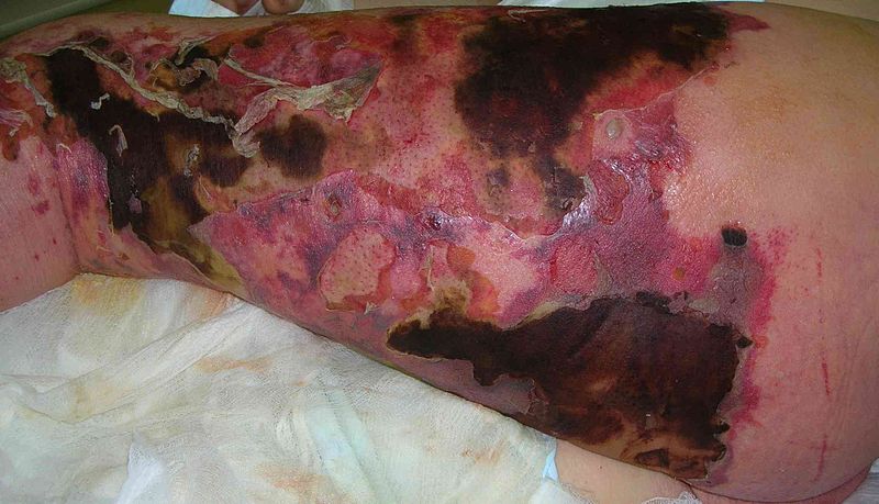
By Nephron - Own work, CC BY-SA 3.0, https://commons.wikimedia.org/w/index.php?curid=19107063

Microscopic Pathology
On microscopic histopathological analysis, the characteristic findings of necrotizing fasciitis are[11]
- Early stages
- Obliterative vasculitis with microangiopathic thrombosis
- Acute inflammation of subcutaneous tissue
- Superficial hyaline necrosis along with edema and inflammation of the dermis and subcutaneous fat
- Dense neutrophil-predominant inflammatory infiltrate
- Late stages
- Noninflammatory intravascular coagulation and hemorrhage
- Myonecrosis
-
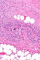
By Piotr Smuszkiewicz, Iwona Trojanowska and Hanna Tomczak - Late diagnosed necrotizing fasciitis as a cause of multiorgan dysfunction syndrome: A case report. Cases Journal 2008, 1:125. doi:10.1186/1757-1626-1-125, CC BY 2.0, https://commons.wikimedia.org/w/index.php?curid=5639655
-
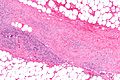
By Piotr Smuszkiewicz, Iwona Trojanowska and Hanna Tomczak - Late diagnosed necrotizing fasciitis as a cause of multiorgan dysfunction syndrome: A case report. Cases Journal 2008, 1:125. doi:10.1186/1757-1626-1-125, CC BY 2.0, https://commons.wikimedia.org/w/index.php?curid=5639655


References
- ↑ 1.0 1.1 Misiakos EP, Bagias G, Patapis P, Sotiropoulos D, Kanavidis P, Machairas A (2014). "Current concepts in the management of necrotizing fasciitis". Front Surg. 1: 36. doi:10.3389/fsurg.2014.00036. PMC 4286984. PMID 25593960.
- ↑ Lynch CM, Pinelli DM, Cruse CW, Spellacy WN, Sinnott JT, Shashy RG (1997). "Maternal death from postpartum necrotizing fasciitis arising in an episiotomy: a case report". Infect Dis Obstet Gynecol. 5 (5): 341–4. doi:10.1155/S1064744997000598. PMC 2364577. PMID 18476182.
- ↑ Morgan MS (2010). "Diagnosis and management of necrotising fasciitis: a multiparametric approach". J Hosp Infect. 75 (4): 249–57. doi:10.1016/j.jhin.2010.01.028. PMID 20542593.
- ↑ Stamenkovic I, Lew PD (1984). "Early recognition of potentially fatal necrotizing fasciitis. The use of frozen-section biopsy". N Engl J Med. 310 (26): 1689–93. doi:10.1056/NEJM198406283102601. PMID 6727947.
- ↑ Elliott DC, Kufera JA, Myers RA (1996). "Necrotizing soft tissue infections. Risk factors for mortality and strategies for management". Ann Surg. 224 (5): 672–83. PMC 1235444. PMID 8916882.
- ↑ Leitch HA, Palepu A, Fernandes CM (2000). "Necrotizing fasciitis secondary to group A streptococcus. Morbidity and mortality still high". Can Fam Physician. 46: 1460–6. PMC 2144855. PMID 10925760.
- ↑ Shiroff AM, Herlitz GN, Gracias VH (2014). "Necrotizing soft tissue infections". J Intensive Care Med. 29 (3): 138–44. doi:10.1177/0885066612463680. PMID 23753218.
- ↑ McHenry CR, Piotrowski JJ, Petrinic D, Malangoni MA (1995). "Determinants of mortality for necrotizing soft-tissue infections". Ann Surg. 221 (5): 558–63, discussion 563-5. PMC 1234638. PMID 7748037.
- ↑ Chelsom J, Halstensen A, Haga T, Høiby EA (1994). "Necrotising fasciitis due to group A streptococci in western Norway: incidence and clinical features". Lancet. 344 (8930): 1111–5. PMID 7934492.
- ↑ Miller LG, Perdreau-Remington F, Rieg G, Mehdi S, Perlroth J, Bayer AS; et al. (2005). "Necrotizing fasciitis caused by community-associated methicillin-resistant Staphylococcus aureus in Los Angeles". N Engl J Med. 352 (14): 1445–53. doi:10.1056/NEJMoa042683. PMID 15814880.
- ↑ 11.0 11.1 Librae pathology(2015) https://librepathology.org/wiki/Necrotizing_fasciitis Accessed on September 2,2016