Liquefactive necrosis
| Liquefactive necrosis | |
 | |
|---|---|
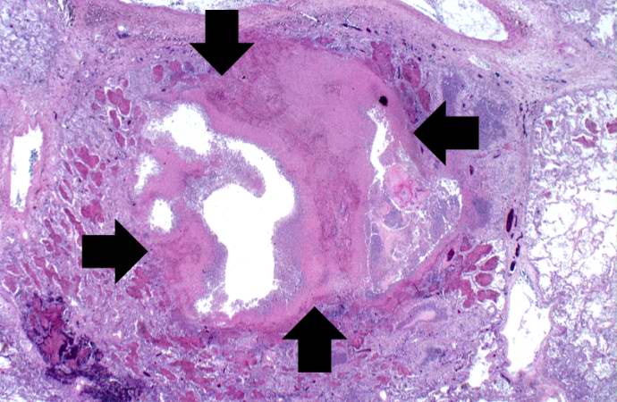
| This is a low-power photomicrograph of lung tissue containing a large abscess. The center of the abscess contains necrotic debris (1) and there is a rim of viable inflammatory cells (arrows) surrounding this abscess. Image courtesy of Professor Peter Anderson DVM PhD and published with permission © PEIR, University of Alabama at Birmingham, Department of Pathology |
Editor-In-Chief: C. Michael Gibson, M.S., M.D. [1]
Liquefactive necrosis (or colliquative necrosis) is a type of necrosis which is characteristic of focal bacterial or fungal infections. In liquefactive necrosis, the affected cell is completely digested by hydrolytic enzymes, resulting in a soft, circumscribed lesion consisting of pus and the fluid remains of necrotic tissue. After the removal of cell debris by white blood cells, a fluid filled space is left. It is generally associated with abscess formation and is commonly found in the central nervous system.
For unclear reasons, hypoxic death of cells within the central nervous system also results in liquefactive necrosis.(Brain Infarction => Emollition) This is a process in which lysosomes turn tissues into soup as a result of lysosomal release of digestive enzymes in the face of bacterial onslaught. Loss of tissue architecture means that the tissue is essentially liquefied.
Pathological Findings: Case #1: Lung: Liquefactive necrosis
Clinical Summary
A 67-year-old male with advanced colon cancer developed obstruction of the bowel and underwent palliative surgery to remove an 8-cm portion of colon containing the obstruction. During the surgery the patient had several episodes of hypotension. After surgery he did not regain consciousness and required ventilator support. Four days after surgery, the patient developed a fever and his white blood cell count was found to be 15,256 cells/cmm. Thus, he was started on broad-spectrum antibiotics. A chest x-ray demonstrated infiltrates in both lungs, which worsened over the next several days. His overall condition continued to deteriorate and he died 12 days after surgery.
Autopsy Findings
At autopsy, metastatic colon cancer was found throughout the abdominal cavity and invading into the liver. The lungs were markedly consolidated and had several focal abscesses that were 2 to 4 cm in diameter. Liquefied material poured out from inside these abscesses when the lungs were sliced.
-
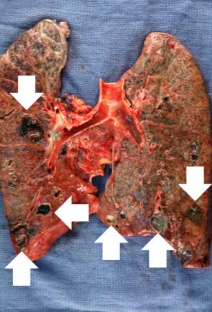
A gross photograph of the lungs from this case. Note the abscesses (arrows) especially in the lower lobes. The entire lung is consolidated.
-
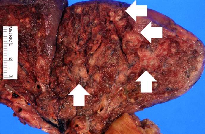
This is a closer view of the lung from this case. In this section of upper lobe there are multiple areas of early abscess formation (arrows). Note the circumscribed whitish-tan lesions. These lesions are filled with white blood cells.


-
This low-power photomicrograph of lung from this case demonstrates one of the abscesses (arrows). Note that the material inside the abscess has been expelled.
-
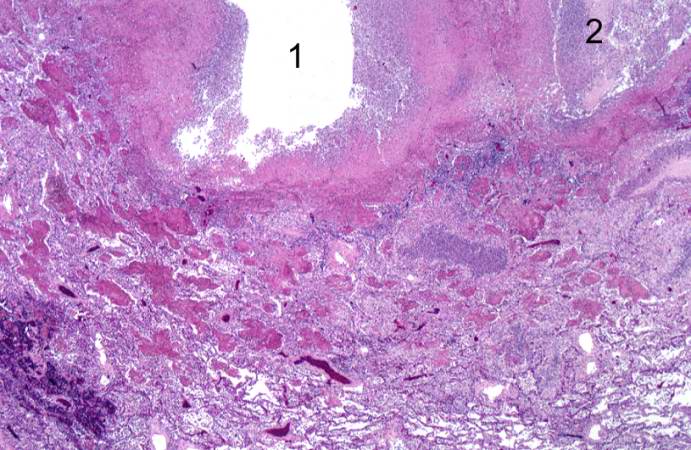
This higher-power photomicrograph of lung demonstrates the edge of the abscess. Note the loss of material from the center of the abscess (1) and loose necrotic material that has not been expelled (2). This material is made up of inflammatory cells (primarily dead white blood cells) and necrotic lung tissue.


-
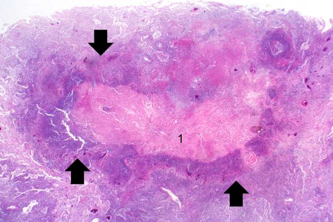
This is a low-power photomicrograph of lung tissue containing a large abscess. The center of the abscess contains necrotic debris (1) and there is a rim of viable inflammatory cells (arrows) surrounding this abscess.
-
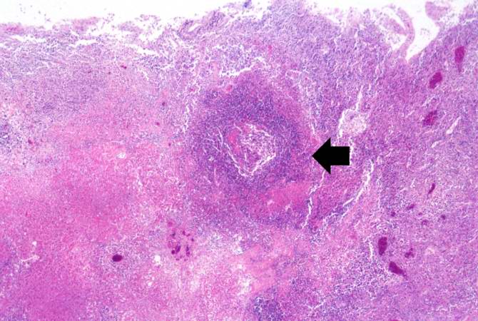
This high-power photomicrograph demonstrates a small abscess (arrow) with a necrotic center.

-
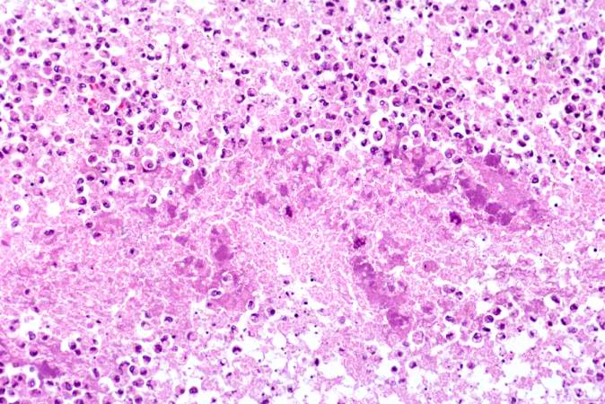
This high-power photomicrograph shows the center of an abscess containing neutrophils and necrotic debris.
-
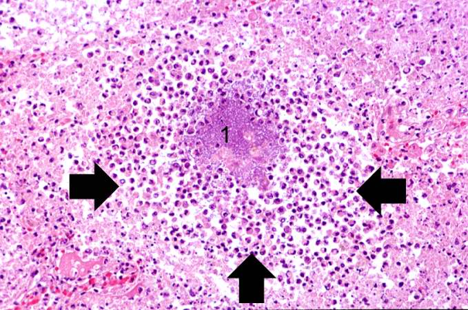
A high-power photomicrograph of lung from this case demonstrates a small abscess full of inflammatory cells (primarily neutrophils) (arrows). There is a bacterial colony in the center of this abscess (1).

