Lyme disease physical examination
|
Lyme disease Microchapters |
|
Diagnosis |
|---|
|
Treatment |
|
Case Studies |
|
Lyme disease physical examination On the Web |
|
American Roentgen Ray Society Images of Lyme disease physical examination |
|
Risk calculators and risk factors for Lyme disease physical examination |
Editor-In-Chief: C. Michael Gibson, M.S., M.D. [1] Associate Editor(s)-in-Chief: Anmol Pitliya, M.B.B.S. M.D.[2]
Overview
The physical examination of Lyme disease is necessary for the diagnosis. Erythema migrans and fever are commonly seen on physical examination in early disease. Disseminated disease is characterized by multiple erythema migrans, neurological, musculoskeletal, and cardiac symptoms.
Physical Examination in Stage 1 - Early localized disease
Appearance of the patient
- Patients are usually well appearing in early stages; may appear fatigue.
Vital signs
- Fever and/or chills
Skin
- Erythema migrans (target like lesions) - Usually single, present at the site of tick bite.
- Regional Lymphadenopathy - Around site of tick bite.
HEENT
Neck
Muscukoskeletal
Physical Examination in Stage 2 - Early disseminated disease
Appearance of the patient
- As the disease progresses patient may appear ill. Patient has severe malaise and fatigue.
Vital signs
- Slow or irregular pulse
Skin
- Erythema migrans (target like lesions) - Usually multiple, multiple lesion are secondary to dissemination; not to be confused with multiple tick bite
- Borrelial lymphocytoma - Cutaneous B-cell pseudolymphoma
- Malar rash
- Diffuse erythema or urticarial
- Regional or generalized lymphadenopathy
HEENT
- Facial deviation
- Dry eyes
- Drooling of saliva
- Blurred vision
Neck
- Neck stiffness
- Neck pain
- Cannon A waves in patients with complete heart block
Cardiovascular
- Tachycardia
- Palpitations
- Cardiac rub
- Dyspnea
- S3 and/or S4 may be present
Abdomen
- Splenomegaly
Neurological
Muscuoskeletal
- Migratory arthralgia
Physical Examination in Stage 3 - Late disseminated disease
Appearance of the patient
- Patients appear ill and fatigue.
Skin
- Acrodermatitis chronica atrophicans
- Localized scleroderma-like lesions
HEENT
- Severe Headache
- Sore throat
- Vision changes
- Odynophagia
- Otalgia
- Tinnitus
- Hearing loss
- Vertigo
- Temporomandibular joint pain
- Dysgeusia
Neck
- Tender neck flexion
- Neck pain
Neurological
- Fasciculations
- Spastic parapareses
- Ataxia
- Neuropsychiatric findings includes dementia, anxiety, depression, anxiety, schizophrenia-like psychosis, bipolar disorder
Muscukoskeletal
- Arthralgias - Commonly involving knee, may be migratory
- Joint tenderness
- Joint swelling
- Joint efffusion
- Enthesitis
- Periositits or joint subluxations below acrodermatitis
Images
-

Common bullseye rash pattern associated with Lyme Disease.
-
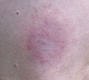
Characteristic "bulls-eye"-like rash caused by Lyme disease.
-
Erythema Chronicum Migrans


.jpg)
-
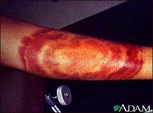
![Right posterior shoulder region of a patient who’d presented with the erythema migrans (EM) rash characteristic of what was diagnosed as Lyme disease, caused by Borrelia burgdorferi. From Public Health Image Library (PHIL). [1]](/images/c/c8/Borrelia07.jpeg)
Right posterior shoulder region of a patient who’d presented with the erythema migrans (EM) rash characteristic of what was diagnosed as Lyme disease, caused by Borrelia burgdorferi. From Public Health Image Library (PHIL). [1]
-
![Left lateral buttock of a patient who’d presented with the erythema migrans (EM) rash characteristic of what was diagnosed as Lyme disease. From Public Health Image Library (PHIL). [1]](/images/b/b4/Borrelia08.jpeg)
Left lateral buttock of a patient who’d presented with the erythema migrans (EM) rash characteristic of what was diagnosed as Lyme disease. From Public Health Image Library (PHIL). [1]
-
![Right hip and waist region of a patient who’d presented with the erythema migrans (EM) rash characteristic of what was diagnosed as Lyme disease. From Public Health Image Library (PHIL). [1]](/images/2/2a/Borrelia10.jpeg)
Right hip and waist region of a patient who’d presented with the erythema migrans (EM) rash characteristic of what was diagnosed as Lyme disease. From Public Health Image Library (PHIL). [1]
-
![Posterior knee with the erythema migrans (EM) rash characteristic of what was diagnosed as Lyme disease, caused by the bacterium, Borrelia burgdorferi. From Public Health Image Library (PHIL). [1]](/images/1/1c/Borrelia11.jpeg)
Posterior knee with the erythema migrans (EM) rash characteristic of what was diagnosed as Lyme disease, caused by the bacterium, Borrelia burgdorferi. From Public Health Image Library (PHIL). [1]
-
![Medial aspect of the right calf of a patient who’d presented with what was diagnosed as Lyme disease. From Public Health Image Library (PHIL). [1]](/images/f/fa/Borrelia14.jpeg)
Medial aspect of the right calf of a patient who’d presented with what was diagnosed as Lyme disease. From Public Health Image Library (PHIL). [1]
-
![Posterior right shoulder region of a patient who’d presented with the erythema migrans (EM) rash characteristic of what was diagnosed as Lyme disease, caused by Borrelia burgdorferi. From Public Health Image Library (PHIL). [1]](/images/6/61/Borrelia15.jpeg)
Posterior right shoulder region of a patient who’d presented with the erythema migrans (EM) rash characteristic of what was diagnosed as Lyme disease, caused by Borrelia burgdorferi. From Public Health Image Library (PHIL). [1]
-
![This Lyme disease patient presented with the signs and symptoms indicative of arthritic changes to his right knee due to a Borrelia burgdorferi bacterial infection. From Public Health Image Library (PHIL). [1]](/images/c/c8/Borrelia17.jpeg)
This Lyme disease patient presented with the signs and symptoms indicative of arthritic changes to his right knee due to a Borrelia burgdorferi bacterial infection. From Public Health Image Library (PHIL). [1]
-
![Pathognomonic erythematous rash in the pattern of a “bull’s-eye”. Patient subsequently contracted Lyme disease. From Public Health Image Library (PHIL). [1]](/images/4/40/Borrelia31.jpeg)
Pathognomonic erythematous rash in the pattern of a “bull’s-eye”. Patient subsequently contracted Lyme disease. From Public Health Image Library (PHIL). [1]
-
![Pathognomonic erythematous rash in the pattern of a “bull’s-eye”. Patient subsequently contracted Lyme disease. From Public Health Image Library (PHIL). [1]](/images/6/69/Borrelia32.jpeg)
Pathognomonic erythematous rash in the pattern of a “bull’s-eye”. Patient subsequently contracted Lyme disease. From Public Health Image Library (PHIL). [1]
-
![Facial palsy caused by an infection by the bacterial spirochete Borrelia burgdorferi. Patient was subsequently diagnosed with Lyme disease. From Public Health Image Library (PHIL). [1]](/images/6/6c/Borrelia36.png)
Facial palsy caused by an infection by the bacterial spirochete Borrelia burgdorferi. Patient was subsequently diagnosed with Lyme disease. From Public Health Image Library (PHIL). [1]
![Right posterior shoulder region of a patient who’d presented with the erythema migrans (EM) rash characteristic of what was diagnosed as Lyme disease, caused by Borrelia burgdorferi. From Public Health Image Library (PHIL). [1]](/index.php/File:Borrelia07.jpeg)
![Left lateral buttock of a patient who’d presented with the erythema migrans (EM) rash characteristic of what was diagnosed as Lyme disease. From Public Health Image Library (PHIL). [1]](/index.php/File:Borrelia08.jpeg)
![Right hip and waist region of a patient who’d presented with the erythema migrans (EM) rash characteristic of what was diagnosed as Lyme disease. From Public Health Image Library (PHIL). [1]](/index.php/File:Borrelia10.jpeg)
![Posterior knee with the erythema migrans (EM) rash characteristic of what was diagnosed as Lyme disease, caused by the bacterium, Borrelia burgdorferi. From Public Health Image Library (PHIL). [1]](/index.php/File:Borrelia11.jpeg)
![Medial aspect of the right calf of a patient who’d presented with what was diagnosed as Lyme disease. From Public Health Image Library (PHIL). [1]](/index.php/File:Borrelia14.jpeg)
![Posterior right shoulder region of a patient who’d presented with the erythema migrans (EM) rash characteristic of what was diagnosed as Lyme disease, caused by Borrelia burgdorferi. From Public Health Image Library (PHIL). [1]](/index.php/File:Borrelia15.jpeg)
![This Lyme disease patient presented with the signs and symptoms indicative of arthritic changes to his right knee due to a Borrelia burgdorferi bacterial infection. From Public Health Image Library (PHIL). [1]](/index.php/File:Borrelia17.jpeg)
![Pathognomonic erythematous rash in the pattern of a “bull’s-eye”. Patient subsequently contracted Lyme disease. From Public Health Image Library (PHIL). [1]](/index.php/File:Borrelia31.jpeg)
![Pathognomonic erythematous rash in the pattern of a “bull’s-eye”. Patient subsequently contracted Lyme disease. From Public Health Image Library (PHIL). [1]](/index.php/File:Borrelia32.jpeg)
![Facial palsy caused by an infection by the bacterial spirochete Borrelia burgdorferi. Patient was subsequently diagnosed with Lyme disease. From Public Health Image Library (PHIL). [1]](/index.php/File:Borrelia36.png)