Glioma CT: Difference between revisions
Jump to navigation
Jump to search
No edit summary |
No edit summary |
||
| Line 9: | Line 9: | ||
Head CT scan may be diagnostic of glioma. | Head CT scan may be diagnostic of glioma. | ||
{| style="border: 0px; font-size: 90%; margin: 3px; width: 600px" align=center | |||
|valign=top| | |||
|+ | |||
! style="background: #4479BA; width: 200px;" | {{fontcolor|#FFF|Type of glioma}} | |||
! style="background: #4479BA; width: 400px;" | {{fontcolor|#FFF|Gross pathological features}} | |||
|- | |||
| style="padding: 5px 5px; background: #DCDCDC; font-weight: bold" | | |||
[[Pilocytic astrocytoma]] | |||
| style="padding: 5px 5px; background: #F5F5F5;" | | |||
:1. Large cystic component with a brightly enhancing mural nodule (67%) | |||
:* Non enhancing cyst wall (21%) | |||
:* Enhancing cyst wall (46%) | |||
:2. Heterogeneous, mixed solid and multiple cysts and central necrosis (16%) | |||
:3. Completely solid (17%) | |||
|- | |||
| style="padding: 5px 5px; background: #DCDCDC;font-weight: bold" | | |||
[[Astrocytoma|Low-grade astrocytoma]] | |||
| style="padding: 5px 5px; background: #F5F5F5;" | | |||
:1. Poorly demarcated tumor | |||
:2. Tumor mass causing enlargement of the involved portion of the brain and blurring of anatomical landmarks | |||
:3. Commonly located in the [[cerebrum|cerebral hemisphere]] | |||
|- | |||
| style="padding: 5px 5px; background: #DCDCDC;font-weight: bold" | | |||
[[Astrocytoma|Anaplastic astrocytoma]] | |||
| style="padding: 5px 5px; background: #F5F5F5;" | | |||
:1. Spongy or gelationous mass | |||
:2. Ill defined borders | |||
:3. Microcysts | |||
:4. [[Calcification]] | |||
:5. Commonly located in [[frontal lobe]], [[temporal lobe]], [[brain stem]], or [[spinal cord]] | |||
|- | |||
| style="padding: 5px 5px; background: #DCDCDC;font-weight: bold" | | |||
[[Glioblastoma multiforme]] | |||
| style="padding: 5px 5px; background: #F5F5F5;" | | |||
:1. Poorly-marginated, diffusely infiltrating tumor | |||
:2. Firm or gelatinous in consistency | |||
:3. Central [[necrosis|necrotic]] core | |||
:4. Commonly located in the [[frontal]] and [[temporal lobe]] | |||
|- | |||
| style="padding: 5px 5px; background: #DCDCDC;font-weight: bold" | | |||
[[Oligodendroglioma]] | |||
| style="padding: 5px 5px; background: #F5F5F5;" | | |||
:1. Well circumscribed tumor | |||
:2. Pinkish-white in color | |||
:3. Mucinous changes | |||
:4. Commonly located in the [[frontal lobe]], followed by [[parietal ]] and [[temporal lobes]] | |||
|- | |||
| style="padding: 5px 5px; background: #DCDCDC;font-weight: bold" | | |||
[[Ependymoma]] | |||
| style="padding: 5px 5px; background: #F5F5F5;" | | |||
:1. Well-differentiated tumor | |||
:2. Exophytic pattern of growth | |||
:3. Commonly located at the [[fourth ventricle]] and [[filum terminale]] | |||
|} | |||
Revision as of 17:51, 22 September 2015
|
Glioma Microchapters |
|
Diagnosis |
|---|
|
Treatment |
|
Case Studies |
|
Glioma CT On the Web |
|
American Roentgen Ray Society Images of Glioma CT |
Editor-In-Chief: C. Michael Gibson, M.S., M.D. [1]; Associate Editor-In-Chief: Cafer Zorkun, M.D., Ph.D. [2], Sujit Routray, M.D. [3]
Overview
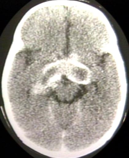
Head CT scan may be diagnostic of glioma.
CT
Head CT scan may be diagnostic of glioma.
| Type of glioma | Gross pathological features |
|---|---|
| |
| |
| |
| |
| |
|
-
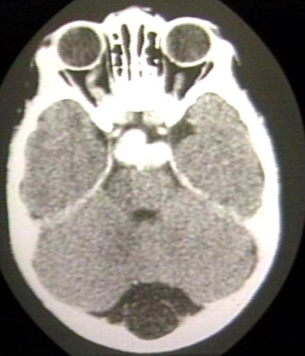
BRAIN: GLIOMA, OPTICOCHIASMATIC; WITH CONTRAST
-
BRAIN: GLIOMA, OPTICOCHIASMATIC; 1 OF 4 WITHOUT CONTRAST
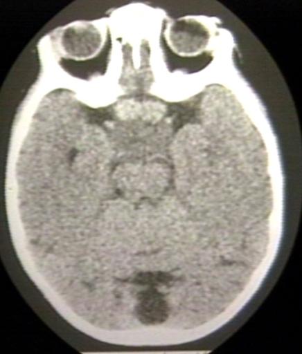
-
BRAIN: GLIOMA, OPTICOCHIASMATIC; 2 OF 4 WITH CONTRAST


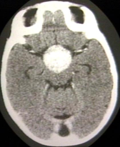
-
BRAIN: GLIOMA, OPTICOCHIASMATIC; 3 OF 4 WITH CONTRAST
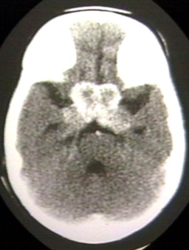
-
BRAIN: GLIOMA, OPTICOCHIASMATIC; 4 OF 4 WITH CONTRAST

