Basal cell carcinoma laboratory tests
|
Basal cell carcinoma Microchapters |
|
Diagnosis |
|---|
|
Case Studies |
|
Basal cell carcinoma laboratory tests On the Web |
|
American Roentgen Ray Society Images of Basal cell carcinoma laboratory tests |
|
Risk calculators and risk factors for Basal cell carcinoma laboratory tests |
Editor-In-Chief: C. Michael Gibson, M.S., M.D. [1]
Overview
To diagnose basal cell carcinomas, a biopsy (where tissue is taken for pathological study) is done using local anesthesia. In small lesions, the tumor is generally removed in its entiriety, while larger ones are biopsied first and surgically removed later if it is confirmed that it is malignant.
Histopathology
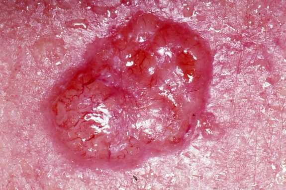
Basal cell carcinoma is a malignant epithelial tumor arising only in skin, from the basal layer of the epidermis or of the pilosebaceous adnexa. Tumor is represented by compact areas, well delineated and invading the dermis, apparent with no connection with the epidermis. Tumor cells resemble normal basal cells (small, monomorphous) are disposed in palisade at the periphery of the tumor nests, but are spindle-shaped and irregular in the middle. Tumor clusters are separated by a reduced stroma with inflammatory infiltrate. 1
-
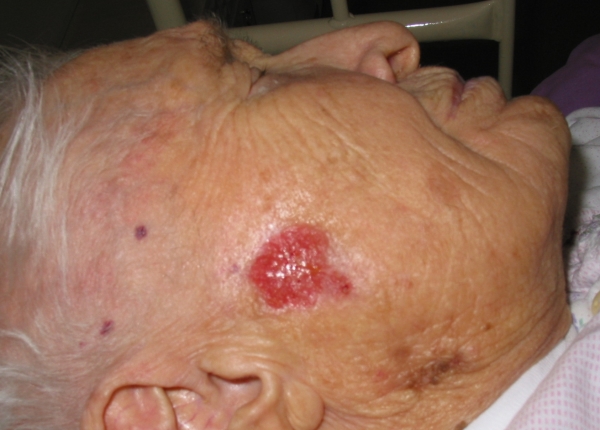
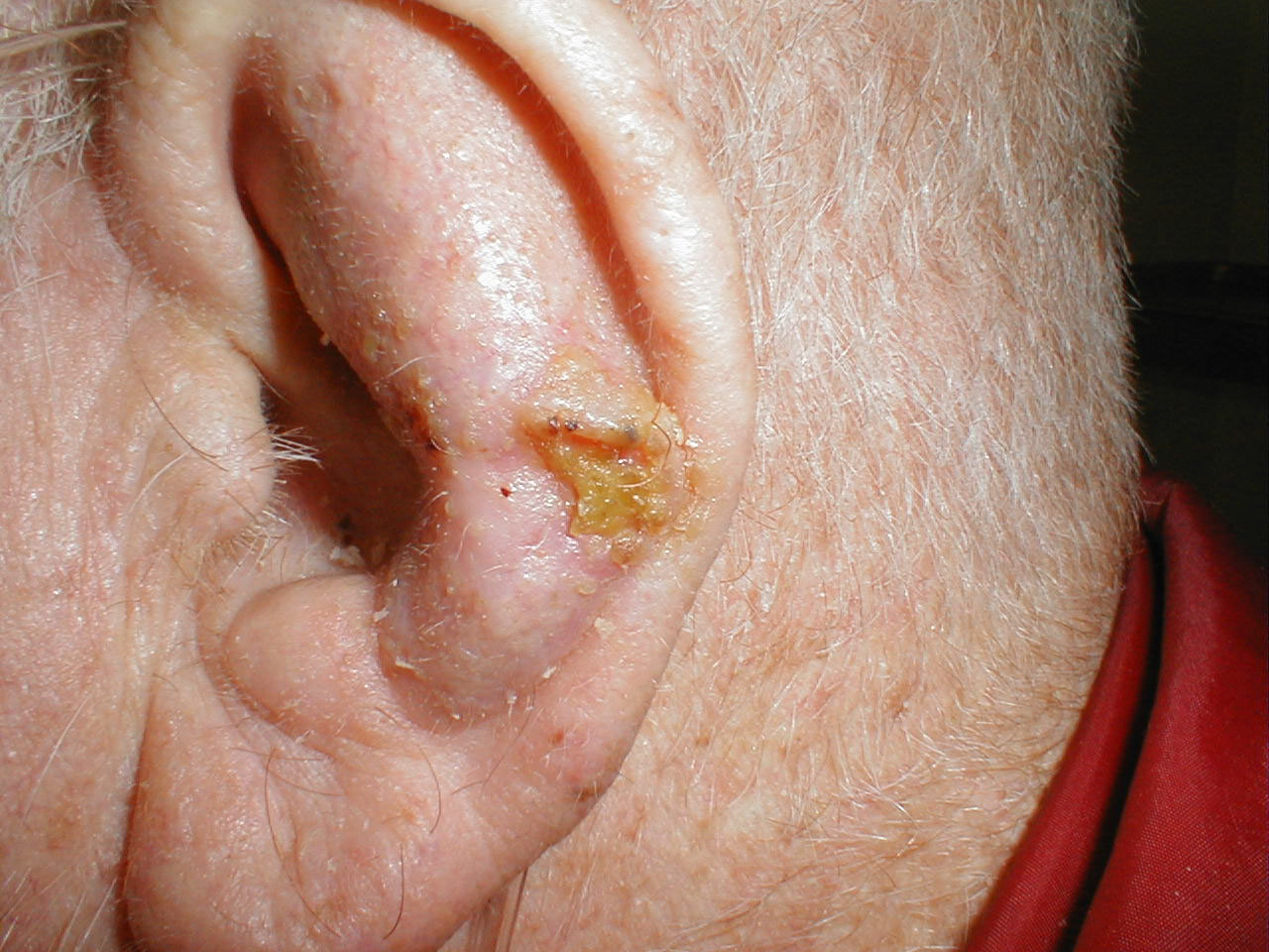
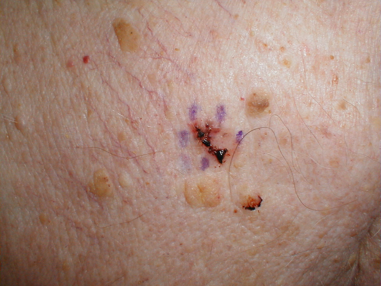
![This elderly patient was noticed to have a basal cell carcinoma of the concha, just behind the tragus[1].](/images/8/8d/Auricle_Basal_Cell_Ca_G_6.jpg)
This elderly patient was noticed to have a basal cell carcinoma of the concha, just behind the tragus[1].
-
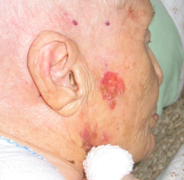
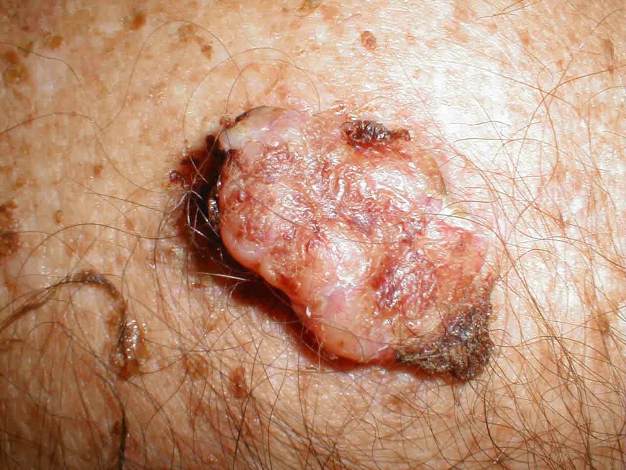
![The tumor was removed and the defect closed with a full thickness skin graft (FTSG) from the neck[2].](/images/0/07/Auricle_Basal_Cell_Ca_FTSG.jpg)
The tumor was removed and the defect closed with a full thickness skin graft (FTSG) from the neck[2].
-
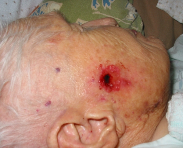
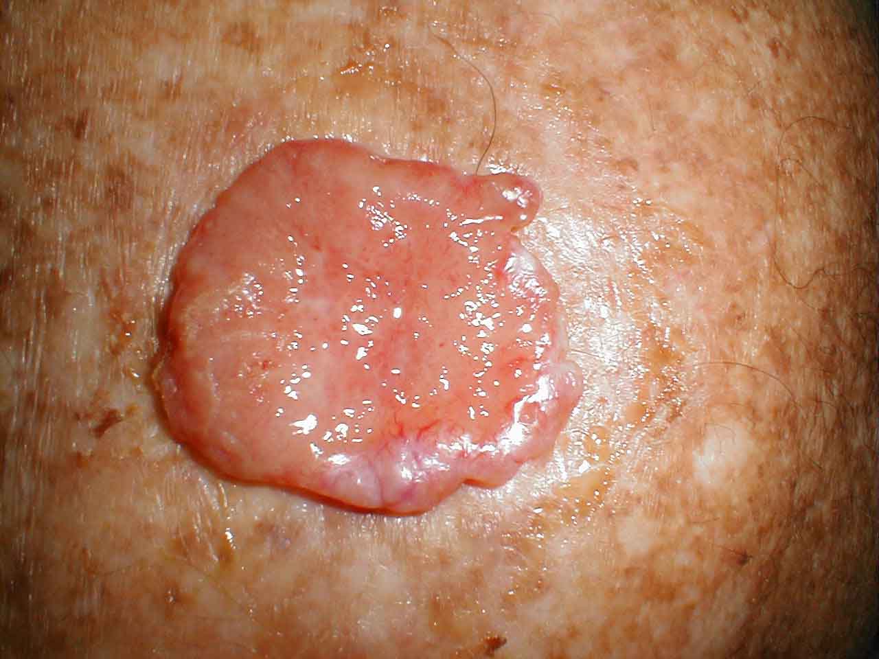
![This photograph was taken three weeks later. The full thickness skin graft has taken well[3].](/images/6/65/FTSG_in_concha_BCC_.jpg)
This photograph was taken three weeks later. The full thickness skin graft has taken well[3].
![This elderly patient was noticed to have a basal cell carcinoma of the concha, just behind the tragus[1].](/index.php/File:Auricle_Basal_Cell_Ca_G_6.jpg)
![The tumor was removed and the defect closed with a full thickness skin graft (FTSG) from the neck[2].](/index.php/File:Auricle_Basal_Cell_Ca_FTSG.jpg)
![This photograph was taken three weeks later. The full thickness skin graft has taken well[3].](/index.php/File:FTSG_in_concha_BCC_.jpg)
-
Basal cell carcinoma
-
Basal cell carcinoma


-
Basal cell carcinoma
-
Basal cell carcinoma


-
Basal cell carcinoma
-
Basal cell carcinoma
-
Basal cell carcinoma


-

Basal cell carcinoma
-
Basal cell carcinoma
-
Basal cell carcinoma (pigmented)


-
Basal cell carcinoma
-
Basal cell carcinoma
-
Basal cell carcinoma


References
Template:Tumors of bone, cartilage, skin, connective, and soft tissue
Template:SIB
de:Basaliom
nl:Basaalcelcarcinoom
fi:Basaliooma