Aortic stenosis electrocardiogram
|
Aortic Stenosis Microchapters |
|
Diagnosis |
|---|
|
Treatment |
|
Percutaneous Aortic Balloon Valvotomy (PABV) or Aortic Valvuloplasty |
|
Transcatheter Aortic Valve Replacement (TAVR) |
|
Case Studies |
|
Aortic stenosis electrocardiogram On the Web |
|
American Roentgen Ray Society Images of Aortic stenosis electrocardiogram |
|
Directions to Hospitals Treating Aortic stenosis electrocardiogram |
|
Risk calculators and risk factors for Aortic stenosis electrocardiogram |
Editor-In-Chief: C. Michael Gibson, M.S., M.D. [1]; Associate Editors-In-Chief: Mohammed A. Sbeih, M.D. [2], Claudia P. Hochberg, M.D. [3], Abdul-Rahman Arabi, M.D. [4], Keri Shafer, M.D. [5], Priyamvada Singh, MBBS [6]; Assistant Editor-In-Chief: Kristin Feeney, B.S. [7]
Overview
Electrocardiogram may be used as a diagnostic tool in the evaluation of aortic stenosis. ECG findings associated with aortic stenosis include left ventricular hypertrophy and heart block.
Electrocardiogram
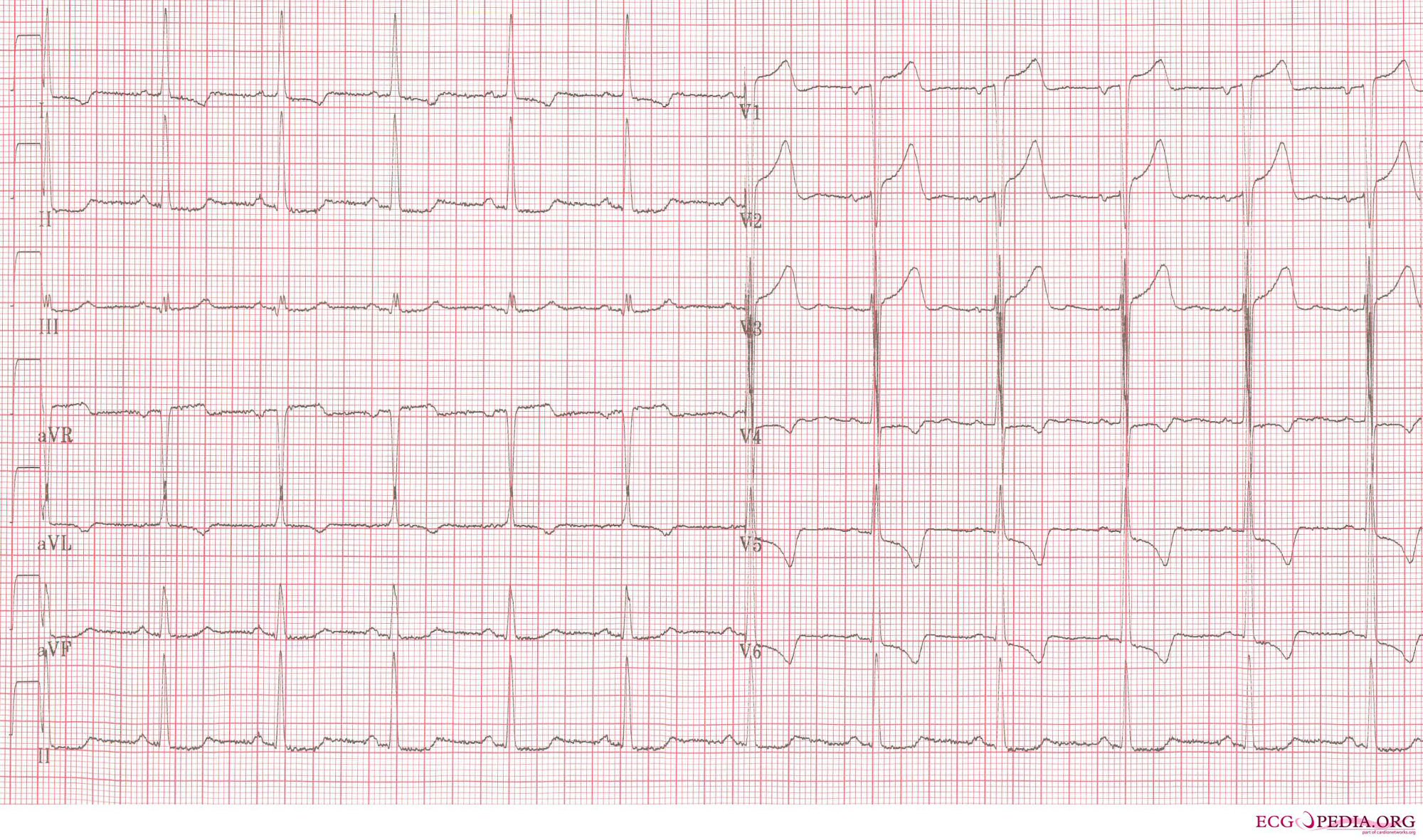
Although aortic stenosis does not lead to any specific findings on the ECG, it still often leads to a number of electrocardiographic abnormalities. ECG manifestations of left ventricular hypertrophy (LVH) are common in aortic stenosis and arise as a result of the stenosis having placed a chronically high pressure load on the left ventricle (with LVH being the expected response to chronic pressure loads on the left ventricle no matter how caused).
As noted below, the calcification process which occurs in aortic stenosis can progress to extend beyond the aortic valve and into the electrical conduction system of the heart. Evidence of this phenomenon may include heart block that is apparent on the ECG but otherwise undetectable.
-
Another example of extreme left ventricular hypertrophy in a patient with severe aortic valve stenosis.
-
ECG of a patient with LVH and subendocardial ischemia leading to positive cardiovascular markers in blood testing.

- Left ventricular hypertrophy; left ventricular strain due to aortic stenosis.


- Aortic Stenosis Hemodynamic Pressure Tracing.
