Aortic stenosis cardiac catheterization: Difference between revisions
| Line 7: | Line 7: | ||
==Cardiac Catheterization== | ==Cardiac Catheterization== | ||
Left heart catheterization is performed to simultaneously assess left ventricular and aortic pressures. | Left heart catheterization is performed to simultaneously assess left ventricular and aortic pressures. A catheter is placed inside the left ventricle, and pressure is simultaneously recorded in the femoral artery. The difference in pressures is the pressure gradient across the aortic valve. | ||
[[Image:Aortic Stenosis - Hemodynamic Pressure Tracing.png|600px]]Simultaneous left ventricular and aortic pressure tracings demonstrate a pressure gradient between the left ventricle and aorta, suggesting aortic stenosis. The left ventricle generates higher pressures than what is transmitted to the aorta. The pressure gradient, caused by aortic stenosis, is represented by the green shaded area. (AO = ascending aorta; LV = left ventricle; ECG = electrocardiogram.) The heart may be [[cardiac catheterization|catheterized]] to directly measure the pressure on both sides of the aortic valve. | [[Image:Aortic Stenosis - Hemodynamic Pressure Tracing.png|600px]]Simultaneous left ventricular and aortic pressure tracings demonstrate a pressure gradient between the left ventricle and aorta, suggesting aortic stenosis. The left ventricle generates higher pressures than what is transmitted to the aorta. The pressure gradient, caused by aortic stenosis, is represented by the green shaded area. (AO = ascending aorta; LV = left ventricle; ECG = electrocardiogram.) The heart may be [[cardiac catheterization|catheterized]] to directly measure the pressure on both sides of the aortic valve. | ||
Revision as of 03:18, 14 April 2012
|
Aortic Stenosis Microchapters |
|
Diagnosis |
|---|
|
Treatment |
|
Percutaneous Aortic Balloon Valvotomy (PABV) or Aortic Valvuloplasty |
|
Transcatheter Aortic Valve Replacement (TAVR) |
|
Case Studies |
|
Aortic stenosis cardiac catheterization On the Web |
|
American Roentgen Ray Society Images of Aortic stenosis cardiac catheterization |
|
Directions to Hospitals Treating Aortic stenosis cardiac catheterization |
|
Risk calculators and risk factors for Aortic stenosis cardiac catheterization |
Editor-In-Chief: C. Michael Gibson, M.S., M.D. [1]; Associate Editors-In-Chief: Mohammed A. Sbeih, M.D. [2]; Assistant Editor-In-Chief: Kristin Feeney, B.S. [3]
Overview
Left and right heart catheterization as well as angiography may be useful in the assessment of the patient prior to aortic valve replacement surgery.
Cardiac Catheterization
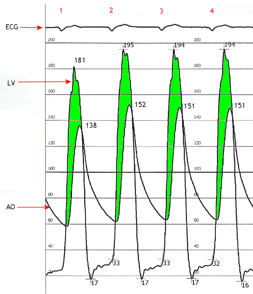
Left heart catheterization is performed to simultaneously assess left ventricular and aortic pressures. A catheter is placed inside the left ventricle, and pressure is simultaneously recorded in the femoral artery. The difference in pressures is the pressure gradient across the aortic valve.
 Simultaneous left ventricular and aortic pressure tracings demonstrate a pressure gradient between the left ventricle and aorta, suggesting aortic stenosis. The left ventricle generates higher pressures than what is transmitted to the aorta. The pressure gradient, caused by aortic stenosis, is represented by the green shaded area. (AO = ascending aorta; LV = left ventricle; ECG = electrocardiogram.) The heart may be catheterized to directly measure the pressure on both sides of the aortic valve.
ACC/AHA Guidelines- Indications for Cardiac Catheterization (DO NOT EDIT) [1]
| “ |
Class I1. Coronary angiography is recommended before AVR in patients with AS at risk for CAD. (Level of Evidence: B) 2. Cardiac catheterization for hemodynamic measurements is recommended for assessment of severity of AS in symptomatic patients when noninvasive tests are inconclusive or when there is a discrepancy between noninvasive tests and clinical findings regarding severity of AS. (Level of Evidence: C) 3. Coronary angiography is recommended before AVR in patients with AS for whom a pulmonary autograft (Ross procedure) is contemplated and if the origin of the coronary arteries was not identified by noninvasive technique. (Level of Evidence: C) Class III1. Cardiac catheterization for hemodynamic measurements is not recommended for the assessment of severity of AS before AVR when noninvasive tests are adequate and concordant with clinical findings. (Level of Evidence: C) 2. Cardiac catheterization for hemodynamic measurements is not recommended for the assessment of LV function and severity of AS in asymptomatic patients. (Level of Evidence: C) |
” |
References
- ↑ Bonow RO, Carabello BA, Chatterjee K, de Leon AC, Faxon DP, Freed MD; et al. (2008). "2008 focused update incorporated into the ACC/AHA 2006 guidelines for the management of patients with valvular heart disease: a report of the American College of Cardiology/American Heart Association Task Force on Practice Guidelines (Writing Committee to revise the 1998 guidelines for the management of patients with valvular heart disease). Endorsed by the Society of Cardiovascular Anesthesiologists, Society for Cardiovascular Angiography and Interventions, and Society of Thoracic Surgeons". J Am Coll Cardiol. 52 (13): e1–142. doi:10.1016/j.jacc.2008.05.007. PMID 18848134.