Proximal tubule
|
WikiDoc Resources for Proximal tubule |
|
Articles |
|---|
|
Most recent articles on Proximal tubule Most cited articles on Proximal tubule |
|
Media |
|
Powerpoint slides on Proximal tubule |
|
Evidence Based Medicine |
|
Clinical Trials |
|
Ongoing Trials on Proximal tubule at Clinical Trials.gov Trial results on Proximal tubule Clinical Trials on Proximal tubule at Google
|
|
Guidelines / Policies / Govt |
|
US National Guidelines Clearinghouse on Proximal tubule NICE Guidance on Proximal tubule
|
|
Books |
|
News |
|
Commentary |
|
Definitions |
|
Patient Resources / Community |
|
Patient resources on Proximal tubule Discussion groups on Proximal tubule Patient Handouts on Proximal tubule Directions to Hospitals Treating Proximal tubule Risk calculators and risk factors for Proximal tubule
|
|
Healthcare Provider Resources |
|
Causes & Risk Factors for Proximal tubule |
|
Continuing Medical Education (CME) |
|
International |
|
|
|
Business |
|
Experimental / Informatics |
Editor-In-Chief: C. Michael Gibson, M.S., M.D. [1]
Please Take Over This Page and Apply to be Editor-In-Chief for this topic: There can be one or more than one Editor-In-Chief. You may also apply to be an Associate Editor-In-Chief of one of the subtopics below. Please mail us [2] to indicate your interest in serving either as an Editor-In-Chief of the entire topic or as an Associate Editor-In-Chief for a subtopic. Please be sure to attach your CV and or biographical sketch.
The proximal tubule is the portion of the duct system of the nephron leading from Bowman's capsule to the loop of Henle.
Structure and appearance
The most distinctive characteristic of the proximal tubule is its brush border (or "striated border").
Brush border cell
The luminal surface of the epithelial cells of this segment of the nephron is covered with densely packed microvilli forming a border readily visible under the light microscope. The microvilli greatly increase the luminal surface area of the cells, presumably facilitating their resorptive function. The infolded membranes forming the microvilli are the site of numerous sodium pumps.
The cytoplasm of the cells is densely packed with mitochondria, which are largely found in the basal region within the infoldings of the basal plasma membrane. The high quantity of mitochondria gives the cells an acidophilic appearance. The mitochondria are needed in order to supply the energy for the active transport of sodium ions out of the proximal tubule. Water passively follows the sodium out of the cell along its concentration gradient.
Cuboidal epithelial cells lining the proximal tubule have extensive lateral interdigitations between neighboring cells, which lend an appearance of having no discrete cell margins when viewed with a light microscope.
Agonal resorption of the contents of the proximal tubular contents after interruption of circulation in the capillaries surrounding the tubule often leads to disturbance of the cellular morphology of the proximal tubule cells, including the ejection of cell nuclei into the tubule lumen.
This has led some observers to describe the lumen of proximal tubules as occluded or "dirty looking", in contrast to the "clean" appearance of distal tubules, which have quite different properties.
Divisions
The proximal tubule as a part of the nephron can be divided into two sections, pars convoluta and pars recta. Differences in cell outlines exist between these segments, and therefore presumably in function too.
Alternatively, regarding ultrastructure, it can be divided into three segments, S1, S2 and S3:
| Segment | Gross divisions | Ultrastructure divisions | Description |
| Proximal tubule | convoluted | S1[1] | Higher cell complexity[1] |
| S2[1] | |||
| straight | |||
| S3[1] | Lower cell complexity[1] |
Pars convoluta
The "Pars convoluta" is the initial convoluted portion.
In relation to the morphology of the kidney as a whole, the convoluted segments of the proximal tubules are confined entirely to the renal cortex.
Some investigators on the basis of particular functional differences have divided the convoluted part into two segments designated '"S1" and "S2".
Pars recta
The "Pars recta" is the following straight (descending) portion.
Straight segments descend into the outer medulla. They terminate at a remarkably uniform level and it is their line of termination that establishes the boundary between the inner and outer stripes of the outer zone of the renal medulla.
As a logical extension of the nomenclature described above, this segment is sometimes designated as "S3".
Absorption
The proximal tubule regulates the pH of the filtrate by exchanging hydrogen ions in the interstitium for bicarbonate ions in the filtrate; furthermore, it is responsible for secreting organic acids, such as creatinine and other bases, into the filtrate.
Fluid in the filtrate entering the proximal convoluted tubule is reabsorbed into the peritubular capillaries. This is driven by sodium transport from the lumen into the blood by the Na+/K+ ATPase in the basolateral membrane of the epithelial cells. Sodium reabsorption is primarily driven by this antiporter. This is the most important transport mechanism in the PCT.
| Substance | % of filtrate reabsorbed | Comments |
| salt and water | approximately two-thirds | Much of the mass movement of water and solutes occurs through the cells, passively across the lumenal membrane via transcellular transport, which is then actively resorbed across the basolateral membrane via the Na/K/ATPase pump. The solutes are absorbed isotonically, in that the osmotic potential of the fluid leaving the proximal tubule is the same as that of the initial glomerular filtrate. |
| organic solutes (primarily glucose and amino acids) | 100% | Glucose, amino acids, inorganic phosphate, and some other solutes are reabsorbed via secondary active transport through cotransport channels driven by the sodium gradient out of the nephron. |
| potassium | approximately 65% | |
| urea | approximately 50% | |
| phosphate | approximately 80% | Parathyroid hormone reduces reabsorption of phosphate in the proximal tubules, but because it also enhances the uptake of phosphate from the intestine and bones into the blood, the responses to PTH cancel each other out, and the serum concentration of phosphate remains approximately the same. |
Secretion
Many types of medication are secreted in the proximal tubule. Further reading: Table of medication secreted in kidney
Pathophysiology in kidney disease
Proximal tubular epithelial cells (PTECs) have a pivotal role in kidney disease.
Acute tubular necrosis occurs when PTECs are directly damaged by toxins such as antibiotics (e.g. gentamicin), pigments (e.g. myoglobin and sepsis (e.g. mediated by lipopolysaccharide from gram negative bacteria). Renal tubular acidosis (proximal type) (Fanconi syndrome) occurs when the PTECs are unable to properly reabsorb glomerular filtrate so that there is increased loss of bicarbonate, glucose, amino acids and phosphate.
PTECs also participate in the progression of tubulointerstitial injury due to glomerulonephritis, ischemia, interstitial nephritis, vascular injury and diabetic nephropathy. In these situations PTECs may be directly affected by protein (e.g. proteinuria in glomerulonephritis), glucose (in diabetes mellitus), or cytokines (e.g. interferon-γ and tumor necrosis factors). There are several ways in which PTECs may respond - by producing cytokines, chemokines and collagen; undergoing epithelial mesenchymal transdifferentiation; necrosis or apoptosis.
See also
Additional images
-
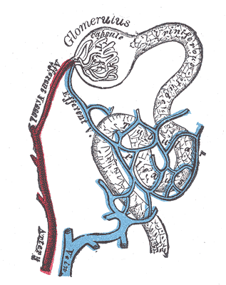 Distribution of blood vessels in cortex of kidney.
 -
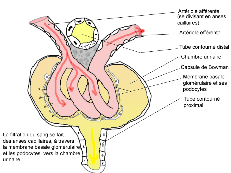 Glomerulus.
 -
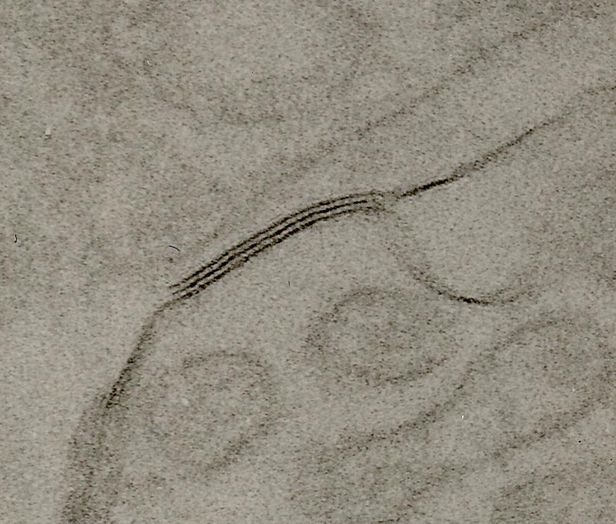 TEM of negatively stained proximal convoluted tubule of Rat kidney tissue at a magnification of ~55,000x and 80KV with Tight junction.


References
External links
- Template:GPnotebook
- Template:UCDavisOrganology - "Mammal, kidney cortex (LM, Medium)"
- Essentials of Human Physiology by Thomas M. Nosek. Section 7/7ch03/7ch03p14. - "The Nephron: Proximal Tubule, Pars Convoluta & Pars Recta"
- Template:EmbryologySwiss