Human papillomavirus physical examination
|
Human papillomavirus Microchapters |
|
Diagnosis |
|
Treatment |
|
Case Studies |
|
Human papillomavirus physical examination On the Web |
|
American Roentgen Ray Society Images of Human papillomavirus physical examination |
|
Risk calculators and risk factors for Human papillomavirus physical examination |
Editor-In-Chief: C. Michael Gibson, M.S., M.D. [1];Associate Editor(s)-in-Chief: Seyedmahdi Pahlavani, M.D. [2]
Overview
Physical examination findings depends on the anatomic region of involvement. The cutaneous lesions are warts and mostly presents as exophytic papules on fingers and lateral surface of the hands. Intraepithelial neoplasia is the most important type of genital involvement and in suspected women, the entire genitalia must be inspected by speculum with acetic acid application in order to find the lesions. Condylomata acuminata is another type of anogenital involvement which presents as a sessile or pedunculated lesion in anogenital area. Respiratory tract may be involved during neonatal passage through birth canal and manifests as stridor and even respiratory distress.
Physical examination
Cutaneous lesions
Warts
They are well demarcated, exophytic, hyperkeratotic papules or plaques with a rough surface that are usually located on the fingers or lateral surface of the hands that could be single or in groups.
-
![Common wart. Adapted from Dermatology Atlas.[1]](/images/8/85/Warts_vulgaris55.jpg) Common wart. Adapted from Dermatology Atlas.[1]
Common wart. Adapted from Dermatology Atlas.[1] -
![Common wart. Adapted from Dermatology Atlas.[1]](/images/8/82/Warts_vulgaris09.jpg) Common wart. Adapted from Dermatology Atlas.[1]
Common wart. Adapted from Dermatology Atlas.[1] -
![Plantar wart. Adapted from Dermatology Atlas.[1]](/images/6/62/Warts_plantaris05.jpg) Plantar wart. Adapted from Dermatology Atlas.[1]
Plantar wart. Adapted from Dermatology Atlas.[1]
![Common wart. Adapted from Dermatology Atlas.[1]](/index.php/File:Warts_vulgaris55.jpg)
![Common wart. Adapted from Dermatology Atlas.[1]](/index.php/File:Warts_vulgaris09.jpg)
![Plantar wart. Adapted from Dermatology Atlas.[1]](/index.php/File:Warts_plantaris05.jpg)
Epidermodysplasia verruciformis
Characterized by the growth of scaly macules and papules, that may become hypertrophic and coalescent particularly on the hands and upper trunk in children.[2]
-
![Epidermodysplasia verruciformis. Adapted from Dermatology Atlas.[1]](/images/1/1a/Epidermodysplasia_verruciformis01.jpg) Epidermodysplasia verruciformis. Adapted from Dermatology Atlas.[1]
Epidermodysplasia verruciformis. Adapted from Dermatology Atlas.[1] -
![Epidermodysplasia verruciformis. Adapted from Dermatology Atlas.[1]](/images/2/28/Epidermodysplasia_verruciformis02.jpg) Epidermodysplasia verruciformis. Adapted from Dermatology Atlas.[1]
Epidermodysplasia verruciformis. Adapted from Dermatology Atlas.[1] -
![Epidermodysplasia verruciformis. Adapted from Dermatology Atlas.[1]](/images/d/d0/Epidermodysplasia_verruciformis03.jpg) Epidermodysplasia verruciformis. Adapted from Dermatology Atlas.[1]
Epidermodysplasia verruciformis. Adapted from Dermatology Atlas.[1] -
![Epidermodysplasia verruciformis. Adapted from Dermatology Atlas.[1]](/images/d/d0/Epidermodysplasia_verruciformis04.jpg) Epidermodysplasia verruciformis. Adapted from Dermatology Atlas.[1]
Epidermodysplasia verruciformis. Adapted from Dermatology Atlas.[1] -
![Epidermodysplasia verruciformis. Adapted from Dermatology Atlas.[1]](/images/5/53/Epidermodysplasia_verruciformis06.jpg) Epidermodysplasia verruciformis. Adapted from Dermatology Atlas.[1]
Epidermodysplasia verruciformis. Adapted from Dermatology Atlas.[1]
![Epidermodysplasia verruciformis. Adapted from Dermatology Atlas.[1]](/index.php/File:Epidermodysplasia_verruciformis01.jpg)
![Epidermodysplasia verruciformis. Adapted from Dermatology Atlas.[1]](/index.php/File:Epidermodysplasia_verruciformis02.jpg)
![Epidermodysplasia verruciformis. Adapted from Dermatology Atlas.[1]](/index.php/File:Epidermodysplasia_verruciformis03.jpg)
![Epidermodysplasia verruciformis. Adapted from Dermatology Atlas.[1]](/index.php/File:Epidermodysplasia_verruciformis04.jpg)
![Epidermodysplasia verruciformis. Adapted from Dermatology Atlas.[1]](/index.php/File:Epidermodysplasia_verruciformis06.jpg)
Anogenital lesions
Intraepithelial neoplasia
Digital palpation of the vagina to assess for thickening or irregularity of the vaginal wall and a thorough colposcopic assessment of the entire vagina must be perform, however physical examination of patients with early stage is usually unremarkable, but after the insertion of a speculum and the application of acetic acid, lesions will appear as raised or flat white, granular epithelium with sharply demarcated borders and may contain areas of vascular punctation.[3]
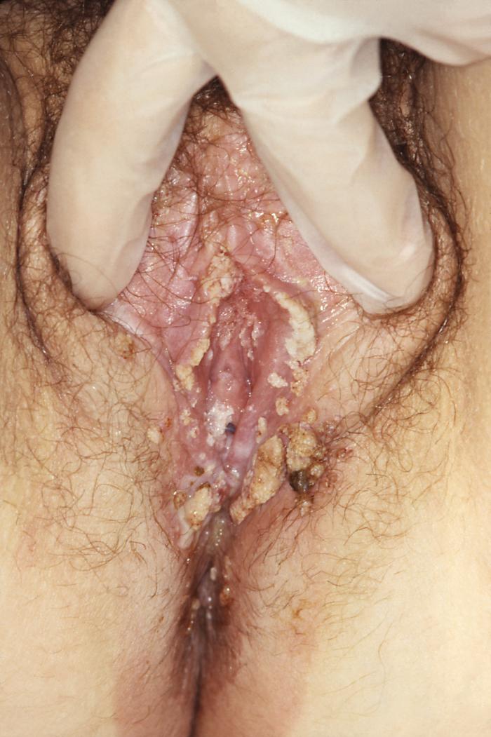
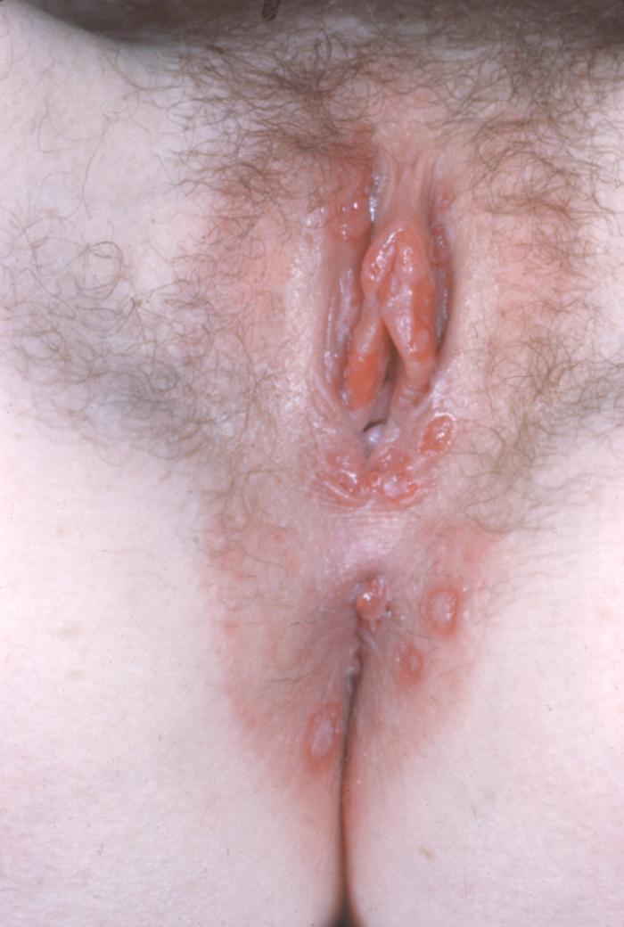
Condylomata acuminata
Its shape ranges from smooth flattened papule to a verrucous, papilliform appearance, may be skin-colored, brown or whitish, may also present as pedunculated or broad-based papillomas up to several centimeters in diameter or as large confluent plaques. Female and male genital area and perianal may be affected.[4]
-
 Condyloma acuminata
 -
 Condyloma acuminata
 -
![Condyloma acuminata. Adapted from Dermatology Atlas.[1]](/images/9/9c/Condyloma_acuminatum01.jpg) Condyloma acuminata. Adapted from Dermatology Atlas.[1]
Condyloma acuminata. Adapted from Dermatology Atlas.[1] -
![Condyloma acuminata. Adapted from Dermatology Atlas.[1]](/images/8/81/Condyloma_acuminatum02.jpg) Condyloma acuminata. Adapted from Dermatology Atlas.[1]
Condyloma acuminata. Adapted from Dermatology Atlas.[1] -
![Condyloma acuminata. Adapted from Dermatology Atlas.[1]](/images/6/6e/Condyloma_acuminatum03.jpg) Condyloma acuminata. Adapted from Dermatology Atlas.[1]
Condyloma acuminata. Adapted from Dermatology Atlas.[1] -
![Condyloma acuminata. Adapted from Dermatology Atlas.[1]](/images/e/ef/Condyloma_acuminatum11.jpg) Condyloma acuminata. Adapted from Dermatology Atlas.[1]
Condyloma acuminata. Adapted from Dermatology Atlas.[1] -
![Condyloma acuminata. Adapted from Dermatology Atlas.[1]](/images/c/c5/Condyloma_acuminatum12.jpg) Condyloma acuminata. Adapted from Dermatology Atlas.[1]
Condyloma acuminata. Adapted from Dermatology Atlas.[1] -
![Condyloma acuminata. Adapted from Dermatology Atlas.[1]](/images/8/8b/Condyloma_acuminatum36.jpg) Condyloma acuminata. Adapted from Dermatology Atlas.[1]
Condyloma acuminata. Adapted from Dermatology Atlas.[1] -
![Condyloma acuminata. Adapted from Dermatology Atlas.[1]](/images/c/c4/Condyloma_acuminatum37.jpg) Condyloma acuminata. Adapted from Dermatology Atlas.[1]
Condyloma acuminata. Adapted from Dermatology Atlas.[1]


![Condyloma acuminata. Adapted from Dermatology Atlas.[1]](/index.php/File:Condyloma_acuminatum01.jpg)
![Condyloma acuminata. Adapted from Dermatology Atlas.[1]](/index.php/File:Condyloma_acuminatum02.jpg)
![Condyloma acuminata. Adapted from Dermatology Atlas.[1]](/index.php/File:Condyloma_acuminatum03.jpg)
![Condyloma acuminata. Adapted from Dermatology Atlas.[1]](/index.php/File:Condyloma_acuminatum11.jpg)
![Condyloma acuminata. Adapted from Dermatology Atlas.[1]](/index.php/File:Condyloma_acuminatum12.jpg)
![Condyloma acuminata. Adapted from Dermatology Atlas.[1]](/index.php/File:Condyloma_acuminatum36.jpg)
![Condyloma acuminata. Adapted from Dermatology Atlas.[1]](/index.php/File:Condyloma_acuminatum37.jpg)
Other mucosal surfaces
Recurrent respiratory papillomatosis
Features benign exophytic laryngeal papillomas. Hoarseness, stridor and respiratory distress are classic triad in this condition.[5]
Conjunctival papillomas
Present with red eye and a fleshy lesion usually arising from the bulbar conjunctiva.
Mucosal Assessment With Acetic Acid
- Application of 3%–5% acetic acid, which might cause affected areas to turn white, has been used by certain providers to detect genital mucosa infected with HPV.[6]
- The routine use of this procedure to detect mucosal changes attributed to HPV infection is not recommended because the results do not influence clinical management.[6]
References
- ↑ 1.00 1.01 1.02 1.03 1.04 1.05 1.06 1.07 1.08 1.09 1.10 1.11 1.12 1.13 1.14 "Dermatology Atlas".
- ↑ Patel T, Morrison LK, Rady P, Tyring S (2010). "Epidermodysplasia verruciformis and susceptibility to HPV". Dis. Markers. 29 (3–4): 199–206. doi:10.3233/DMA-2010-0733. PMC 3835378. PMID 21178278.
- ↑ Boonlikit S, Noinual N (2010). "Vaginal intraepithelial neoplasia: a retrospective analysis of clinical features and colpohistology". J. Obstet. Gynaecol. Res. 36 (1): 94–100. doi:10.1111/j.1447-0756.2009.01108.x. PMID 20178533.
- ↑ Bennett, John (2015). Mandell, Douglas, and Bennett's principles and practice of infectious diseases. Philadelphia, PA: Elsevier/Saunders. ISBN 9781455748013.
- ↑ Venkatesan NN, Pine HS, Underbrink MP (2012). "Recurrent respiratory papillomatosis". Otolaryngol. Clin. North Am. 45 (3): 671–94, viii–ix. doi:10.1016/j.otc.2012.03.006. PMC 3682415. PMID 22588043.
- ↑ 6.0 6.1 Workowski KA, Bachmann LH, Chan PA, Johnston CM, Muzny CA, Park I; et al. (2021). "Sexually Transmitted Infections Treatment Guidelines, 2021". MMWR Recomm Rep. 70 (4): 1–187. doi:10.15585/mmwr.rr7004a1. PMC 8344968 Check
|pmc=value (help). PMID 34292926 Check|pmid=value (help).