Glomerulus
|
WikiDoc Resources for Glomerulus |
|
Articles |
|---|
|
Most recent articles on Glomerulus |
|
Media |
|
Evidence Based Medicine |
|
Clinical Trials |
|
Ongoing Trials on Glomerulus at Clinical Trials.gov Clinical Trials on Glomerulus at Google
|
|
Guidelines / Policies / Govt |
|
US National Guidelines Clearinghouse on Glomerulus
|
|
Books |
|
News |
|
Commentary |
|
Definitions |
|
Patient Resources / Community |
|
Patient resources on Glomerulus Discussion groups on Glomerulus Patient Handouts on Glomerulus Directions to Hospitals Treating Glomerulus Risk calculators and risk factors for Glomerulus
|
|
Healthcare Provider Resources |
|
Causes & Risk Factors for Glomerulus |
|
Continuing Medical Education (CME) |
|
International |
|
|
|
Business |
|
Experimental / Informatics |
Editor-In-Chief: C. Michael Gibson, M.S., M.D. [1]
Associate Editor-In-Chief: Cafer Zorkun, M.D., Ph.D. [2]
Overview
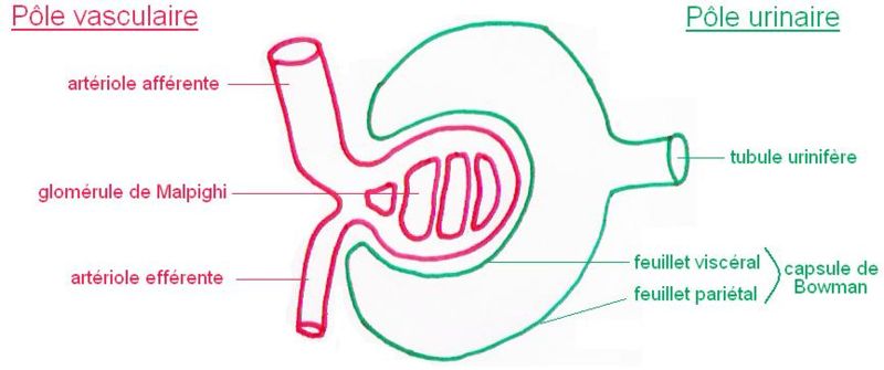
A glomerulus is a capillary tuft surrounded by Bowman's capsule in nephrons of the vertebrate kidney. It receives its blood supply from an afferent arteriole of the renal circulation. Unlike most other capillary beds, the glomerulus drains into an efferent arteriole rather than a venule. The resistance of the arterioles results in high pressure in the glomerulus aiding the process of ultrafiltration where fluids and soluble materials in the blood are forced out of the capillaries and into Bowman's capsule.
A glomerulus and its surrounding Bowman's capsule constitute a renal corpuscle, the basic filtration unit of the kidney. The rate at which blood is filtered through all of the glomeruli is the glomerular filtration rate (GFR), measurements of which are often used to determine renal function.
Afferent circulation
The afferent arteriole that supplies the glomerulus is a branch off of an interlobular artery in the cortex.
Layers
If a substance can pass through the endothelial cells, glomerular basement membrane, and podocytes, then it is known as ultrafiltrate, and it enters proximal tubule. Otherwise, it returns through the efferent circulation, discussed below.
Endothelial cells
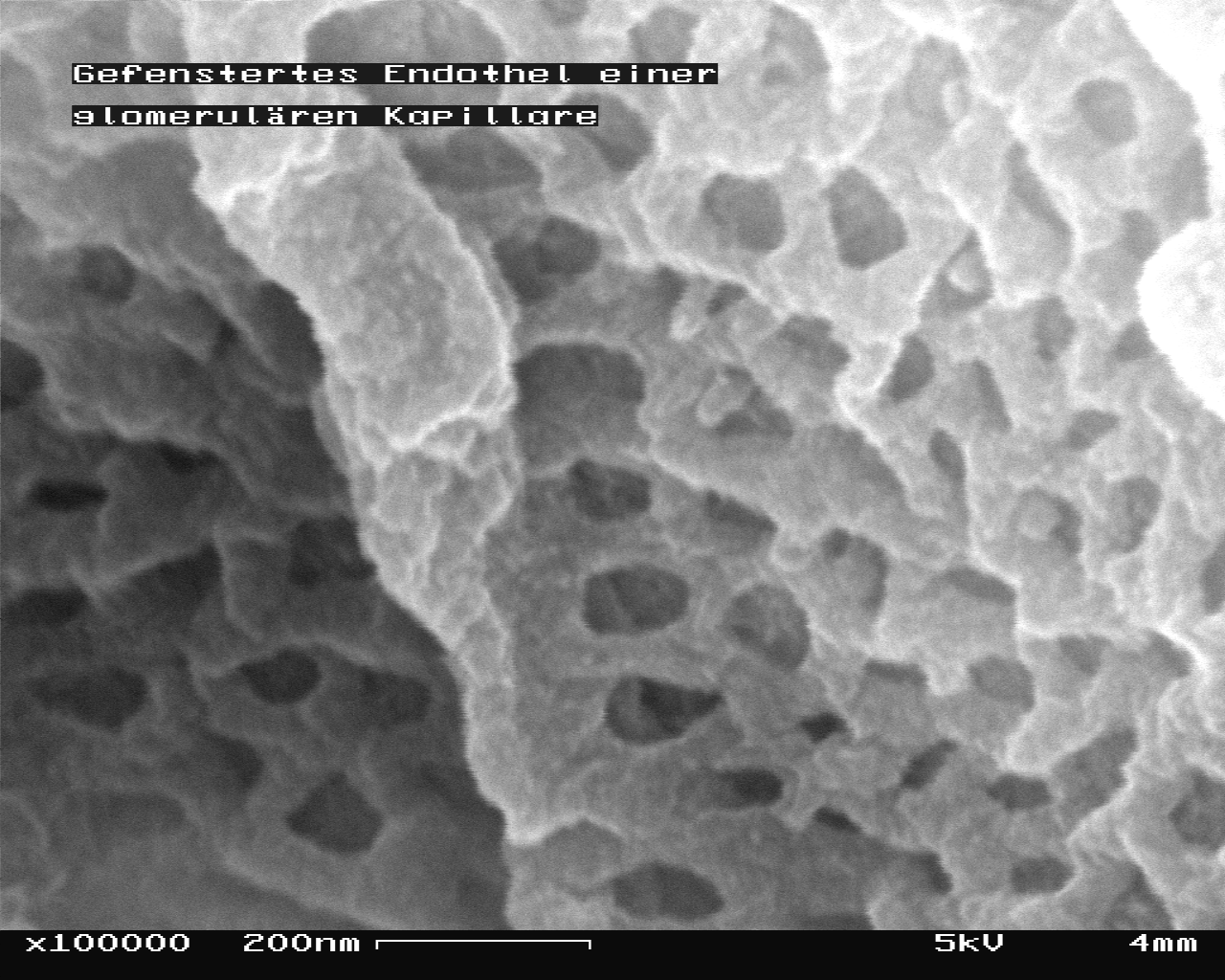
The endothelial cells of the glomerulus contain numerous pores (fenestrae) that, unlike those of other fenestrated capillaries, are not spanned by diaphragms. The cells have openings which are so large that nearly anything smaller than a red blood cell passes through that layer.
Because of this, the endothelial cells lining the glomerulus are not usually considered part of the renal filtration barrier.
Glomerular basement membrane
The glomerular endothelium sits on a very thick (100-200 nm) glomerular basement membrane. It is not only uncharacteristically thick compared to most other basement membranes (40-50 nm), but it is also rich in negatively charged glycosaminoglycans such as heparan sulfate.
The negatively-charged basement membrane repels negatively-charged proteins from the blood, helping to prevent their passage into Bowman's space.
Podocytes
Podocytes line the other side of the glomerular basement membrane and form part of the lining of Bowman's space. Podocytes form a tight interdigitating network of foot processes (pedicels) that control the filtration of proteins from the capillary lumen into Bowman's space.
The space between adjacent podocyte foot processes is spanned by a slit diaphragm formed by several proteins including podocin and nephrin. In addition, foot processes have a negatively-charged coat (glycocalyx) that limits the filtration of negatively-charged molecules, such as serum albumin.
The podocytes are sometimes considered the "visceral layer of Bowman's capsule", rather than part of the glomerulus.
Intraglomerular mesangial cell
Intraglomerular mesangial cells are found in the interstitium between endothelial cells of the glomerulus. They are not part of the filtration barrier but are specialized pericytes that participate indirectly in filtration.
Efferent circulation
Blood is carried out of the glomerulus by an efferent arteriole instead of a venule, as is observed in most other capillary systems. This provides tighter control over the bloodflow through the glomerulus, since arterioles can be dilated and constricted more readily than venules, owing to arterioles' larger smooth muscle layer (tunica media).
Efferent arterioles of juxtamedullary nephrons (ie, the 15% of nephrons closest to the medulla) send straight capillary branches that deliver isotonic blood to the renal medulla. Along with the loop of Henle, these vasa recta play a crucial role in the establishment of the nephron's countercurrent exchange system.
The efferent arteriole, into which the glomerulus delivers blood, empties into an interlobular vein.
Juxtaglomerular cells
The walls of the afferent arteriole contain specialized smooth muscle cells that synthesize renin. These juxtaglomerular cells play a major role in the renin-angiotensin system, which helps regulate blood volume and pressure.
Additional images
-
Malphigian corpuscle
-
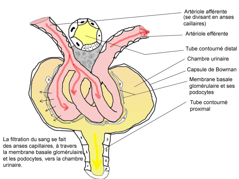
1 Glomerulus, 2 proximal tubule, 3 distal tubule
-
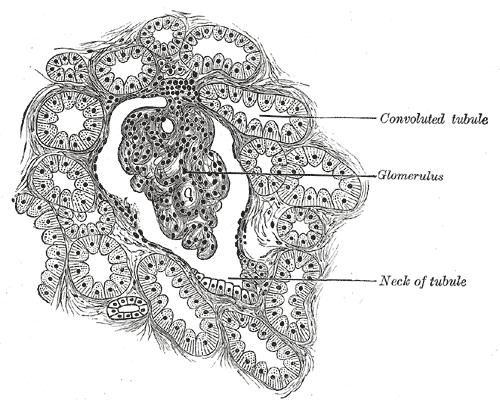
Glomerulus.
-
Glomerulus.
-
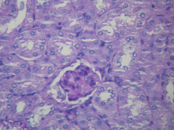
Section of cortex of human kidney.
-
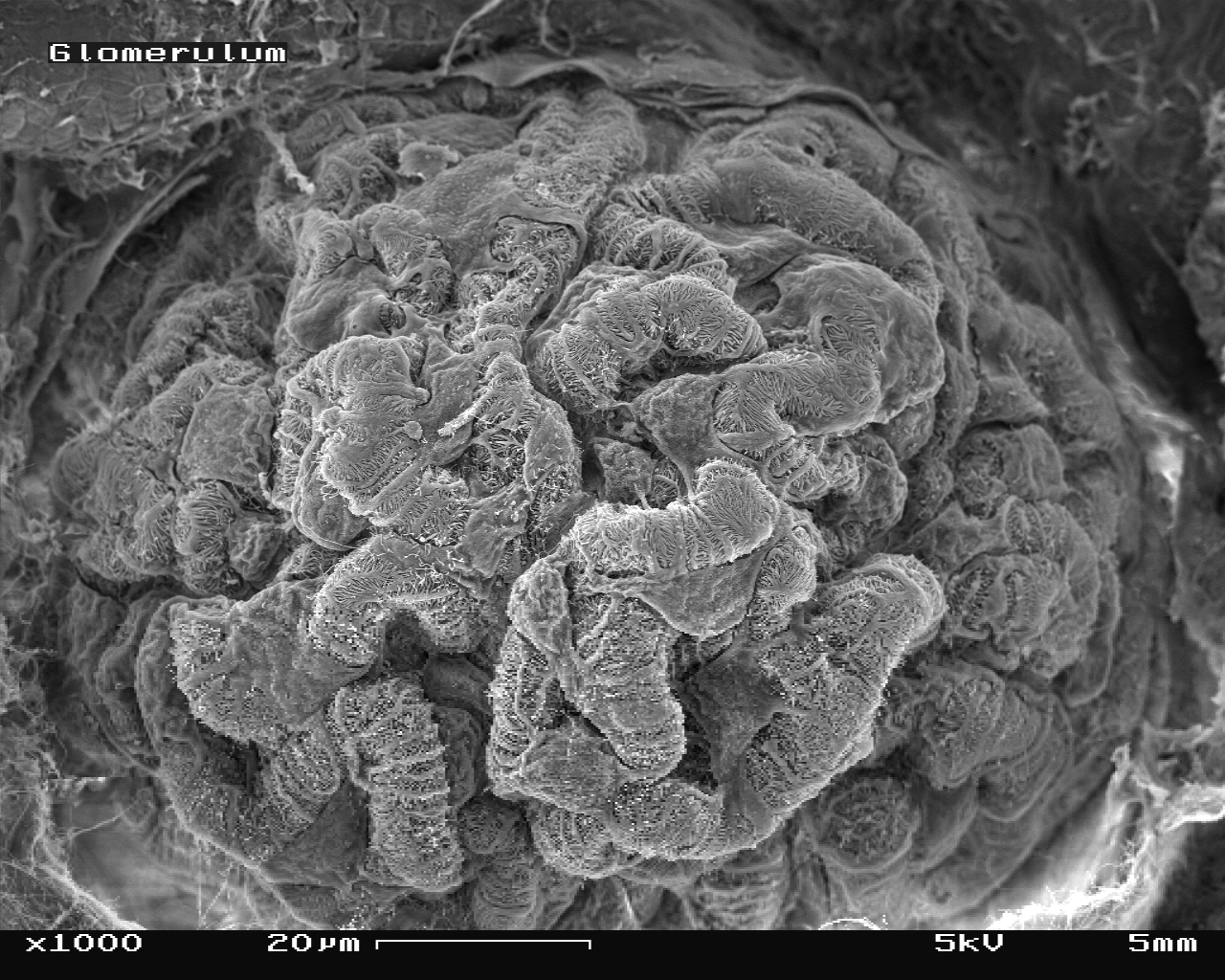
Mouse glomerulus in the Scanning Electron Microscope ("SEM"), magnification 1,000x
-
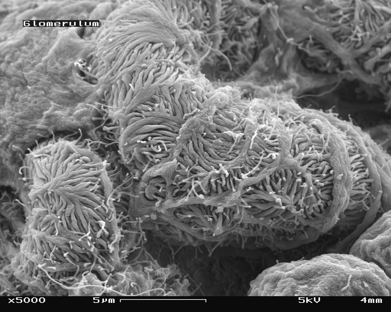
Mouse glomerulus in the SEM, magnification 5,000x
-
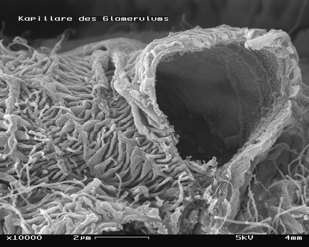
Mouse glomerulus in the SEM with one capillary broken, magnification 10,000x
-
View on the inside of broken capillary with fenestrae visible, magnification 100,000x
External links
- Image and article at FGCU
- Histology at KUMC epithel-epith02 "Kidney (Glomerulus)"
- Template:UCDavisOrganology - "Mammal, kidney cortex (LM, Medium)"
- Histology image: 16010loa – Histology Learning System at Boston University
- {http://www.uncnephropathology.org UNC Nephropathology}
