Heparan sulfate
|
WikiDoc Resources for Heparan sulfate |
|
Articles |
|---|
|
Most recent articles on Heparan sulfate Most cited articles on Heparan sulfate |
|
Media |
|
Powerpoint slides on Heparan sulfate |
|
Evidence Based Medicine |
|
Clinical Trials |
|
Ongoing Trials on Heparan sulfate at Clinical Trials.gov Trial results on Heparan sulfate Clinical Trials on Heparan sulfate at Google
|
|
Guidelines / Policies / Govt |
|
US National Guidelines Clearinghouse on Heparan sulfate NICE Guidance on Heparan sulfate
|
|
Books |
|
News |
|
Commentary |
|
Definitions |
|
Patient Resources / Community |
|
Patient resources on Heparan sulfate Discussion groups on Heparan sulfate Patient Handouts on Heparan sulfate Directions to Hospitals Treating Heparan sulfate Risk calculators and risk factors for Heparan sulfate
|
|
Healthcare Provider Resources |
|
Causes & Risk Factors for Heparan sulfate |
|
Continuing Medical Education (CME) |
|
International |
|
|
|
Business |
|
Experimental / Informatics |
Overview
Heparan sulfate (HS) is a linear polysaccharide found in all animal tissues. It occurs as a proteoglycan (PG) in which two or three HS chains are attached in close proximity to cell surface or extracellular matrix proteins.[1][2] It is in this form that HS binds to a variety of protein ligands and regulates a wide variety of biological activities, including developmental processes, angiogenesis, blood coagulation and tumour metastasis.
Proteoglycans
The major cell membrane HSPGs are the transmembrane syndecans and the glycosylphosphatidylinositol (GPI) anchored glypicans. Other minor forms of membrane HSPG include betaglycan[3] and the V-3 isoform of CD44 present on keratinocytes and activated monocytes.[4]
In the extracellular matrix, especially basement membranes, the multi-domain perlecan, agrin and collagen XVIII core proteins are the main HS-bearing species.
HS structure and differences from heparin
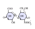
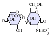
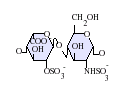
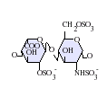
Heparan sulfate is a member of the glycosaminoglycan family of carbohydrates and is very closely related in structure to heparin. Both consist of a variably sulfated repeating disaccharide unit. The main disaccharide units that occur in heparan sulfate and heparin are shown below.
The most common disaccharide unit within heparan sulfate is composed of a glucuronic (GlcA) linked to N-acetyl glucosamine (GlcNAc) typically making up around 50% of the total disaccharide units. Compare this to heparin where IdoA(2S)-GlcNS(6S) makes up 85% of heparins from beef lung and about 75% of those from porcine intestinal mucosa. Problems arise when defining hybrid GAGs that contain both 'heparin-like' and 'HS-like' structures. It has been suggested that a GAG should qualify as heparin only if its content of N-sulfate groups largely exceeds that of N-acetyl groups and the concentration of O-sulfate groups exceeds those of N-sulfate.[5]
Not shown below are the rare disaccharides containing a 3-O-sulfated glucosamine (GlcNS(3S,6S) or a free amine group (GlcNH3+). Under physiological conditions the ester and amide sulfate groups are deprotonated and attract positively charged counterions to form a salt. It is in this form that HS is thought to exist at the cell surface.
-
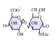
GlcA-GlcNAc -
GlcA-GlcNS -
IdoA-GlcNS -
IdoA(2S)-GlcNS -

IdoA-GlcNS(6S) -
IdoA(2S)-GlcNS(6S)


-GlcNS.png)
.png)
-GlcNS(6S).png)
Abbreviations
- GlcA = β-L-glucuronic acid
- IdoA = α-L-iduronic acid
- IdoA(2S) = 2-O-sulfo-α-L-iduronic acid
- GlcNAc = 2-deoxy-2-acetamido-α-D-glucopyranosyl
- GlcNS = 2-deoxy-2-sulfamido-α-D-glucopyranosyl
- GlcNS(6S) = 2-deoxy-2-sulfamido-α-D-glucopyranosyl-6-O-sulfate
HS biosynthesis
Many different cell types produce HS chains with many different primary structures. Therefore there is room for a great deal of variability in the way HS chains are synthesised. However, essential to the formation of HS regardless of primary sequence is a range of biosynthetic enzymes. These enzymes consist of multiple glycosyltransferases, sulphotransferases and an epimerase. These same enzymes also synthesise heparin a related polysacchride.
Many of these enzymes have now been purified, molecularly cloned and their expression patterns studied. From this and early work on the fundamental stages of HS/heparin biosynthesis using a mouse mastocytoma cell free system a lot is known about the order of enzyme reactions and specificity.[6]
Chain initiation
HS synthesis initiates with the transfer of xylose from UDP-xylose by xylotransferase (XT) to specific serine residues within the protein core. Attachment of two galactose (Gal) residues by galactosyltransferases I and II (GalTI and GalTII) and glucuronic acid (GlcA) by glucuronosyltransferase I (GlcATI) completes the formation of a core protein linkage tetrasaccharide
ßGlcA-1,3-ßGal-1,3-ßGal-1,4-ßXyl.
Xylose attachment to the core protein is thought to occur in the endoplasmic reticulum (ER) with further assembly of the linkage region and the remainder of the chain occurring in the golgi apparatus.
The pathways for HS/heparin or chondroitin sulphate (CS) and dermatan sulphate (DS) biosynthesis diverge after the formation of this common linkage structure. The next enzyme to act GlcNAcT-I or GalNAcT-I direct synthesis either to HS/heparin or CS/DS respectively.
Chain elongation
After attachment of the first N-acetyl glucosamine (GlcNAc) residue elongation of the tetrasacchride linker is continued by the stepwise addition of GlcA and GlcNAc residues. These are transferred from their respective UDP-sugar nucleotides. This is carried out by one or more related enzymes whose genes are members of the exostoses (EXT) gene family of tumour supressors.
Mutations at the EXT1-3 gene loci in humans leads to an in-ability of cells to produce HS and to the development of the disease Multiple Hereditary Exostoses (MHE).
MHE is characterized by cartilage-capped tumours, known as osteochondromas or exostoses, which develop primarily on the long bones of affected individuals from early childhood until puberty. Although exostoses are in themselves benign, surgery may be required to alleviate secondary complications such as joint pain and restricted movement.
For further information on this disease see the dedicated web site here
Chain modification
As the chain polymerises it undergoes a series of modification reactions carried out by four classes of sulfotransferases and an epimerase. The availability of the sulfate donor PAPS is crucial to the activity of the sulfotransferases.[7]
N-deacetylation/N-sulphation
The first polymer modification is the N-deacetylation/N-sulphation of GlcNAc residues into GlcNS. This is a prerequisite for all subsequent modification reactions and is carried out by one or more members of a family of four GlcNAc N-deacetylase/N-sulfotransferase enzymes (NDSTs). In early studies it was shown that modifying enzymes could recognize and act on any N-acetylated residue in the forming polymer.[8] Therefore the modification of GlcNAc residues should occur randomly throughout the chain. However, in HS N-sulphated residues are mainly grouped together and separated by regions of N-acetylation where GlcNAc remains unmodified.
Generation of GlcNH2
Due to the N-deacetylase and N-sulfotransferase being carried out by the same enzyme N-sulphation is normally tightly coupled to N-desulphation. GlcNH2 residues resulting from apparent uncoupling of the two activities have been found in heparin and some species of HS.[9]
Epimerisation and 2-O-sulphation
Epimerisation is catalysed by one enzyme, the GlcA C5 epimerase or heparosan-N-sulfate-glucuronate 5-epimerase (EC 5.1.3.17). This enzyme epimerises GlcA to iduronic acid (IdoA). Substrate recognition requires that the GlcN residue linked to the non-reducing side of a potential GlcA target be N-sulphated. Uronosyl-2-O-sulphotransferase (2OST) sulphates the resulting IdoA residues.
6-O-sulphation
Three glucosaminyl 6-O-transferases (6OSTs) have been identified that result in the formation of GlcNS(6S) adjacent to sulphated or non-sulphated IdoA. GlcNAc(6S) is also found in mature HS chains.
3-O-sulphation
At least five glucosaminyl 3-O-sulfotransferases (3OSTs) exist and result in the formation of the rare monosacchide GlcNS(3S,6S).
Ligand binding
Interferon-γ
The cell surface receptor binding region of Interferon-γ overlaps with the HS binding region, near the proteins C-terminal. Binding of HS blocks the receptor binding site and as a result, protein-HS complexes are inactive.[10]
The HS-binding properties of a number of other proteins are also being studied:
- Antithrombin III
- Hepatocyte Growth Factor
- Interleukin-8
- Vascular Endothelial Growth Factor
- Wnt/Wingless
- Endostatin
References
- ↑ Gallagher, J.T., Lyon, M. (2000). "Molecular structure of Heparan Sulfate and interactions with growth factors and morphogens". In Iozzo, M, V. Proteoglycans: structure, biology and molecular interactions. Marcel Dekker Inc. New York, New York. pp. 27–59.
- ↑ Iozzo, R. V. (1998). "Matrix proteoglycans: from molecular design to cellular function". Annu. Rev. Biochem. 67: 609–652. PMID 9759499.
- ↑ Andres, J. L.; et al. (1992). "Binding of two growth factor families to separate domains of the proteoglycan betaglycan". J. Biol. Chem. 267: 5927–5930. PMID 1556106.
- ↑ Jackson, D. G.; et al. (1995). "Proteoglycan forms of lymphocyte homing receptor CD44 are alternatively spliced variants containing the V-3 exon". J. Cell. Biol. 128: 673–685. PMID 7532175.
- ↑ Gallagher, J. T. Walker, A. (1985). "Molecular distinctions between Heparan Sulphate and Heparin: Analysis of sulphation patterns indicates Heparan Sulphate and Heparin are separate families of N-sulphated polysaccharides". Biochem. J. 230: 665–674. PMID 2933029.
- ↑ Lindahl, U.; et al. (1998). "Regulated diversity of Heparan Sulfate". J. Biol. Chem. 273: 24979–24982. PMID 9737951.
- ↑ Silbert, J. E. (1967). "Formation of a sulfate glycosaminoglycan with a microsomal preparation from mast cells". J. Biol. Chem. 242: 5146–5152.
- ↑ Höök, M.; et al. (1975). "Biosynthesis of heparin. Studies on the microsomal sulfation process". J. Biol. Chem. 250: 6065–6071. PMID 807579.
- ↑ Toida, T.; et al. (1997). "Structural differences and the presence of unsubstituted amino groups in heparan sulphates from different tissues and species". Biochem. J. 322(Pt2): 499–506.
- ↑ Sadir, R.; et al. (1998). "The heparan sulphate binding sequence of interferon-γ increased the on rate of the interferon-γ / interferon-γ receptor complex formation". J. Biol. Chem. 273: 10919–10925.
Further Reading
- HajMohammadi et. al (2003). "Normal levels of anticoagulant heparan sulfate are not essential for normal hemostasis". J Clin Invest. 111(7): 989–999. PMID 152578.
- Ettenson DS, Koo EW, Januzzi JL, Edelman ER. (2000). "Endothelial heparan sulfate is necessary but not sufficient for control of vascular smooth muscle cell growth". J Cell Physiol. 184(1): 93–100. PMID 10825238.
- Levy-Adam F, Feld S, Suss-Toby E, Vlodavsky I, Ilan N. (2008). "Heparanase facilitates cell adhesion and spreading by clustering of cell surface heparan sulfate proteoglycans". PLoS ONE. 3(6): e2319. PMID 18545691.