Coronary artery aneurysm
|
Coronary Angiography | |
|
General Principles | |
|---|---|
|
Anatomy & Projection Angles | |
|
Normal Anatomy | |
|
Anatomic Variants | |
|
Projection Angles | |
|
Epicardial Flow & Myocardial Perfusion | |
|
Epicardial Flow | |
|
Myocardial Perfusion | |
|
Lesion Complexity | |
|
ACC/AHA Lesion-Specific Classification of the Primary Target Stenosis | |
|
Lesion Morphology | |
Editor-In-Chief: C. Michael Gibson, M.S., M.D. [1]; Associate Editor(s)-in-Chief: Cafer Zorkun, M.D., Ph.D. [2]; Varun Kumar, M.B.B.S. [3]
Synonyms and keywords: CAA, coronary artery ectasia
Overview
Coronary artery aneurysm is an abnormal dilatation of a coronary artery segment over 1.5 times the diameter of normal adjacent segment.[1] An aneurysm can be classified as either saccular (wider than it is long) or fusiform (elongated). Coronary artery aneurysm must be differentiated from coronary artery ectasia; in fact, coronary artery ectasia is a localized arterial dilatation that is graded according to the size of the dilatation in comparison to the normal artery diameter located anywhere in the culprit artery. A coronary artery ectasia is considered as an aneurysm when the coronary artery dilatation is 1.5 times the normal artery diameter located anywhere in the culprit artery.
Grading System
A localized arterial widening (dilatation) that usually manifests itself as a bulge. Its presence may lead to weakening of the wall and eventual rupture.
Grade 0
None – no ectasia present.
Grade 1
Ectasia – visual assessment of ectasia >1 & < 1.5 times the normal artery diameter located anywhere in the culprit artery.
Grade 2
Aneurysm – visual assessment of an aneurysm > 1.5 times the normal artery diameter located anywhere in the culprit artery.
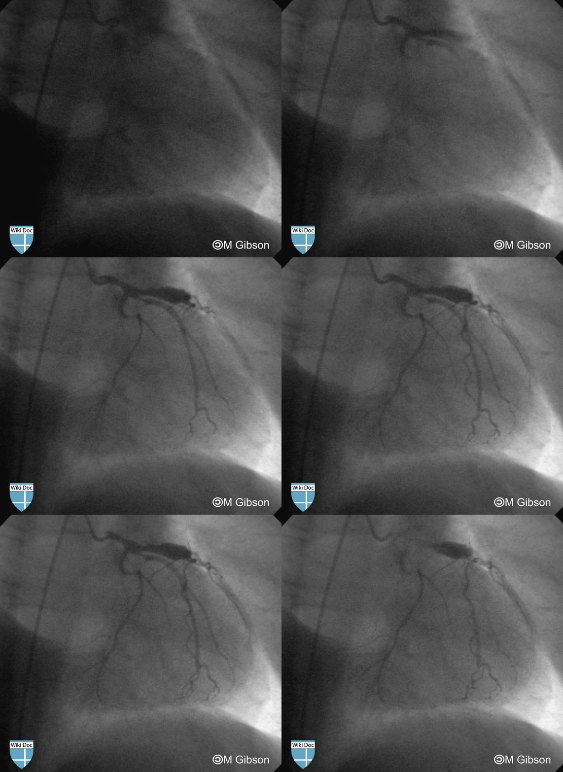
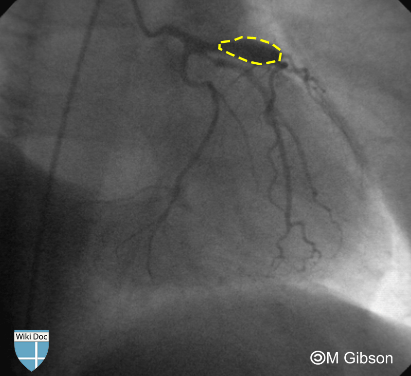
Shown below are an animated image and a static image depicting an aneurysm in the left coronary artery. Outlined in yellow in the image on the right is the localized aneurysm which is a dilatation of the coronary artery segment >1.5 times the diameter of the adjacent segments on the LCA.
Causes
- Atherosclerosis[2]
- Collagen vascular diseases
- Coronary catheterization
- Kawasaki disease[3]
- Vasculitides
Epidemiology and Demographics
- The incidence of CAA ranges widely between 0.3-5.3% within angiographic series with a mean incidence of 1.65%.[4] A highest incidence of 10-12% was reported in a study from India. Perhaps reflecting a specific genetic and/or environmental predisposition.[5]
- CAA most commonly occurs in right coronary artery accounting for 40-87% followed by left anterior descending artery and left circumflex artery.[6][7]
- CAA of left main artery or triple-vessel CAA are rare.[6]
Prognosis
Coronary artery aneurysm has a good prognosis.[8]
Diagnosis
It is often found coincidentally on coronary angiography.[8]
References
- ↑ Jarcho S (1969). "Bougon on coronary aneurysm (1812)". Am J Cardiol. 24 (4): 551–3. PMID 4897732.
- ↑ Nichols L, Lagana S, Parwani A (2008). "Coronary artery aneurysm: a review and hypothesis regarding etiology". Arch. Pathol. Lab. Med. 132 (5): 823–8. PMID 18466032. Unknown parameter
|month=ignored (help) - ↑ Fukazawa R, Ikegam E, Watanabe M; et al. (2007). "Coronary artery aneurysm induced by Kawasaki disease in children show features typical senescence". Circ. J. 71 (5): 709–15. PMID 17456996. Unknown parameter
|month=ignored (help) - ↑ Hartnell GG, Parnell BM, Pridie RB (1985). "Coronary artery ectasia. Its prevalence and clinical significance in 4993 patients". Br Heart J. 54 (4): 392–5. PMC 481917. PMID 4052280.
- ↑ Sharma SN, Kaul U, Sharma S, Wasir HS, Manchanda SC, Bahl VK; et al. (1990). "Coronary arteriographic profile in young and old Indian patients with ischaemic heart disease: a comparative study". Indian Heart J. 42 (5): 365–9. PMID 2086442.
- ↑ 6.0 6.1 Villines TC, Avedissian LS, Elgin EE (2005). "Diffuse nonatherosclerotic coronary aneurysms: an unusual cause of sudden death in a young male and a literature review". Cardiol Rev. 13 (6): 309–11. PMID 16230889.
- ↑ Swaye PS, Fisher LD, Litwin P, Vignola PA, Judkins MP, Kemp HG; et al. (1983). "Aneurysmal coronary artery disease". Circulation. 67 (1): 134–8. PMID 6847792.
- ↑ 8.0 8.1 Pahlavan PS, Niroomand F (2006). "Coronary artery aneurysm: a review". Clin Cardiol. 29 (10): 439–43. PMID 17063947. Unknown parameter
|month=ignored (help)

