Bone or cartilage mass imaging
|
Bone or Cartilage Mass Microchapters |
|
Diagnosis |
|---|
|
Case Studies |
|
Bone or cartilage mass imaging On the Web |
|
American Roentgen Ray Society Images of Bone or cartilage mass imaging |
|
Risk calculators and risk factors for Bone or cartilage mass imaging |
Editor-In-Chief: C. Michael Gibson, M.S., M.D. [1]Associate Editor(s)-in-Chief: Maria Fernanda Villarreal, M.D. [2]
Overview
Conventional radiography is the method of choice for the diagnosis of bone and cartilage tumors. The evaluation of bone and cartilage tumors will depend on 7 characteristics: periosteal reaction, opacity and mineralization, location, size, margins, cortical involvement, and soft-tissue component.[1]
Imaging
Plain Radiograph
Conventional radiography is the method of choice for the diagnosis of bone and cartilage tumors. The evaluation of bone and cartilage tumors will depend on 7 characteristics: location, margins, opacity and mineralization, size, periosteal reaction, cortical involvement, and soft-tissue component.[1]
Plain Radiograph | |||||||||||||||||||||||||||||
| What type of bone is involved? | |||||||||||||||||||||||||||||
| Long bone | Flat bone | ||||||||||||||||||||||||||||
Where is the lesion located? ❑ Epiphysis ❑ Diaphysis ❑ Metaphysis ❑ Apophysis | |||||||||||||||||||||||||||||
What is the pattern involved? ❑ Osteoblastic ❑ Osteolytic ❑ Mixed | |||||||||||||||||||||||||||||
What is the transversal location? ❑ Medullary ❑ Cortical ❑ Juxtacortical | |||||||||||||||||||||||||||||
What type of margin is involved? ❑ Well defined ❑ Ill-defined ❑ Sclerotic | |||||||||||||||||||||||||||||
Is there a periosteal reaction? ❑ Yes ❑ No | |||||||||||||||||||||||||||||
What is the size? | |||||||||||||||||||||||||||||
Likely benign? | Likely malignant? | ||||||||||||||||||||||||||||
Location
- Bone and cartilage tumors can be divided by location into 3 different categories, such as:
- Location in relation to the skeleton
- Location in relation to the physis (long bones)
- Location in relation to the transverse bone
Bone and cartilage tumors location by different parts of the skeleton, include:
- Axial skeleton
- Appendicular skeleton
- Long bones
- Flat bones
- Pelvis bone
- Lacrimal bone
- Nasal bone
Bone and cartilage tumors location in relation to the physis, include:
Bone and cartilage tumors location in relation to the transverse bone, include:
- Medullary
- Cortical
- Juxtacortical
Margin
The margin evaluation of bone and cartilage tumors, is divided into 3 categories:
- Transition zone
- Narrow
- Wide
- Margin characteristics
- Well-defined
- Ill-defined
- Sclerotic
- Patterns of bone destruction (appearance)
- Moth-eaten (myeloma, metastases, Ewing's sarcoma)
- Geographic (non-ossifying fibroma, chondromyxoid fibroma, and eosinophilic granuloma)
- Permeated (round cell lesions)
Opacity and mineralization
- Bone and cartilage tumors opacity depends on the stimulation of osteoclasts or osteoblasts in the bone
- Bone and cartilage tumors can be characterized by the tumor opacity into 3 different categories, including:
- Lytic lesions
- Sclerotic lesions
- Mixed lesions
- Bone and cartilage tumors can be characterized by 2 patterns of mineralization:
- Osseous
- Fluffy
- Cloud-like
- Chondral
- Punctate
- Flocculent
- Arclike
Periosteal reaction
- Periosteal reaction is a non-specific radiographic feature, that occurs with periosteal irritation
- Periosteal reactions may be broadly characterized by pattern and tumor nature (benign/aggressive)
- Useful to characterize a bone lesion
- Common periosteal reactions, include:
- Single layer
- Multilayered (onion-skin)
- Solid
- Spiculated
- Perpendicular (hair-on-end)
- Divergent (sunburst)
- Sloping (velvet)
- Disorganised/complex
- Codman triangle
Size
- In some cases, bone tumor size may be helpful to establish the diagnosis (eg. osteoblastoma (>1.5 cm) vs osteoid osteoma (<1.5 cm))
- Size can range from 0.1 cm - 10 cm
- In general, large size tumors are more likely to be malignant, whereas small size tumors tend to be related with benign origin.
Cortical involvement
- In some cases, bone and cartilage tumors lesions can specifically arise within the cortex, in such cases the evaluation will depend on:
- Type of erosion
- Endosteal scalloping
- "Soap bubble” lesions
Soft-tissue component
- Involvement of the soft-tissue is suggestive of a malignant process.
CT
- Bone CT scan may be helpful in the diagnosis of bone and cartilage tumors
- The majority of bone and cartilage tumors require further evaluation with CT scan
- Features of bone CT scan, include:[2]
- Characterization of sclerotic or mixed (lytic/sclerotic) lesions
- Imaging method of choice for follow-up of malignant tumors
- Characterization of occult bone destruction
MRI
- Musculoskeletal MRI may be helpful in the diagnosis of bone and cartilage tumors, common features include:[3]
- Evaluation of the local extent of a malignant process
- Useful for tumor staging
- Evaluation of soft tissue extension
- Extension within the bone marrow compartment and tumoral tissue infiltration
- Soft-tissue edema
- Narrow the differential diagnosis of bone and cartilage tumors, especially when there are signs of aggressiveness
- Contrast-enhanced musculoskeletal MRI can help demonstrate vascularized parts of the tumor
- Useful evaluating lesions involving the cortical or medullary region, and determine whether they penetrate or invade the region
- Useful to assess the response of chemotherapy in malignant bone tumors
- The use of FS/STIR sequences may be helpful to confirm the presence or absence of fat in a lesion (useful to differentiate hemangioma from lipoma)
Gallery
Plain Radiograph
-
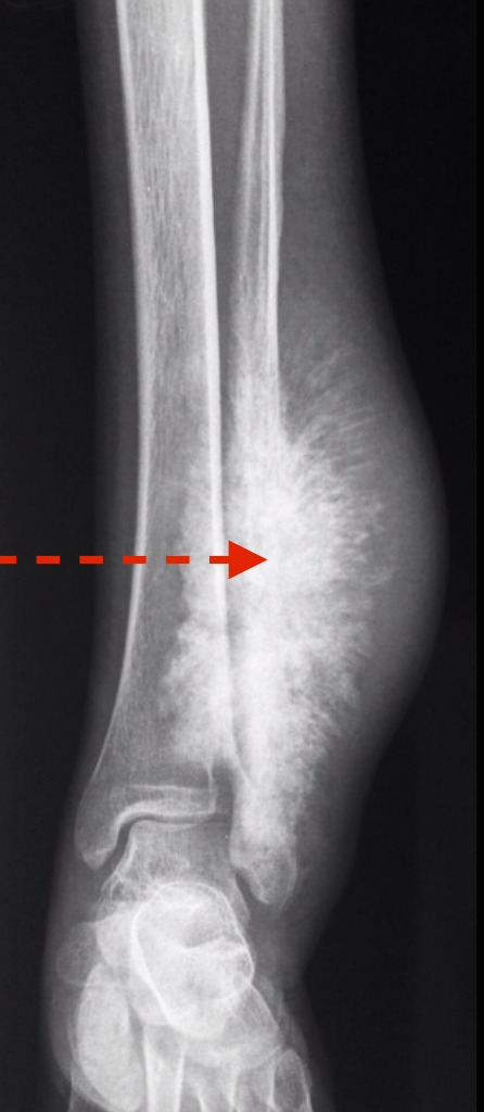
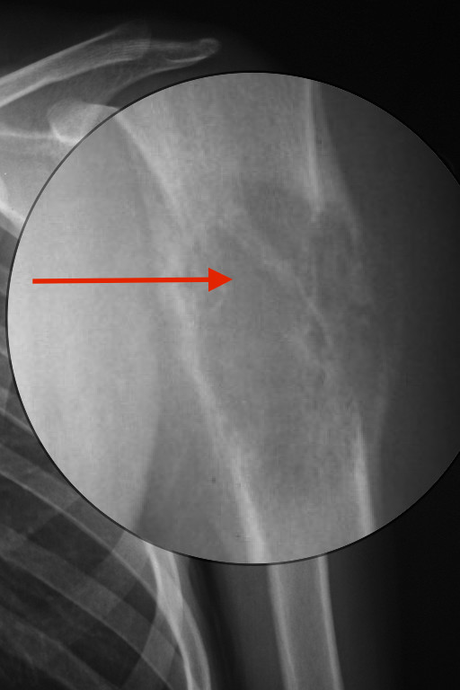 Osteosarcoma with Codman triangles: the tumor is essentially lytic and destructive with irregular, permeative margins, and soft tissue extension (red arrow). Codman triangles (reactive periosteal new bone formation around the edges) are very prominent
Adapted from Creative Commons 3.0 -
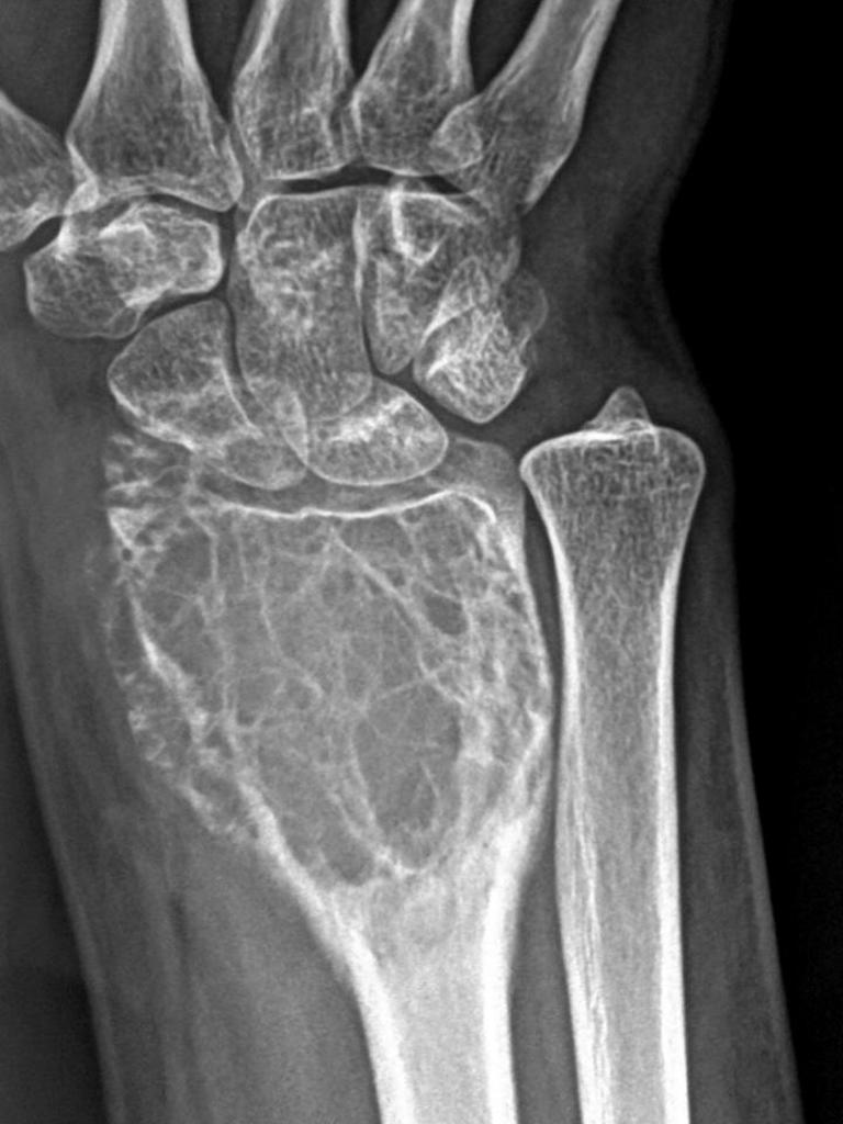 Giant cell tumor: located on distal radius
Adapted from Radiopedia -
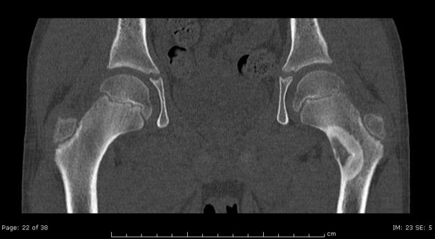
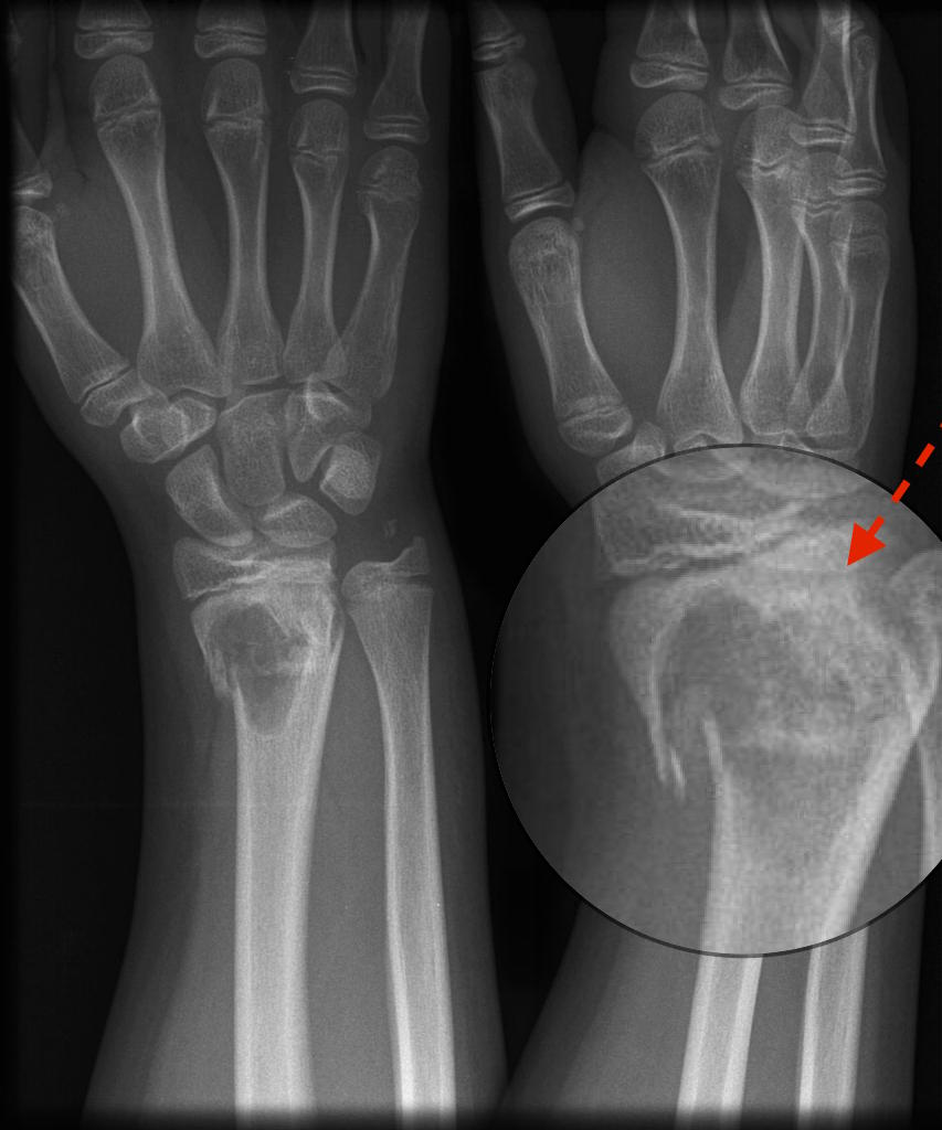 Pathological fracture: located in the metaphyseal region
Adapted from Radiopedia -
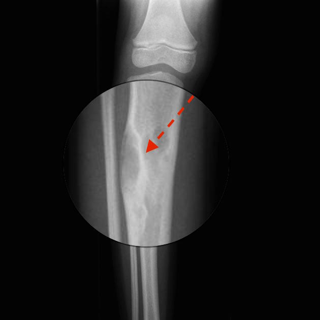 Rind sign: a lesion surrounded by a layer of thick, sclerotic reactive bone (rind) and is suggestive of fibrous dysplasia
Adapted from Radiopedia -
 Sunburst appearance: a type of periosteal reaction giving the appearance of a sunburst secondary to an aggressive periostitis. Present in aggresive tumors, such as: osteosarcoma, Ewing sarcoma, and osteoblastic metastases (e.g. prostate, lung or breast cancer)
Adapted from Radiopedia -
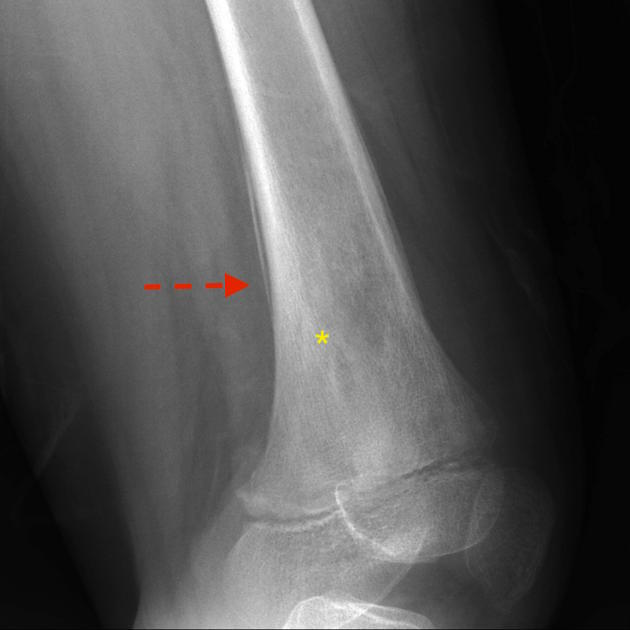 Onion skin sign : Also known as "multilayered periosteal reaction" demonstrates multiple concentric parallel layers of new bone adjacent to the cortex, reminiscent of the layers on an onion. Associated with osteosarcoma,Ewing sarcoma, and acute osteomyelitis (*) Parallel layers of new bone
Adapted from Radiopedia -
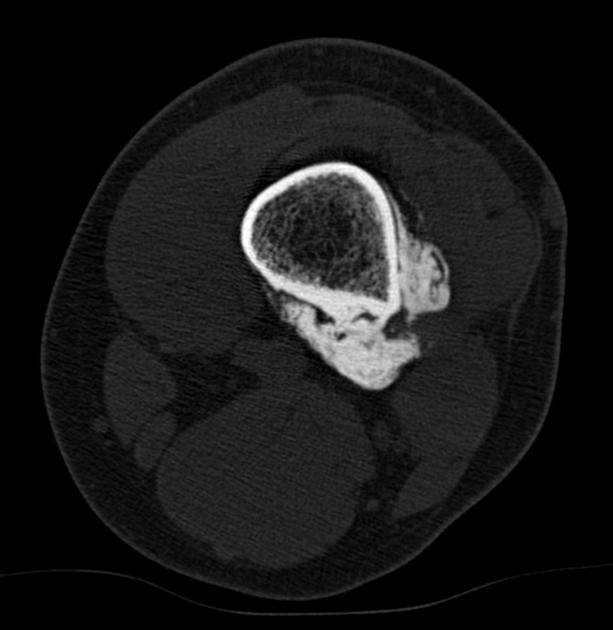
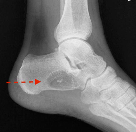 Cockade sign: Classic appearance of an intraosseous lipoma of the calcaneus which presents as a well-defined lytic lesion with a central calcification resembling a cockade
Adapted from Radiopedia -
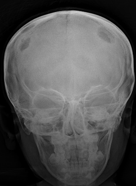 Geographic skull sign: radiographic appearance which is seen at eosinophilic granuloma. Destructive lytic bone lesion, edges of which may be bevelled, scalloped or confluent
Adapted from Radiopedia


CT
-
 String sign : appearance a radiolucent cleavage plane between portions of the tumor and cortex of the affected bone
Adapted from Radiopedia -
 Osteoblastoma: Internal matrix mineralisation is better appreciated on CT
Adapted from Radiopedia


MRI
-
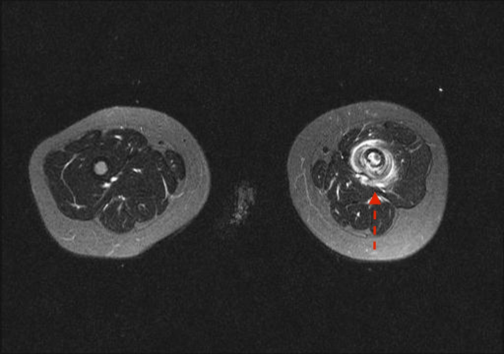 MRI- Onion skin sign
Adapted from Radiopedia -
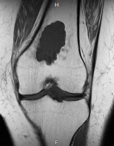 MRI-Enchondroma : Well circumscribed somewhat lobulated masses replacing marrow
Adapted from Radiopedia


References
- ↑ 1.0 1.1 Miller TT (2008). "Bone tumors and tumorlike conditions: analysis with conventional radiography". Radiology. 246 (3): 662–74. doi:10.1148/radiol.2463061038. PMID 18223119.
- ↑ American College of Radiology (2011) ACR Appropriateness Criteria. Follow-up of Malignant or Aggressive Musculoskeletal Tumors. Available via http://www.acr.org/~/media/ACR/Documents/AppCriteria/Diagnostic/FollowupMalignantOrAggressiveMusculoskeletalTumors.pdf.
- ↑ Nascimento D, Suchard G, Hatem M, de Abreu A (2014). "The role of magnetic resonance imaging in the evaluation of bone tumours and tumour-like lesions". Insights Imaging. 5 (4): 419–40. doi:10.1007/s13244-014-0339-z. PMC 4141345. PMID 25005774.