Superior vena cava syndrome CT: Difference between revisions
Hardik Patel (talk | contribs) No edit summary |
No edit summary |
||
| (11 intermediate revisions by 4 users not shown) | |||
| Line 1: | Line 1: | ||
__NOTOC__ | __NOTOC__ | ||
{{Superior vena cava syndrome}} | {{Superior vena cava syndrome}} | ||
{{CMG}} | {{CMG}}{{AE}}{{MV}} | ||
==Overview== | ==Overview== | ||
On enhanced [[Computed tomography|CT scan]], findings include location and severity of the [[superior vena cava obstruction]], superimposed [[thrombosis]], a [[mediastinal mass]] or [[lymphadenopathy]], collateral [[vessels]], and associated [[Lung mass|lung masses]]. [[Computed tomography|CT scan]] is the [[imaging]] modality of choice. | |||
==CT== | ==CT== | ||
* [[Computed tomography|CT scan]] is the [[imaging]] modality of choice for the diagnosis of [[superior vena cava syndrome]]. | |||
* On [[Computed tomography|CT scan]], [[superior vena cava syndrome]] is characterized by:<ref name="radio">Superior Vena Cava Syndrome.Dr Amir Rezaee and Radswiki et al. Radiopedia http://radiopaedia.org/articles/superior-vena-cava-obstruction Accessed on January 13, 2016</ref> | |||
:*Location and severity of the [[superior vena cava obstruction]] | |||
:*Superimposed [[thrombosis]] | |||
:*[[Mediastinal mass]] or [[lymphadenopathy]] | |||
:*Collateral vessels and associated [[Lung mass|lung masses]] | |||
==Gallery== | |||
<div align="left"> | <div align="left"> | ||
<gallery heights="175" widths="175"> | <gallery heights="175" widths="175"> | ||
Image:Svc syndrome collaterals.jpg|Collaterals on [[CT]] scan in a patient with | Image:Svc syndrome collaterals.jpg|Collaterals on [[CT]] scan in a patient with superior vena cava syndrome | ||
</gallery> | |||
</div> | |||
<div align="left"> | |||
<gallery heights="175" widths="175"> | |||
Image:INVASION svcs Ct.png|Lung cancer with vena cava invasion. | |||
</gallery> | </gallery> | ||
</div> | </div> | ||
==References== | ==References== | ||
{{Reflist| | {{Reflist|1}} | ||
[[Category:Disease]] | [[Category:Disease]] | ||
[[Category:Hematology]] | [[Category:Hematology]] | ||
[[Category:Cardiology]] | [[Category:Cardiology]] | ||
[[Category:Emergency medicine]] | [[Category:Emergency medicine]] | ||
[[Category:Intensive care medicine]] | [[Category:Intensive care medicine]] | ||
{{WH}} | {{WH}} | ||
{{WS}} | {{WS}} | ||
[[Category:Up-To-Date]] | |||
[[Category:Oncology]] | |||
[[Category:Medicine]] | |||
[[Category:Hematology]] | |||
[[Category:Vascular medicine]] | |||
[[Category:Surgery]] | |||
Latest revision as of 14:10, 12 April 2019
|
Superior Vena Cava Syndrome Microchapters |
|
Differentiating Superior Vena Cava Syndrome from Other Diseases |
|---|
|
Diagnosis |
|
Treatment |
|
Case Studies |
|
Superior vena cava syndrome CT On the Web |
|
American Roentgen Ray Society Images of Superior vena cava syndrome CT |
|
Directions to Hospitals Treating Superior vena cava syndrome |
|
Risk calculators and risk factors for Superior vena cava syndrome CT |
Editor-In-Chief: C. Michael Gibson, M.S., M.D. [1]Associate Editor(s)-in-Chief: Maria Fernanda Villarreal, M.D. [2]
Overview
On enhanced CT scan, findings include location and severity of the superior vena cava obstruction, superimposed thrombosis, a mediastinal mass or lymphadenopathy, collateral vessels, and associated lung masses. CT scan is the imaging modality of choice.
CT
- CT scan is the imaging modality of choice for the diagnosis of superior vena cava syndrome.
- On CT scan, superior vena cava syndrome is characterized by:[1]
- Location and severity of the superior vena cava obstruction
- Superimposed thrombosis
- Mediastinal mass or lymphadenopathy
- Collateral vessels and associated lung masses
Gallery
-
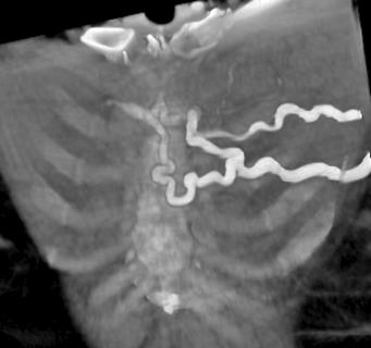
Collaterals on CT scan in a patient with superior vena cava syndrome

-

Lung cancer with vena cava invasion.

References
- ↑ Superior Vena Cava Syndrome.Dr Amir Rezaee and Radswiki et al. Radiopedia http://radiopaedia.org/articles/superior-vena-cava-obstruction Accessed on January 13, 2016