Pericarditis EKG examples
Editor-In-Chief: C. Michael Gibson, M.S., M.D. [1]
For the main page on pericarditis, click here.
EKG Examples
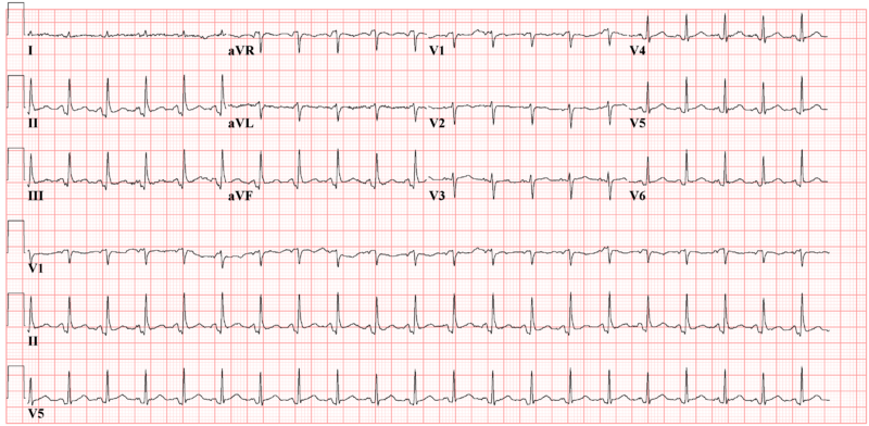
Shown below is an EKG image with PTa depression (depression between the end of the P wave and the beginning of the QRS complex), but no ST elevation depicting acute pericarditis.

Copyleft image obtained courtesy of ECGpedia,http://en.ecgpedia.org/wiki/File:De-Ptadepressieecg.png
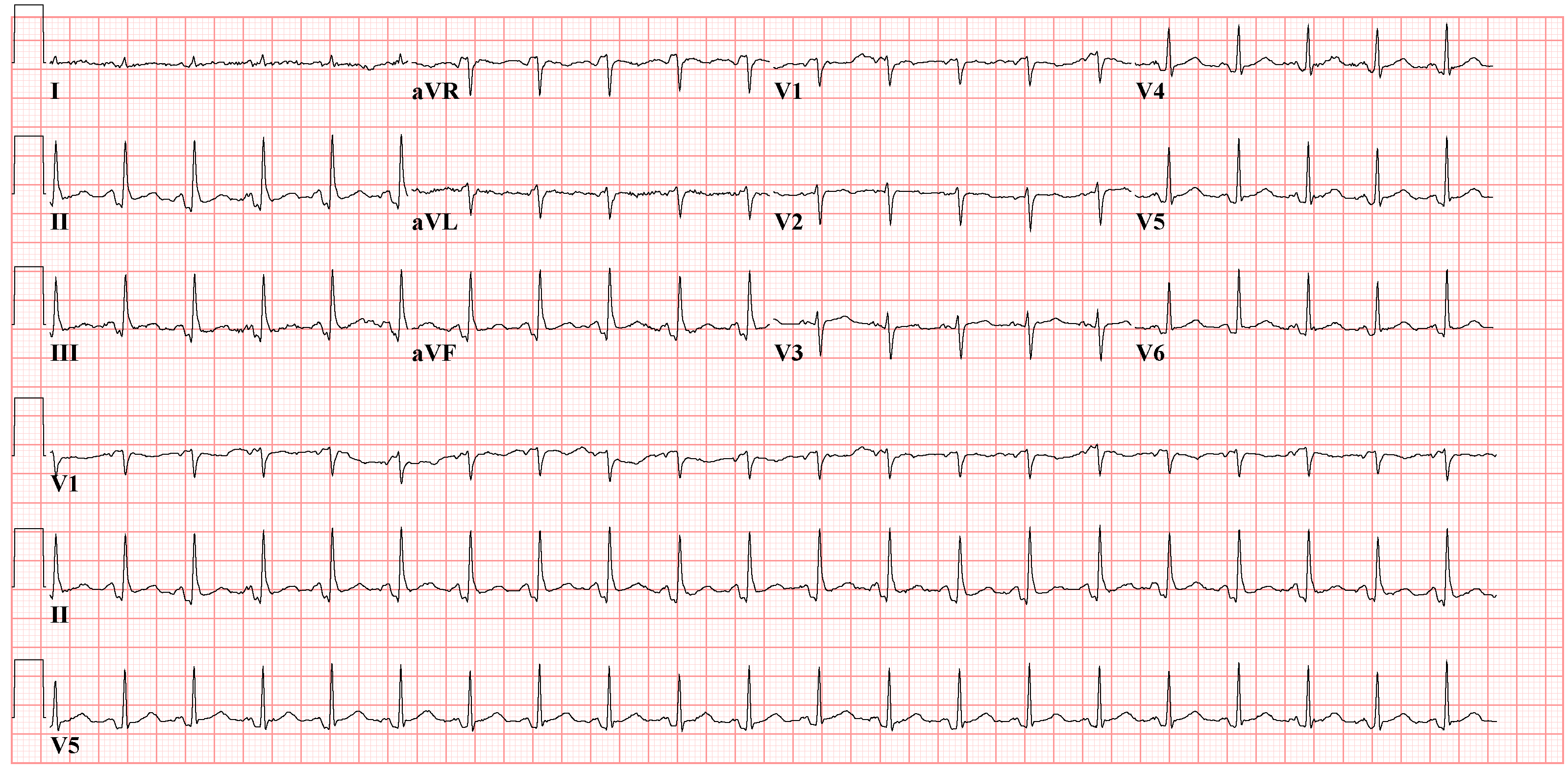
Shown below is an EKG with PTa depression depicting acute pericarditis.

Copyleft image obtained courtesy of ECGpedia, http://en.ecgpedia.org/wiki/Main_Page
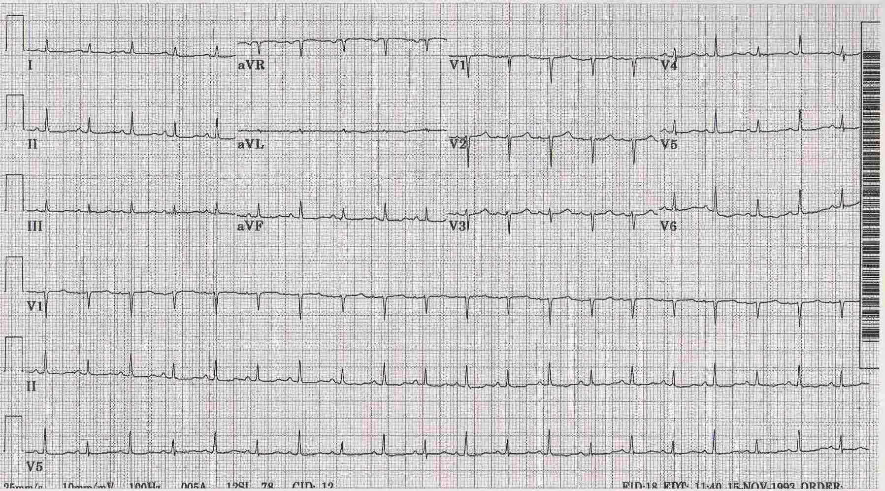
Shown below is an EKG from a case of pericarditis with effusion depicting electrical alterans.

Copyleft image obtained courtesy of ECGpedia,http://en.ecgpedia.org/wiki/Main_Page