Pancreatic cancer CT

Jump to navigation Jump to search

Pancreatic cancer Microchapters

Home

Patient Information

Overview

Historical Perspective

Classification

Pathophysiology

Causes

Differentiating Pancreatic Cancer from other Diseases

Epidemiology and Demographics

Risk Factors

Screening

Natural History, Complications and Prognosis

Diagnosis

Staging

Diagnostic study of choice

History and Symptoms

Physical Examination

Laboratory Findings

Chest X Ray

CT

MRI

Ultrasound

Other Imaging Findings

Other Diagnostic Studies

Treatment

Medical Therapy

Surgery

Primary Prevention

Secondary Prevention

Cost-Effectiveness of Therapy

Future or Investigational Therapies

Case Studies

Case #1

Pancreatic cancer CT On the Web

Most recent articles

Most cited articles

Review articles

CME Programs

Powerpoint slides

Images

American Roentgen Ray Society Images of Pancreatic cancer CT

All Images
X-rays
Echo & Ultrasound
CT Images
MRI

Ongoing Trials at Clinical Trials.gov

US National Guidelines Clearinghouse

NICE Guidance

FDA on Pancreatic cancer CT

CDC on Pancreatic cancer CT

Pancreatic cancer CT in the news

Blogs on Pancreatic cancer CT

Directions to Hospitals Treating Pancreatic cancer

Risk calculators and risk factors for Pancreatic cancer CT

Editor-In-Chief: C. Michael Gibson, M.S., M.D. [1]; Associate Editor-In-Chief: Cafer Zorkun, M.D., Ph.D. [2]

Overview

CT

An x-ray machine linked to a computer takes a series of detailed pictures. The x-ray machine is shaped like a donut with a large hole. The patient lies on a bed that passes through the hole. As the bed moves slowly through the hole, the machine takes many x-rays. The computer puts the x-rays together to create pictures of the pancreas and other organs and blood vessels in the abdomen. This scan can help identify the location of the cancer.

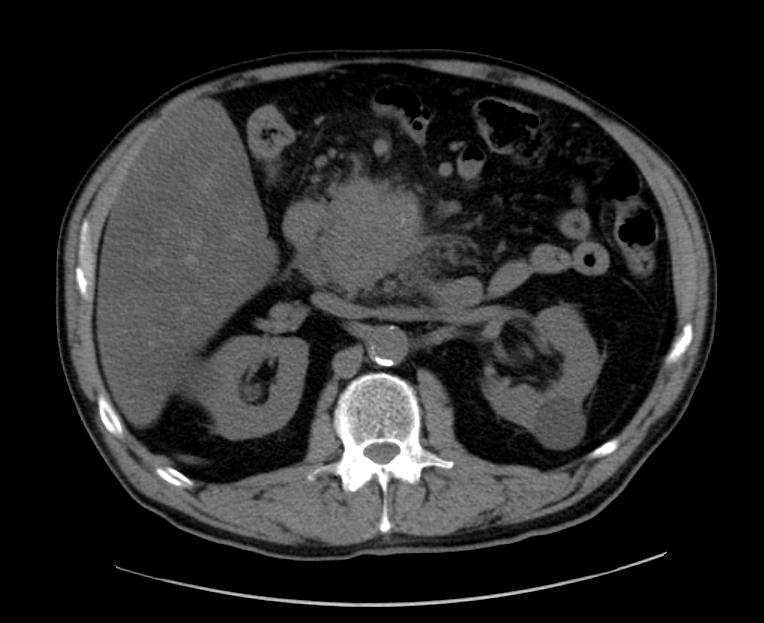
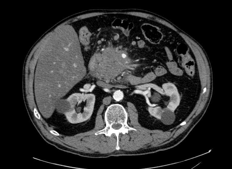
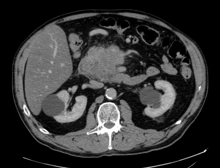
Pancreatic adenocarcinoma

Images courtesy of RadsWiki

References

[[Category:Disease] ]


Template:WikiDoc Sources