Osteoma: Difference between revisions
No edit summary |
No edit summary |
||
| (9 intermediate revisions by one other user not shown) | |||
| Line 1: | Line 1: | ||
__NOTOC__ | __NOTOC__ | ||
'''For patient information, click [[{{PAGENAME}} (patient information)|here]]''' | '''For patient information, click [[{{PAGENAME}} (patient information)|here]]''' | ||
<br>'''For more information about osteoid osteoma that is not associated with sino-orbital osteoma, see [[osteoid osteoma]]''' | <br>'''For more information about osteoid osteoma that is not associated with sino-orbital osteoma, see [[osteoid osteoma]]''' | ||
| Line 8: | Line 9: | ||
==Overview== | ==Overview== | ||
Osteoma (also known as ''Osteomata'') is a slow growing [[benign]] [[tumor]] of [[bone]], occurring most commonly in the [[craniofacial]] skeletal structures, primarily in the [[nasal]] and [[Paranasal sinus|paranasal]] (75-90%) sinuses. Osteoma arises from [[bone]] overgrowth, which is normally composed of [[connective tissue]]. Osteomas are slow growing [[tumors]] composed of compact or mature [[trabecular bone]] limited to [[craniofacial]] [[bones]]. Osteoma may be incidentally identified as a mass in the [[skull]], [[mandible]], or as the underlying cause of [[sinusitis]] or [[mucocele]] formation within the [[paranasal sinuses]]. When they are multiple, [[Gardner's syndrome|Gardner syndrome]] should be considered. Osteoma represents the most common [[benign]] [[neoplasm]] of the [[nose]] and [[paranasal sinuses]]. The causes remain uncertain, but commonly accepted theories propose [[embryologic]], [[Trauma|traumatic]], or [[Infection|infectious]] causes. Osteomas are usually [[asymptomatic]]. [[Excision]] may be performed if osteoma is responsible for [[symptoms]]. | |||
==Historical Perspective== | ==Historical Perspective== | ||
*In 1898, the description of craniofacial osteoma was first reported by Paul Schulze.<ref>{{cite book | last = Peabody | first = Terrance | title = Orthopaedic oncology : primary and metastatic tumors of the skeletal system | publisher = Springer | location = Cham | year = 2014 | isbn = 9783319073224 }}</ref> | *In 1898, the description of [[craniofacial]] osteoma was first reported by Paul Schulze.<ref>{{cite book | last = Peabody | first = Terrance | title = Orthopaedic oncology : primary and metastatic tumors of the skeletal system | publisher = Springer | location = Cham | year = 2014 | isbn = 9783319073224 }}</ref> | ||
*In 1951, Eldon J. Gardner (1909–1989) a geneticist first described the occurrence of multiple osteomas in hereditary familial adenomatous polyposis (FAP). | *In 1951, Eldon J. Gardner (1909–1989) a [[geneticist]] first described the occurrence of multiple osteomas in [[hereditary]] [[familial adenomatous polyposis]] (FAP). | ||
*In 2014, The Lancet published an article named "''Did René Descartes have a giant ethmoidal sinus osteoma?''" the authenticity has been confirmed by anthropological and historical investigations to be true.<ref name="pmid25307842">{{cite journal |vauthors=Charlier P, Froesch P, Benmoussa N, Froment A, Shorto R, Huynh-Charlier I |title=Did René Descartes have a giant ethmoidal sinus osteoma? |journal=Lancet |volume=384 |issue=9951 |pages=1348 |year=2014 |pmid=25307842 |doi=10.1016/S0140-6736(14)61816-X |url=}}</ref> | *In 2014, The Lancet published an article named "''Did René Descartes have a giant [[Ethmoid sinus|ethmoidal sinus]] osteoma?''" the authenticity has been confirmed by anthropological and historical investigations to be true.<ref name="pmid25307842">{{cite journal |vauthors=Charlier P, Froesch P, Benmoussa N, Froment A, Shorto R, Huynh-Charlier I |title=Did René Descartes have a giant ethmoidal sinus osteoma? |journal=Lancet |volume=384 |issue=9951 |pages=1348 |year=2014 |pmid=25307842 |doi=10.1016/S0140-6736(14)61816-X |url=}}</ref> | ||
==Classification== | ==Classification== | ||
| Line 19: | Line 21: | ||
===Enneking (MSTS) Staging System=== | ===Enneking (MSTS) Staging System=== | ||
*The Enneking surgical staging system (also known as the MSTS system) for benign [[Musculoskeletal system|musculoskeletal]] [[Tumor|tumors]] based on [[radiographic]] characteristics of the tumor host margin.<ref name="pmid20333492">{{cite journal| author=Jawad MU, Scully SP| title=In brief: classifications in brief: enneking classification: benign and malignant tumors of the musculoskeletal system. | journal=Clin Orthop Relat Res | year= 2010 | volume= 468 | issue= 7 | pages= 2000-2 | pmid=20333492 | doi=10.1007/s11999-010-1315-7 | pmc=2882012 | url=https://www.ncbi.nlm.nih.gov/entrez/eutils/elink.fcgi?dbfrom=pubmed&tool=sumsearch.org/cite&retmode=ref&cmd=prlinks&id=20333492 }} </ref> | *The Enneking surgical staging system (also known as the MSTS system) for benign [[Musculoskeletal system|musculoskeletal]] [[Tumor|tumors]] based on [[radiographic]] characteristics of the [[tumor]] host margin.<ref name="pmid20333492">{{cite journal| author=Jawad MU, Scully SP| title=In brief: classifications in brief: enneking classification: benign and malignant tumors of the musculoskeletal system. | journal=Clin Orthop Relat Res | year= 2010 | volume= 468 | issue= 7 | pages= 2000-2 | pmid=20333492 | doi=10.1007/s11999-010-1315-7 | pmc=2882012 | url=https://www.ncbi.nlm.nih.gov/entrez/eutils/elink.fcgi?dbfrom=pubmed&tool=sumsearch.org/cite&retmode=ref&cmd=prlinks&id=20333492 }} </ref> | ||
*It is widely accepted and routinely used classification. | *It is widely accepted and routinely used classification. | ||
| Line 37: | Line 39: | ||
| style="padding: 5px 5px; background: #F5F5F5;" | Aggressive: Indistinct borders | | style="padding: 5px 5px; background: #F5F5F5;" | Aggressive: Indistinct borders | ||
|} | |} | ||
==Pathophysiology== | ==Pathophysiology== | ||
| Line 43: | Line 44: | ||
*The possibility of a reactive mechanism, triggered by trauma or infection, has been suggested.<ref name="pmid15111819">{{cite journal| author=Bilkay U, Erdem O, Ozek C, Helvaci E, Kilic K, Ertan Y et al.| title=Benign osteoma with Gardner syndrome: review of the literature and report of a case. | journal=J Craniofac Surg | year= 2004 | volume= 15 | issue= 3 | pages= 506-9 | pmid=15111819 | doi= | pmc= | url=https://www.ncbi.nlm.nih.gov/entrez/eutils/elink.fcgi?dbfrom=pubmed&tool=sumsearch.org/cite&retmode=ref&cmd=prlinks&id=15111819 }} </ref> | *The possibility of a reactive mechanism, triggered by trauma or infection, has been suggested.<ref name="pmid15111819">{{cite journal| author=Bilkay U, Erdem O, Ozek C, Helvaci E, Kilic K, Ertan Y et al.| title=Benign osteoma with Gardner syndrome: review of the literature and report of a case. | journal=J Craniofac Surg | year= 2004 | volume= 15 | issue= 3 | pages= 506-9 | pmid=15111819 | doi= | pmc= | url=https://www.ncbi.nlm.nih.gov/entrez/eutils/elink.fcgi?dbfrom=pubmed&tool=sumsearch.org/cite&retmode=ref&cmd=prlinks&id=15111819 }} </ref> | ||
*Osteoma arises from bone overgrowth, which is normally composed of [[connective tissue]].<ref name="pmid25767729">{{cite journal |vauthors=Abdel Tawab HM, Kumar V R, Tabook SM |title=Osteoma presenting as a painless solitary mastoid swelling |journal=Case Rep Otolaryngol |volume=2015 |issue= |pages=590783 |year=2015 |pmid=25767729 |pmc=4341844 |doi=10.1155/2015/590783 |url=}}</ref> | *Osteoma arises from bone overgrowth, which is normally composed of [[connective tissue]].<ref name="pmid25767729">{{cite journal |vauthors=Abdel Tawab HM, Kumar V R, Tabook SM |title=Osteoma presenting as a painless solitary mastoid swelling |journal=Case Rep Otolaryngol |volume=2015 |issue= |pages=590783 |year=2015 |pmid=25767729 |pmc=4341844 |doi=10.1155/2015/590783 |url=}}</ref> | ||
*Osteomas are slow growing tumors composed of compact or mature [[trabecular bone]] limited to craniofacial bones. | *Osteomas are slow growing [[tumors]] composed of compact or mature [[trabecular bone]] limited to craniofacial bones. | ||
*Very rarely osteomas of the facial bones may be associated with Gardner's syndrome. | *Very rarely osteomas of the facial bones may be associated with Gardner's syndrome. | ||
*Osteomas have a particular frequency distribution within the [[paranasal sinuses]]: frontal sinuses 80%, ethmoid air cells 15%, maxillary sinuses 5% and sphenoid sinus rare. | *Osteomas have a particular frequency distribution within the [[paranasal sinuses]]: frontal sinuses 80%, ethmoid air cells 15%, maxillary sinuses 5% and sphenoid sinus rare. | ||
| Line 54: | Line 55: | ||
==Differentiating ((Page name)) from Other Diseases== | ==Differentiating ((Page name)) from Other Diseases== | ||
Osteoma must be differentiated from other diseases that cause sinus or facial pain, [[headache]], and changes to or loss of sense of smell, such as other osteogenic | Osteoma must be differentiated from other diseases that cause sinus or facial pain, [[headache]], and changes to or loss of sense of smell, such as other osteogenic [[tumors]], [[fibrous dysplasia]], and [[chronic sinusitis]].<ref name="pmid19780030">{{cite journal |vauthors=Erdogan N, Demir U, Songu M, Ozenler NK, Uluç E, Dirim B |title=A prospective study of paranasal sinus osteomas in 1,889 cases: changing patterns of localization |journal=Laryngoscope |volume=119 |issue=12 |pages=2355–9 |year=2009 |pmid=19780030 |doi=10.1002/lary.20646 |url=}}</ref><ref name="pmid18154576">{{cite journal| author=Larrea-Oyarbide N, Valmaseda-Castellón E, Berini-Aytés L, Gay-Escoda C| title=Osteomas of the craniofacial region. Review of 106 cases. | journal=J Oral Pathol Med | year= 2008 | volume= 37 | issue= 1 | pages= 38-42 | pmid=18154576 | doi=10.1111/j.1600-0714.2007.00590.x | pmc= | url=https://www.ncbi.nlm.nih.gov/entrez/eutils/elink.fcgi?dbfrom=pubmed&tool=sumsearch.org/cite&retmode=ref&cmd=prlinks&id=18154576 }} </ref> | ||
{| style="border: 0px; font-size: 90%; margin: 3px; width: 1000px" align="center" | |||
{| style="border: 0px; font-size: 90%; margin: 3px; width: 1000px" align=center | | valign="top" | | ||
|valign=top| | |||
|+ | |+ | ||
! style="background: #4479BA; width: 200px;" | {{fontcolor|#FFF|Differential Diagnosis}} | ! style="background: #4479BA; width: 200px;" | {{fontcolor|#FFF|Differential Diagnosis}} | ||
| Line 64: | Line 64: | ||
! style="background: #4479BA; width: 300px;" | {{fontcolor|#FFF|Differentiating Features}} | ! style="background: #4479BA; width: 300px;" | {{fontcolor|#FFF|Differentiating Features}} | ||
|- | |- | ||
| style="padding: 5px 5px; background: #DCDCDC; font-weight: bold; text-align:center;"| [[Fibrous dysplasia]] | | style="padding: 5px 5px; background: #DCDCDC; font-weight: bold; text-align:center;" | [[Fibrous dysplasia]] | ||
| style="padding: 5px 5px; background: #F5F5F5;"| | | style="padding: 5px 5px; background: #F5F5F5;" | | ||
*Benign, often an incidental finding, affects the same group of patients, and symptoms include facial pain and headache | *Benign, often an incidental finding, affects the same group of patients, and symptoms include facial pain and headache | ||
| | | style="padding: 5px 5px; background: #F5F5F5;" | | ||
*In fibrous dysplasia, differentiating features include: More common presentation is on ribs: 28%, no gender predilection, and complete resection is usually not possible | *In fibrous dysplasia, differentiating features include: More common presentation is on ribs: 28%, no gender predilection, and complete resection is usually not possible | ||
|- | |- | ||
| style="padding: 5px 5px; background: #DCDCDC; font-weight: bold; text-align:center;"| [[Osteoblastoma]] | | style="padding: 5px 5px; background: #DCDCDC; font-weight: bold; text-align:center;" | [[Osteoblastoma]] | ||
| style="padding: 5px 5px; background: #F5F5F5;"| | | style="padding: 5px 5px; background: #F5F5F5;" | | ||
*Benign, incidental, and male predilection | *Benign, incidental, and male predilection | ||
| style="padding: 5px 5px; background: #F5F5F5;"| | | style="padding: 5px 5px; background: #F5F5F5;" | | ||
*In osteoblastoma, differentiating features include: normally affect the axial skeleton, lesions are typically larger than 2 cm, and surgical excision is often the treatment of choice | *In osteoblastoma, differentiating features include: normally affect the axial skeleton, lesions are typically larger than 2 cm, and surgical excision is often the treatment of choice | ||
|- | |- | ||
| style="padding: 5px 5px; background: #DCDCDC; font-weight: bold; text-align:center;"| Adamantinomas | | style="padding: 5px 5px; background: #DCDCDC; font-weight: bold; text-align:center;" | [[Adamantinoma|Adamantinomas]] | ||
| style="padding: 5px 5px; background: #F5F5F5;"| | | style="padding: 5px 5px; background: #F5F5F5;" | | ||
*Benign, slow growing, and similar clinical onset | *Benign, slow growing, and similar clinical onset | ||
| style="padding: 5px 5px; background: #F5F5F5;"| | | style="padding: 5px 5px; background: #F5F5F5;" | | ||
*In adamantinomas, differentiating features include: locally aggressive tumor, common in the 3rd to 5th decades of life, and location is usually confined to the jaw | *In adamantinomas, differentiating features include: locally aggressive tumor, common in the 3rd to 5th decades of life, and location is usually confined to the jaw | ||
|- | |- | ||
|style="padding: 5px 5px; background: #DCDCDC; font-weight: bold; text-align:center;" | [[Chronic sinusitis]] | | style="padding: 5px 5px; background: #DCDCDC; font-weight: bold; text-align:center;" | [[Chronic sinusitis]] | ||
|style="padding: 5px 5px; background: #F5F5F5;"| | | style="padding: 5px 5px; background: #F5F5F5;" | | ||
*Affects same group of population (young to middle aged adults) and the clinical presentation is similar | *Affects same group of population (young to middle aged adults) and the clinical presentation is similar | ||
|style="padding: 5px 5px; background: #F5F5F5;"| | | style="padding: 5px 5px; background: #F5F5F5;" | | ||
*In chronic sinusitis, differentiating features include: fever, previous history of acute sinusitis, lack of facial deformation or imaging findings compatible with osteoma | *In chronic sinusitis, differentiating features include: fever, previous history of acute sinusitis, lack of facial deformation or imaging findings compatible with osteoma | ||
|} | |} | ||
| Line 102: | Line 102: | ||
==Screening== | ==Screening== | ||
Screening for multiple osteomas is recommended among patients with [[family history]] or/and a confirmed diagnosis of Gardner syndrome. [[Thyroid]] exam and annual ultrasound, should be performed starting at age 10 to 12 years.<ref name="pmid24093640">{{cite journal |vauthors=Septer S, Slowik V, Morgan R, Dai H, Attard T |title=Thyroid cancer complicating familial adenomatous polyposis: mutation spectrum of at-risk individuals |journal=Hered Cancer Clin Pract |volume=11 |issue=1 |pages=13 |year=2013 |pmid=24093640 |pmc=3854022 |doi=10.1186/1897-4287-11-13 |url=}}</ref> | Screening for multiple osteomas is recommended among patients with [[family history]] or/and a confirmed diagnosis of [[Gardner's syndrome|Gardner syndrome]]. [[Thyroid]] exam and annual [[ultrasound]], should be performed starting at age 10 to 12 years.<ref name="pmid24093640">{{cite journal |vauthors=Septer S, Slowik V, Morgan R, Dai H, Attard T |title=Thyroid cancer complicating familial adenomatous polyposis: mutation spectrum of at-risk individuals |journal=Hered Cancer Clin Pract |volume=11 |issue=1 |pages=13 |year=2013 |pmid=24093640 |pmc=3854022 |doi=10.1186/1897-4287-11-13 |url=}}</ref> | ||
==Natural History, Complications, and Prognosis== | ==Natural History, Complications, and Prognosis== | ||
| Line 109: | Line 109: | ||
**[[Proptosis]] | **[[Proptosis]] | ||
**Facial deformity | **Facial deformity | ||
**Airway obstruction | **[[Airway obstruction]] | ||
**Sensory loss | **[[Sensory loss]] | ||
**[[Anosmia]] | **[[Anosmia]] | ||
**[[Visual loss]] | **[[Visual loss]] | ||
*Prognosis is generally excellent,the lesion does not recur after surgical excision and it is not associated with malignant change.<ref name="pmid17577321">{{cite journal| author=Wijn MA, Keller JJ, Giardiello FM, Brand HS| title=Oral and maxillofacial manifestations of familial adenomatous polyposis. | journal=Oral Dis | year= 2007 | volume= 13 | issue= 4 | pages= 360-5 | pmid=17577321 | doi=10.1111/j.1601-0825.2006.01293.x | pmc= | url=https://www.ncbi.nlm.nih.gov/entrez/eutils/elink.fcgi?dbfrom=pubmed&tool=sumsearch.org/cite&retmode=ref&cmd=prlinks&id=17577321 }} </ref> | *Prognosis is generally excellent,the lesion does not recur after surgical [[excision]] and it is not associated with [[Malignant|malignant change]].<ref name="pmid17577321">{{cite journal| author=Wijn MA, Keller JJ, Giardiello FM, Brand HS| title=Oral and maxillofacial manifestations of familial adenomatous polyposis. | journal=Oral Dis | year= 2007 | volume= 13 | issue= 4 | pages= 360-5 | pmid=17577321 | doi=10.1111/j.1601-0825.2006.01293.x | pmc= | url=https://www.ncbi.nlm.nih.gov/entrez/eutils/elink.fcgi?dbfrom=pubmed&tool=sumsearch.org/cite&retmode=ref&cmd=prlinks&id=17577321 }} </ref> | ||
==Diagnosis== | ==Diagnosis== | ||
===Diagnostic Study of Choice=== | ===Diagnostic Study of Choice=== | ||
Biopsy is the diagnostic study of choice for the diagnosis of osteoma. | |||
*Gross appearance of osteoma include: | |||
**Osteomas have a spongy to densely appearance, conformed of in a polypoid and lobullated shape.[1] | |||
**The median size tumor size is 3.0 cm (range 0.5-8 cm). | |||
**Osteomas have a smooth surface and composed of dense [[compact bone]] (ivory osteoma), [[trabecular bone]] (mature osteoma, or both patterns). | |||
{| align="right" | |||
| | |||
[[File:Histology osteoma.jpg|200px|thumb|Histology of osteoma.[https://commons.wikimedia.org/wiki/File:Osteoma_--_high_mag.jpg Source: Case courtesy of Nephron [CC BY-SA 4.0 (https://creativecommons.org/licenses/by-sa/4.0) or GFDL (http://www.gnu.org/copyleft/fdl.html)], from Wikimedia Commons]]] | |||
|} | |||
*Histological appearance includes: | |||
**Presence of dense compact mature bone in paucicellular fibrous stroma. | |||
**Large trabeculae of mature lamellar bone can be also be seen. | |||
===History and Symptoms=== | ===History and Symptoms=== | ||
| Line 136: | Line 135: | ||
**[[Headache]] | **[[Headache]] | ||
**[[Nasal congestion]] | **[[Nasal congestion]] | ||
**Facial pain | **Facial [[pain]] | ||
**Facial tenderness | **Facial [[tenderness]] | ||
**Loss of the sense of smell | **[[Anosmia|Loss of the sense of smell]] | ||
===Physical Examination=== | ===Physical Examination=== | ||
{| align="right" | |||
| | |||
[[File:Xray osteoma.gif|200px|thumb|X-ray showing osteoma of the frontal sinus.[https://radiopaedia.org/cases/ivory-osteoma?lang=us Source: Case courtesy of Dr Ahmed Abdrabou, Radiopaedia.org, rID: 42250]]] | |||
|} | |||
*Patients with osteoid osteoma usually appears well. | *Patients with osteoid osteoma usually appears well. | ||
*Common physical examination findings of osteoid osteoma include:<ref name="pmid4207295">{{cite journal| author=Fu YS, Perzin KH| title=Non-epithelial tumors of the nasal cavity, paranasal sinuses, and nasopharynx. A clinicopathologic study. II. Osseous and fibro-osseous lesions, including osteoma, fibrous dysplasia, ossifying fibroma, osteoblastoma, giant cell tumor, and osteosarcoma. | journal=Cancer | year= 1974 | volume= 33 | issue= 5 | pages= 1289-305 | pmid=4207295 | doi= | pmc= | url=https://www.ncbi.nlm.nih.gov/entrez/eutils/elink.fcgi?dbfrom=pubmed&tool=sumsearch.org/cite&retmode=ref&cmd=prlinks&id=4207295 }} </ref> | *Common physical examination findings of osteoid osteoma include:<ref name="pmid4207295">{{cite journal| author=Fu YS, Perzin KH| title=Non-epithelial tumors of the nasal cavity, paranasal sinuses, and nasopharynx. A clinicopathologic study. II. Osseous and fibro-osseous lesions, including osteoma, fibrous dysplasia, ossifying fibroma, osteoblastoma, giant cell tumor, and osteosarcoma. | journal=Cancer | year= 1974 | volume= 33 | issue= 5 | pages= 1289-305 | pmid=4207295 | doi= | pmc= | url=https://www.ncbi.nlm.nih.gov/entrez/eutils/elink.fcgi?dbfrom=pubmed&tool=sumsearch.org/cite&retmode=ref&cmd=prlinks&id=4207295 }} </ref> | ||
**Facial tenderness | **Facial [[tenderness]] | ||
**Nasal obstruction and discharge | **Nasal obstruction and [[discharge]] | ||
**Tapping over a sinus area produces dull sound | **Tapping over a [[sinus]] area produces dull sound | ||
**Physical deformity over mastoid or facial area | **Physical deformity over [[mastoid]] or facial area | ||
===Laboratory Findings=== | ===Laboratory Findings=== | ||
| Line 155: | Line 158: | ||
===X-ray=== | ===X-ray=== | ||
*Three views of affected bone or joint are recommended.<ref>{{cite book | last = Schajowicz | first = Fritz | title = Tumors and Tumorlike Lesions of Bone : Pathology, Radiology, and Treatment | publisher = Springer Berlin Heidelberg | location = Berlin, Heidelberg | year = 1994 | isbn = 9783642499562 }}</ref> | |||
*Radiological findings for osteoma include: | |||
**Well circumscribed mass | |||
**Varying amounts of central lucency | |||
**Lobulated mass occupying [[frontal]] or [[maxillary sinus]] | |||
===Echocardiography or Ultrasound=== | ===Echocardiography or Ultrasound=== | ||
{| align="right" | |||
| | |||
[[File:CT osteoma.gif|200px|thumb|CT showing osteoma of the mandible.[https://radiopaedia.org/cases/ivory-osteoma?lang=us Source: Case courtesy of Dr Ahmed Abdrabou, Radiopaedia.org, rID: 42250]]] | |||
|} | |||
There are no echocardiography/ultrasound findings associated with osteoma. | There are no echocardiography/ultrasound findings associated with osteoma. | ||
===CT scan=== | ===CT scan=== | ||
*CT findings of osteoma include:<ref>{{cite book | last = Schajowicz | first = Fritz | title = Tumors and Tumorlike Lesions of Bone : Pathology, Radiology, and Treatment | publisher = Springer Berlin Heidelberg | location = Berlin, Heidelberg | year = 1994 | isbn = 9783642499562 }}</ref> | |||
**Well-circumscribed mass of variable density | |||
**Ground-glass appearance | |||
**Exophytical mass growing out of a [[sinus]] | |||
===MRI=== | ===MRI=== | ||
*MRI findings of osteoma include:<ref>{{cite book | last = Schajowicz | first = Fritz | title = Tumors and Tumorlike Lesions of Bone : Pathology, Radiology, and Treatment | publisher = Springer Berlin Heidelberg | location = Berlin, Heidelberg | year = 1994 | isbn = 9783642499562 }}</ref> | |||
**Low signal on all sequences. | |||
**Mature osteomas may demonstrate some marrow signal, but are also predominantly low on all sequences | |||
===Other Imaging Findings=== | ===Other Imaging Findings=== | ||
| Line 194: | Line 186: | ||
===Other Diagnostic Studies=== | ===Other Diagnostic Studies=== | ||
===Nasal Endoscopy=== | |||
Nasal endoscopy findings include:<ref name="pmid19894552">{{cite journal |vauthors=Li Y, Zhang L, Zhou B, Han D |title=[Resection of frontal ethmoid sinus osteomas with nasal endoscopy] |language=Chinese |journal=Lin Chung Er Bi Yan Hou Tou Jing Wai Ke Za Zhi |volume=23 |issue=14 |pages=628–30 |year=2009 |pmid=19894552 |doi= |url=}}</ref> | |||
*Direct visualization of the nasal passages structures, and [[sinuses]]. | |||
*Tumor location, size, and adjacent structure evaluation. | |||
==Treatment== | ==Treatment== | ||
===Medical Therapy=== | ===Medical Therapy=== | ||
There is no treatment for | There is no medical treatment for osteoma; the mainstay of therapy is surgery.<ref name="pmid25580337">{{cite journal| author=Gorini E, Mullace M, Migliorini L, Mevio E| title=Osseous choristoma of the tongue: a review of etiopathogenesis. | journal=Case Rep Otolaryngol | year= 2014 | volume= 2014 | issue= | pages= 373104 | pmid=25580337 | doi=10.1155/2014/373104 | pmc=4279709 | url=https://www.ncbi.nlm.nih.gov/entrez/eutils/elink.fcgi?dbfrom=pubmed&tool=sumsearch.org/cite&retmode=ref&cmd=prlinks&id=25580337 }} </ref> | ||
===Surgery=== | ===Surgery=== | ||
Surgery is the mainstay of treatment for osteoma.<ref name="pmid24900131">{{cite journal| author=Kim WH, Kim DW, Kim CG, Kim MH| title=Additional Detection of Multiple Osteomas in a Patient with Gardner's Syndrome by Bone SPECT/CT. | journal=Nucl Med Mol Imaging | year= 2013 | volume= 47 | issue= 4 | pages= 297-8 | pmid=24900131 | doi=10.1007/s13139-013-0225-5 | pmc=4035179 | url=https://www.ncbi.nlm.nih.gov/entrez/eutils/elink.fcgi?dbfrom=pubmed&tool=sumsearch.org/cite&retmode=ref&cmd=prlinks&id=24900131 }} </ref><ref name="pmid17926587">{{cite journal| author=Alexander AA, Patel AA, Odland R| title=Paranasal sinus osteomas and Gardner's syndrome. | journal=Ann Otol Rhinol Laryngol | year= 2007 | volume= 116 | issue= 9 | pages= 658-62 | pmid=17926587 | doi=10.1177/000348940711600906 | pmc= | url=https://www.ncbi.nlm.nih.gov/entrez/eutils/elink.fcgi?dbfrom=pubmed&tool=sumsearch.org/cite&retmode=ref&cmd=prlinks&id=17926587 }} </ref> | |||
'''Indication''' | |||
*For symptomatic lesions, local [[excision]] is performed. | |||
'''Types of Surgery''' | |||
*Medial maxillectomy with a lateral rhinotomy | |||
*[[Craniofacial]] resection | |||
*[[Endoscopy|Endoscopic]] resection | |||
'''Recurrence''' | |||
Rare recurrence may occur after several years. | |||
===Primary Prevention=== | ===Primary Prevention=== | ||
There are no established measures for the primary prevention of | There are no established measures for the primary prevention of osteoma. | ||
===Secondary Prevention=== | ===Secondary Prevention=== | ||
There are no established measures for the secondary prevention of | There are no established measures for the secondary prevention of osteoma. | ||
==References== | ==References== | ||
Latest revision as of 17:22, 3 April 2019
For patient information, click here
For more information about osteoid osteoma that is not associated with sino-orbital osteoma, see osteoid osteoma
|
Osteoma Microchapters |
|
Diagnosis |
|---|
|
Treatment |
|
Case Studies |
|
Osteoma On the Web |
|
American Roentgen Ray Society Images of Osteoma |
Editor-In-Chief: C. Michael Gibson, M.S., M.D. [1]; Associate Editor(s)-in-Chief: Rohan A. Bhimani, M.B.B.S., D.N.B., M.Ch.[2]
Synonyms and keywords: Osteoma; Osteomata; Osteoncus; Ivory osteoma; Mature osteoma; Mixed osteoma; Homoplastic osteoma; Heteroplastic osteoma; Osteomas; Ivory exostosis; Sino-orbital osteoma; Sino-nasal osteoma; Paranasal sinus osteoma; Skull vault osteoma; Mandibular osteoma
Overview
Osteoma (also known as Osteomata) is a slow growing benign tumor of bone, occurring most commonly in the craniofacial skeletal structures, primarily in the nasal and paranasal (75-90%) sinuses. Osteoma arises from bone overgrowth, which is normally composed of connective tissue. Osteomas are slow growing tumors composed of compact or mature trabecular bone limited to craniofacial bones. Osteoma may be incidentally identified as a mass in the skull, mandible, or as the underlying cause of sinusitis or mucocele formation within the paranasal sinuses. When they are multiple, Gardner syndrome should be considered. Osteoma represents the most common benign neoplasm of the nose and paranasal sinuses. The causes remain uncertain, but commonly accepted theories propose embryologic, traumatic, or infectious causes. Osteomas are usually asymptomatic. Excision may be performed if osteoma is responsible for symptoms.
Historical Perspective
- In 1898, the description of craniofacial osteoma was first reported by Paul Schulze.[1]
- In 1951, Eldon J. Gardner (1909–1989) a geneticist first described the occurrence of multiple osteomas in hereditary familial adenomatous polyposis (FAP).
- In 2014, The Lancet published an article named "Did René Descartes have a giant ethmoidal sinus osteoma?" the authenticity has been confirmed by anthropological and historical investigations to be true.[2]
Classification
Osteoma can be classified based on imaging findings.
Enneking (MSTS) Staging System
- The Enneking surgical staging system (also known as the MSTS system) for benign musculoskeletal tumors based on radiographic characteristics of the tumor host margin.[3]
- It is widely accepted and routinely used classification.
| Stages | Description |
|---|---|
| 1 | Latent: Well demarcated borders |
| 2 | Active: Indistinct borders |
| 3 | Aggressive: Indistinct borders |
Pathophysiology
- The exact etiology of osteoma is unknown.[4]
- The possibility of a reactive mechanism, triggered by trauma or infection, has been suggested.[5]
- Osteoma arises from bone overgrowth, which is normally composed of connective tissue.[6]
- Osteomas are slow growing tumors composed of compact or mature trabecular bone limited to craniofacial bones.
- Very rarely osteomas of the facial bones may be associated with Gardner's syndrome.
- Osteomas have a particular frequency distribution within the paranasal sinuses: frontal sinuses 80%, ethmoid air cells 15%, maxillary sinuses 5% and sphenoid sinus rare.
Genetics
- The hallmark of multiple osteomas is a mutation in the APC gene, that results in the Gardner syndrome.[7]
Causes
- The cause of osteoma has not been identified.[8]
Differentiating ((Page name)) from Other Diseases
Osteoma must be differentiated from other diseases that cause sinus or facial pain, headache, and changes to or loss of sense of smell, such as other osteogenic tumors, fibrous dysplasia, and chronic sinusitis.[9][10]
| Differential Diagnosis | Similar Features | Differentiating Features |
|---|---|---|
| Fibrous dysplasia |
|
|
| Osteoblastoma |
|
|
| Adamantinomas |
|
|
| Chronic sinusitis |
|
|
Epidemiology and Demographics
- The prevalence of osteoma is approximately 3000 per 100,000 individuals worldwide.[11]
- The incidence of osteoma remains unknown.
- Patients of all age groups may develop osteoma.
- The average patient age varies from 25 to 35 years.
- The mean age of the patients with osteoma is is 37 years.[9]
- Men are more commonly affected than women, with a 3:1 ratio.[12]
- There is no racial predilection to osteoma.
Risk Factors
There are no established risk factors for osteoma.[6]
Screening
Screening for multiple osteomas is recommended among patients with family history or/and a confirmed diagnosis of Gardner syndrome. Thyroid exam and annual ultrasound, should be performed starting at age 10 to 12 years.[13]
Natural History, Complications, and Prognosis
- If left untreated, osteoma progression occurs slowly and is then followed by facial distortion.[14]
- Common complications of osteoma include:[15]
- Proptosis
- Facial deformity
- Airway obstruction
- Sensory loss
- Anosmia
- Visual loss
- Prognosis is generally excellent,the lesion does not recur after surgical excision and it is not associated with malignant change.[15]
Diagnosis
Diagnostic Study of Choice
Biopsy is the diagnostic study of choice for the diagnosis of osteoma.
- Gross appearance of osteoma include:
- Osteomas have a spongy to densely appearance, conformed of in a polypoid and lobullated shape.[1]
- The median size tumor size is 3.0 cm (range 0.5-8 cm).
- Osteomas have a smooth surface and composed of dense compact bone (ivory osteoma), trabecular bone (mature osteoma, or both patterns).
 |
- Histological appearance includes:
- Presence of dense compact mature bone in paucicellular fibrous stroma.
- Large trabeculae of mature lamellar bone can be also be seen.
History and Symptoms
- Small osteomas are asymptomatic and usually incidental findings.
- Common symotoms of osteoma include:[16][17]
- Headache
- Nasal congestion
- Facial pain
- Facial tenderness
- Loss of the sense of smell
Physical Examination
 |
- Patients with osteoid osteoma usually appears well.
- Common physical examination findings of osteoid osteoma include:[18]
- Facial tenderness
- Nasal obstruction and discharge
- Tapping over a sinus area produces dull sound
- Physical deformity over mastoid or facial area
Laboratory Findings
There are no diagnostic laboratory findings associated with osteoma.
Electrocardiogram
There are no ECG findings associated with osteoma.
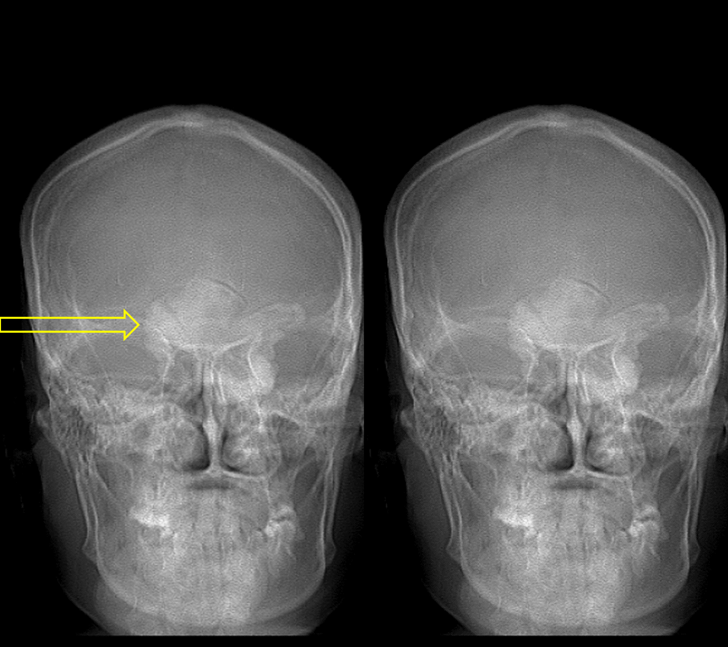
X-ray
- Three views of affected bone or joint are recommended.[19]
- Radiological findings for osteoma include:
- Well circumscribed mass
- Varying amounts of central lucency
- Lobulated mass occupying frontal or maxillary sinus
Echocardiography or Ultrasound
 |
There are no echocardiography/ultrasound findings associated with osteoma.
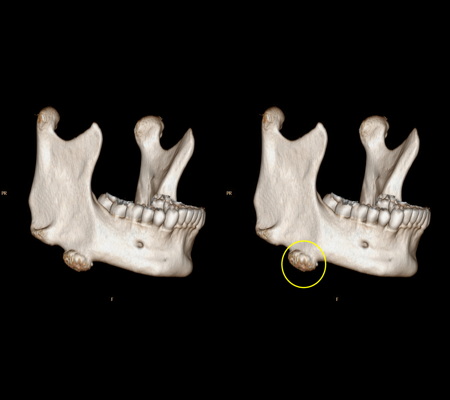
CT scan
- CT findings of osteoma include:[20]
- Well-circumscribed mass of variable density
- Ground-glass appearance
- Exophytical mass growing out of a sinus
MRI
- MRI findings of osteoma include:[21]
- Low signal on all sequences.
- Mature osteomas may demonstrate some marrow signal, but are also predominantly low on all sequences
Other Imaging Findings
There are no other imaging findings associated with osteoma.
Other Diagnostic Studies
Nasal Endoscopy
Nasal endoscopy findings include:[22]
- Direct visualization of the nasal passages structures, and sinuses.
- Tumor location, size, and adjacent structure evaluation.
Treatment
Medical Therapy
There is no medical treatment for osteoma; the mainstay of therapy is surgery.[23]
Surgery
Surgery is the mainstay of treatment for osteoma.[24][25]
Indication
- For symptomatic lesions, local excision is performed.
Types of Surgery
- Medial maxillectomy with a lateral rhinotomy
- Craniofacial resection
- Endoscopic resection
Recurrence Rare recurrence may occur after several years.
Primary Prevention
There are no established measures for the primary prevention of osteoma.
Secondary Prevention
There are no established measures for the secondary prevention of osteoma.
References
- ↑ Peabody, Terrance (2014). Orthopaedic oncology : primary and metastatic tumors of the skeletal system. Cham: Springer. ISBN 9783319073224.
- ↑ Charlier P, Froesch P, Benmoussa N, Froment A, Shorto R, Huynh-Charlier I (2014). "Did René Descartes have a giant ethmoidal sinus osteoma?". Lancet. 384 (9951): 1348. doi:10.1016/S0140-6736(14)61816-X. PMID 25307842.
- ↑ Jawad MU, Scully SP (2010). "In brief: classifications in brief: enneking classification: benign and malignant tumors of the musculoskeletal system". Clin Orthop Relat Res. 468 (7): 2000–2. doi:10.1007/s11999-010-1315-7. PMC 2882012. PMID 20333492.
- ↑ Athwal P, Stock H (2014). "Osteoid osteoma: a pictorial review". Conn Med. 78 (4): 233–5. PMID 24830123.
- ↑ Bilkay U, Erdem O, Ozek C, Helvaci E, Kilic K, Ertan Y; et al. (2004). "Benign osteoma with Gardner syndrome: review of the literature and report of a case". J Craniofac Surg. 15 (3): 506–9. PMID 15111819.
- ↑ 6.0 6.1 Abdel Tawab HM, Kumar V R, Tabook SM (2015). "Osteoma presenting as a painless solitary mastoid swelling". Case Rep Otolaryngol. 2015: 590783. doi:10.1155/2015/590783. PMC 4341844. PMID 25767729. Vancouver style error: name (help)
- ↑ Bisgaard ML, Bülow S (2006). "Familial adenomatous polyposis (FAP): genotype correlation to FAP phenotype with osteomas and sebaceous cysts". Am J Med Genet A. 140 (3): 200–4. doi:10.1002/ajmg.a.31010. PMID 16411234.
- ↑ Kaplan I, Calderon S, Buchner A (1994). "Peripheral osteoma of the mandible: a study of 10 new cases and analysis of the literature". J Oral Maxillofac Surg. 52 (5): 467–70. PMID 8169708.
- ↑ 9.0 9.1 Erdogan N, Demir U, Songu M, Ozenler NK, Uluç E, Dirim B (2009). "A prospective study of paranasal sinus osteomas in 1,889 cases: changing patterns of localization". Laryngoscope. 119 (12): 2355–9. doi:10.1002/lary.20646. PMID 19780030.
- ↑ Larrea-Oyarbide N, Valmaseda-Castellón E, Berini-Aytés L, Gay-Escoda C (2008). "Osteomas of the craniofacial region. Review of 106 cases". J Oral Pathol Med. 37 (1): 38–42. doi:10.1111/j.1600-0714.2007.00590.x. PMID 18154576.
- ↑ Earwaker J (1993). "Paranasal sinus osteomas: a review of 46 cases". Skeletal Radiol. 22 (6): 417–23. PMID 8248815.
- ↑ Boysen M (1978). "Osteomas of the paranasal sinuses". J Otolaryngol. 7 (4): 366–70. PMID 691104.
- ↑ Septer S, Slowik V, Morgan R, Dai H, Attard T (2013). "Thyroid cancer complicating familial adenomatous polyposis: mutation spectrum of at-risk individuals". Hered Cancer Clin Pract. 11 (1): 13. doi:10.1186/1897-4287-11-13. PMC 3854022. PMID 24093640.
- ↑ Sayan NB, Uçok C, Karasu HA, Günhan O (2002). "Peripheral osteoma of the oral and maxillofacial region: a study of 35 new cases". J Oral Maxillofac Surg. 60 (11): 1299–301. PMID 12420263.
- ↑ 15.0 15.1 Wijn MA, Keller JJ, Giardiello FM, Brand HS (2007). "Oral and maxillofacial manifestations of familial adenomatous polyposis". Oral Dis. 13 (4): 360–5. doi:10.1111/j.1601-0825.2006.01293.x. PMID 17577321.
- ↑ GARDNER EJ, PLENK HP (1952). "Hereditary pattern for multiple osteomas in a family group". Am. J. Hum. Genet. 4 (1): 31–6. PMC 1716387. PMID 14933371.
- ↑ Smith ME, Calcaterra TC (1989). "Frontal sinus osteoma". Ann Otol Rhinol Laryngol. 98 (11): 896–900. doi:10.1177/000348948909801111. PMID 2817682.
- ↑ Fu YS, Perzin KH (1974). "Non-epithelial tumors of the nasal cavity, paranasal sinuses, and nasopharynx. A clinicopathologic study. II. Osseous and fibro-osseous lesions, including osteoma, fibrous dysplasia, ossifying fibroma, osteoblastoma, giant cell tumor, and osteosarcoma". Cancer. 33 (5): 1289–305. PMID 4207295.
- ↑ Schajowicz, Fritz (1994). Tumors and Tumorlike Lesions of Bone : Pathology, Radiology, and Treatment. Berlin, Heidelberg: Springer Berlin Heidelberg. ISBN 9783642499562.
- ↑ Schajowicz, Fritz (1994). Tumors and Tumorlike Lesions of Bone : Pathology, Radiology, and Treatment. Berlin, Heidelberg: Springer Berlin Heidelberg. ISBN 9783642499562.
- ↑ Schajowicz, Fritz (1994). Tumors and Tumorlike Lesions of Bone : Pathology, Radiology, and Treatment. Berlin, Heidelberg: Springer Berlin Heidelberg. ISBN 9783642499562.
- ↑ Li Y, Zhang L, Zhou B, Han D (2009). "[Resection of frontal ethmoid sinus osteomas with nasal endoscopy]". Lin Chung Er Bi Yan Hou Tou Jing Wai Ke Za Zhi (in Chinese). 23 (14): 628–30. PMID 19894552.
- ↑ Gorini E, Mullace M, Migliorini L, Mevio E (2014). "Osseous choristoma of the tongue: a review of etiopathogenesis". Case Rep Otolaryngol. 2014: 373104. doi:10.1155/2014/373104. PMC 4279709. PMID 25580337.
- ↑ Kim WH, Kim DW, Kim CG, Kim MH (2013). "Additional Detection of Multiple Osteomas in a Patient with Gardner's Syndrome by Bone SPECT/CT". Nucl Med Mol Imaging. 47 (4): 297–8. doi:10.1007/s13139-013-0225-5. PMC 4035179. PMID 24900131.
- ↑ Alexander AA, Patel AA, Odland R (2007). "Paranasal sinus osteomas and Gardner's syndrome". Ann Otol Rhinol Laryngol. 116 (9): 658–62. doi:10.1177/000348940711600906. PMID 17926587.