Lung cancer CT: Difference between revisions

Jump to navigation Jump to search
No edit summary
Line 26: Line 26:
*A positive result for a tumor using a CT scan is typically followed up with a [[biopsy]] for confirmation.


{| class="wikitable"
|
|
|-
|
|
|-
|
|
|}
{| align="left"
{| align="left"
|- valign="top"
|- valign="top"

Revision as of 17:27, 15 February 2018

Lung cancer Microchapters

Home

Patient Information

Overview

Historical Perspective

Classification

Pathophysiology

Causes

Differentiating Lung cancer from other Diseases

Epidemiology and Demographics

Risk Factors

Screening

Natural History, Complications and Prognosis

Diagnosis

Diagnostic study of choice

History and Symptoms

Physical Examination

Laboratory Findings

Electrocardiogram

X-ray

Echocardiography and Ultrasound

CT scan

MRI

Other Imaging Findings

Other Diagnostic Studies

Treatment

Medical Therapy

Surgery

Primary Prevention

Secondary Prevention

Cost-Effectiveness of Therapy

Future or Investigational Therapies

Case Studies

Case #1

Lung cancer CT On the Web

Most recent articles

Most cited articles

Review articles

CME Programs

Powerpoint slides

Images

American Roentgen Ray Society Images of Lung cancer CT

All Images
X-rays
Echo & Ultrasound
CT Images
MRI

Ongoing Trials at Clinical Trials.gov

US National Guidelines Clearinghouse

NICE Guidance

FDA on Lung cancer CT

CDC on Lung cancer CT

Lung cancer CT in the news

Blogs on Lung cancer CT

Directions to Hospitals Treating Lung cancer

Risk calculators and risk factors for Lung cancer CT

Editor-In-Chief: C. Michael Gibson, M.S., M.D. [1] Associate Editor(s)-in-Chief: Dildar Hussain, MBBS [2] Saarah T. Alkhairy M.D Kim-Son H. Nguyen M.D.

Overview

CT scans help stage the lung cancer. A CT scan of the abdomen and brain can help visualize the common sights of metastases: adrenal glands, liver, and brain. CT scans diagnose lung cancer by providing anatomical detail to locate the tumor, demonstrating proximity to nearby structures, and deciphering whether lymph nodes are enlarged in the mediastinum.

CT Scan

  • CT scans help stage the lung cancer. A CT scan of the abdomen and brain can help visualize the common sights of metastases: adrenal glands, liver, and brain.
  • The benefits of CT Scans in lung cancer patients are the following:[2]
    • Provides anatomical detail to locate the tumor
    • Demonstrates proximity to nearby structures
    • Deciphers whether lymph nodes are enlarged in the mediastinum
  • Unfortunately, research has shown that there are a number of false positives associated with CT scanning because a CT scan on its own cannot determine malignancy.
  • A positive result for a tumor using a CT scan is typically followed up with a biopsy for confirmation.
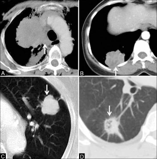
Common radiological appearances of lung cancer. Centrally located mass with mediastinal invasion (arrow, A), peripherally situated mass abutting the pleura (arrow, B), mass with smooth, lobulated margins (arrow, C) and with spiculated, irregular margins (arrow, D), via <https://www.ncbi.nlm.nih.gov/pmc/articles/PMC4419420/figure/F1/>This is an open-access article distributed under the terms of the Creative Commons Attribution-Noncommercial-Share Alike 3.0 Unported, which permits unrestricted use, distribution, and reproduction in any medium, provided the original work is properly cited.[1]
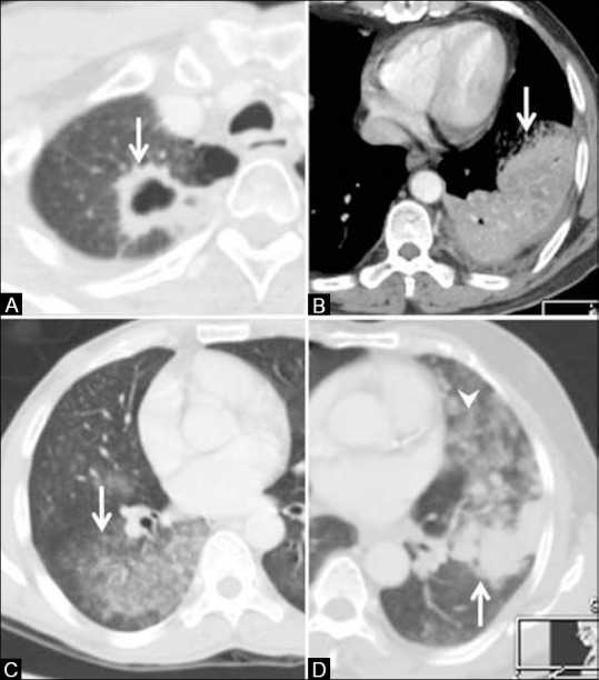
Lung cancers with atypical radiological pattern. Squamous cell cancer presenting as a cavitating mass (arrow, A). Adenocarcinoma presenting as dense consolidation (arrow, B). Bronchoalveolar carcinoma (adenocarcinoma in situ) presenting as ground-glass opacity (arrow, C) and mixed density, solid (arrow), and ground-glass nodules (arrowhead) in D via <https://www.ncbi.nlm.nih.gov/pmc/articles/PMC4419420/figure/F2/>This is an open-access article distributed under the terms of the Creative Commons Attribution-Noncommercial-Share Alike 3.0 Unported, which permits unrestricted use, distribution, and reproduction in any medium, provided the original work is properly cited.[1]
Stage T1 and T2 tumors. Stage T1 tumor due to size <3 cm (arrow, A). Stage T2 endobronchial tumor (arrowhead) causing pneumonitis restricted to the upper lobe (arrow) in B. T2a tumor >3 cm but <5 cm (arrow, C). T2b tumor >5 cm but <7 cm (arrow in D) via <https://www.ncbi.nlm.nih.gov/pmc/articles/PMC4419420/figure/F3/>This is an open-access article distributed under the terms of the Creative Commons Attribution-Noncommercial-Share Alike 3.0 Unported, which permits unrestricted use, distribution, and reproduction in any medium, provided the original work is properly cited.[1]
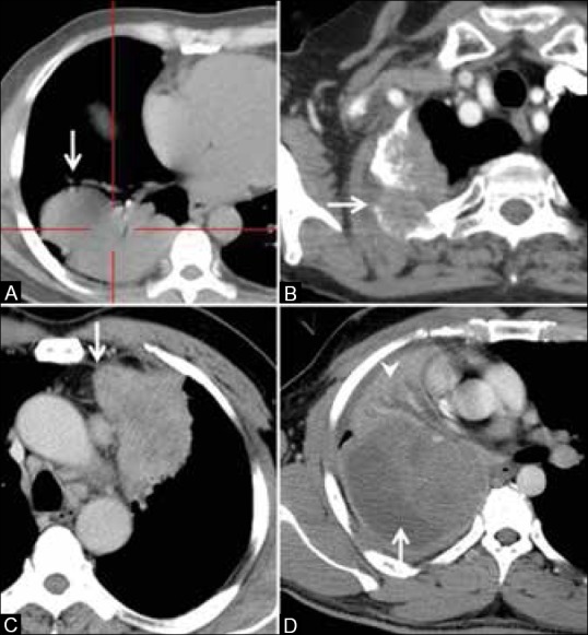
Stage T3 tumors. T3 tumor due to size >7 cm in size (arrow, A), eroding the ribs (arrow, B), infiltrating the mediastinal pleura but not the vessels (arrow, C), and causing atelectasis of the entire lung (arrowhead, D via<https://www.ncbi.nlm.nih.gov/pmc/articles/PMC4419420/figure/F4/>This is an open-access article distributed under the terms of the Creative Commons Attribution-Noncommercial-Share Alike 3.0 Unported, which permits unrestricted use, distribution, and reproduction in any medium, provided the original work is properly cited.[1]
Stage T4 tumors. T4 tumor due to invasion of pulmonary artery (arrow, A), descending aorta (arrow, B), vertebral body (arrow, C), superior vena cava with thrombus (arrow, D)via<https://www.ncbi.nlm.nih.gov/pmc/articles/PMC4419420/figure/F5/>This is an open-access article distributed under the terms of the Creative Commons Attribution-Noncommercial-Share Alike 3.0 Unported, which permits unrestricted use, distribution, and reproduction in any medium, provided the original work is properly cited.[1]


References

  1. 1.0 1.1 1.2 1.3 1.4 1.5 Purandare, NilenduC; Rangarajan, Venkatesh (2015). "Imaging of lung cancer: Implications on staging and management". Indian Journal of Radiology and Imaging. 25 (2): 109. doi:10.4103/0971-3026.155831. ISSN 0971-3026.
  2. Gerard A. Silvestri, Lynn T. Tanoue, Mitchell L. Margolis, John Barker, Frank Detterbeck.11/30/11.The Noninvasive Staging of Non Small-cell Lung Cancer. Chestpubs. http://chestjournal.chestpubs.org/content/123/1_suppl/147S.full/

Template:Tumors


Template:WikiDoc Sources