Langerhans cell histiocytosis CT: Difference between revisions
No edit summary |
No edit summary |
||
| (9 intermediate revisions by 2 users not shown) | |||
| Line 3: | Line 3: | ||
{{CMG}} {{AE}} {{HL}} | {{CMG}} {{AE}} {{HL}} | ||
==Overview== | ==Overview== | ||
CT scan may be helpful in the diagnosis of Langerhans cell histiocytosis. Findings on CT scan suggestive of Langerhans cell histiocytosis include multiple osteolytic lesions causing full thickness [[bone]] destruction. | |||
==CT Scan== | ==CT Scan== | ||
* Head CT scan may be helpful in the diagnosis of Langerhans cell histiocytosis.<ref name="radio"> Langerhans cell histiocytosis. Radiopeadia (2015) http://radiopaedia.org/articles/langerhans-cell-histiocytosis Accessed on February, 3 2016</ref><ref name="pmid23907805">{{cite journal| author=Khung S, Budzik JF, Amzallag-Bellenger E, Lambilliote A, Soto Ares G, Cotten A et al.| title=Skeletal involvement in Langerhans cell histiocytosis. | journal=Insights Imaging | year= 2013 | volume= 4 | issue= 5 | pages= 569-79 | pmid=23907805 | doi=10.1007/s13244-013-0271-7 | pmc=PMC3781243 | url=http://www.ncbi.nlm.nih.gov/entrez/eutils/elink.fcgi?dbfrom=pubmed&tool=sumsearch.org/cite&retmode=ref&cmd=prlinks&id=23907805 }} </ref> | |||
< | * Findings on head CT scan suggestive of Langerhans cell histiocytosis include:<ref name="pmid23109216">{{cite journal |vauthors=Haupt R, Minkov M, Astigarraga I, Schäfer E, Nanduri V, Jubran R, Egeler RM, Janka G, Micic D, Rodriguez-Galindo C, Van Gool S, Visser J, Weitzman S, Donadieu J |title=Langerhans cell histiocytosis (LCH): guidelines for diagnosis, clinical work-up, and treatment for patients till the age of 18 years |journal=Pediatr Blood Cancer |volume=60 |issue=2 |pages=175–84 |date=February 2013 |pmid=23109216 |pmc=4557042 |doi=10.1002/pbc.24367 |url=}}</ref><ref name="pmid8008491">{{cite journal |vauthors=Hermans R, De Foer B, Smet MH, Leysen J, Feenstra L, Fossion E, Baert AL |title=Eosinophilic granuloma of the head and neck: CT and MRI features in three cases |journal=Pediatr Radiol |volume=24 |issue=1 |pages=33–6 |date=1994 |pmid=8008491 |doi= |url=}}</ref> | ||
:* Multiple osteolytic lesions | |||
:* Full thickness [[bone]] destruction | |||
:* “Button sequestrum” sign | |||
==Gallery== | |||
<gallery> | |||
Image: | |||
Langerhans CT scan.jpeg|Head CT scan illustrating multiple osteolytic lesions of Langerhans cell histiocytosis | |||
Image: | |||
Langerhans CT scan 2.jpg|Head CT scan illustrating multiple osteolytic lesions of Langerhans cell histiocytosis | |||
Image: | |||
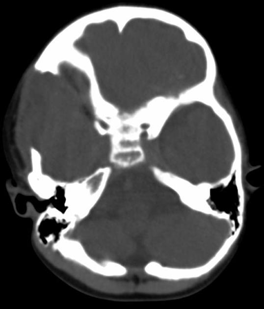
Langerhans CT scan 3.jpg|Axial CT scan illustrating full thickness bone destruction of Langerhans cell histiocytosis | |||
</gallery> | |||
==References== | ==References== | ||
{{Reflist|2}} | {{Reflist|2}} | ||
| Line 15: | Line 27: | ||
[[Category:Autoimmune diseases]] | [[Category:Autoimmune diseases]] | ||
[[Category:Hematology]] | [[Category:Hematology]] | ||
[[Category:Up-To-Date]] | |||
[[Category:Oncology]] | |||
[[Category:Medicine]] | |||
[[Category:Dermatology]] | |||
[[Category:Surgery]] | |||
Latest revision as of 02:32, 28 March 2019
|
Langerhans cell histiocytosis Microchapters |
|
Differentiating Langerhans cell histiocytosis from other Diseases |
|---|
|
Diagnosis |
|
Treatment |
|
Case Studies |
|
Langerhans cell histiocytosis CT On the Web |
|
American Roentgen Ray Society Images of Langerhans cell histiocytosis CT |
|
Directions to Hospitals Treating Langerhans cell histiocytosis |
|
Risk calculators and risk factors for Langerhans cell histiocytosis CT |
Editor-In-Chief: C. Michael Gibson, M.S., M.D. [1] Associate Editor(s)-in-Chief: Haytham Allaham, M.D. [2]
Overview
CT scan may be helpful in the diagnosis of Langerhans cell histiocytosis. Findings on CT scan suggestive of Langerhans cell histiocytosis include multiple osteolytic lesions causing full thickness bone destruction.
CT Scan
- Head CT scan may be helpful in the diagnosis of Langerhans cell histiocytosis.[1][2]
- Findings on head CT scan suggestive of Langerhans cell histiocytosis include:[3][4]
- Multiple osteolytic lesions
- Full thickness bone destruction
- “Button sequestrum” sign
Gallery
-
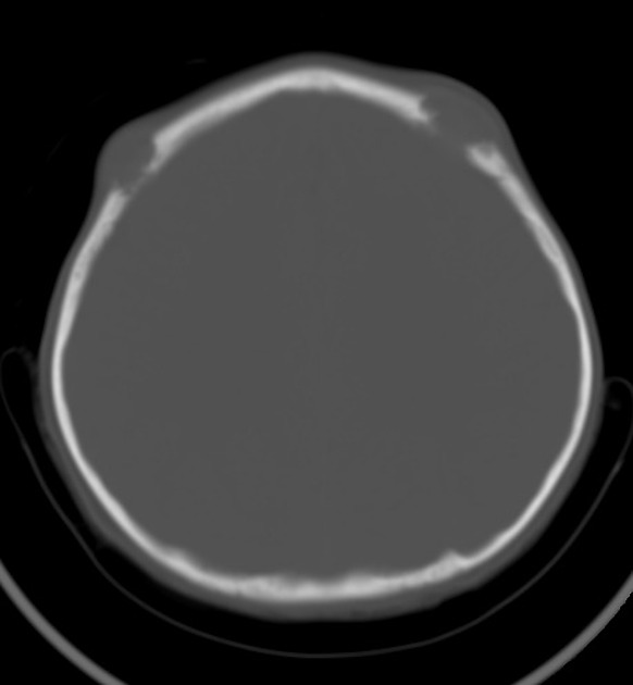
Head CT scan illustrating multiple osteolytic lesions of Langerhans cell histiocytosis
-
Head CT scan illustrating multiple osteolytic lesions of Langerhans cell histiocytosis
-

Axial CT scan illustrating full thickness bone destruction of Langerhans cell histiocytosis


References
- ↑ Langerhans cell histiocytosis. Radiopeadia (2015) http://radiopaedia.org/articles/langerhans-cell-histiocytosis Accessed on February, 3 2016
- ↑ Khung S, Budzik JF, Amzallag-Bellenger E, Lambilliote A, Soto Ares G, Cotten A; et al. (2013). "Skeletal involvement in Langerhans cell histiocytosis". Insights Imaging. 4 (5): 569–79. doi:10.1007/s13244-013-0271-7. PMC 3781243. PMID 23907805.
- ↑ Haupt R, Minkov M, Astigarraga I, Schäfer E, Nanduri V, Jubran R, Egeler RM, Janka G, Micic D, Rodriguez-Galindo C, Van Gool S, Visser J, Weitzman S, Donadieu J (February 2013). "Langerhans cell histiocytosis (LCH): guidelines for diagnosis, clinical work-up, and treatment for patients till the age of 18 years". Pediatr Blood Cancer. 60 (2): 175–84. doi:10.1002/pbc.24367. PMC 4557042. PMID 23109216.
- ↑ Hermans R, De Foer B, Smet MH, Leysen J, Feenstra L, Fossion E, Baert AL (1994). "Eosinophilic granuloma of the head and neck: CT and MRI features in three cases". Pediatr Radiol. 24 (1): 33–6. PMID 8008491.