Hypopharyngeal cancer mri: Difference between revisions
Jump to navigation
Jump to search
No edit summary |
No edit summary |
||
| Line 29: | Line 29: | ||
|} | |} | ||
<gallery>Image:Post-cricoid-carcinoma_(1).jpg |Post cricoid carcinoma MRI</gallery> | <gallery>Image:Post-cricoid-carcinoma_(1).jpg |Post cricoid carcinoma MRI</gallery><ref name=aaa>Hypopharyngeal cancer. Image courtesy of Radswiki [http://www.radiopaedia.org Radiopaedia](original file [http://radiopaedia.org/articles/hypopharyngeal-squamous-cell-carcinoma"here"]).[http://radiopaedia.org/licence Creative Commons BY-SA-NC]</ref> | ||
==References== | ==References== | ||
{{reflist| | {{reflist|1}} | ||
{{WH}} | {{WH}} | ||
Revision as of 20:12, 5 October 2015
|
Hypopharyngeal cancer Microchapters |
|
Diagnosis |
|---|
|
Treatment |
|
Case Studies |
|
Hypopharyngeal cancer mri On the Web |
|
American Roentgen Ray Society Images of Hypopharyngeal cancer mri |
|
Risk calculators and risk factors for Hypopharyngeal cancer mri |
Editor-In-Chief: C. Michael Gibson, M.S., M.D. [1] Associate Editor(s)-in-Chief: Faizan Sheraz, M.D. [2]
Overview
MRI
MRI has the ability to be superior to CT in local staging and assessing perineural spread.
| MRI Component | Features |
|---|---|
|
|
|
|
|
|
-
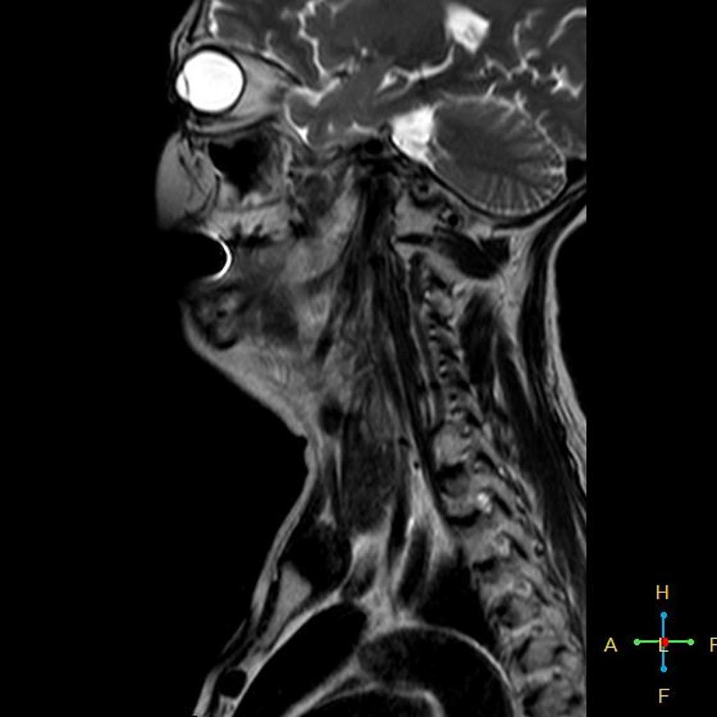
Post cricoid carcinoma MRI
.jpg)
References
- ↑ Hypopharyngeal cancer. Image courtesy of Radswiki Radiopaedia(original file "here").Creative Commons BY-SA-NC