Hemolytic anemia laboratory findings: Difference between revisions
| Line 15: | Line 15: | ||
* Absolute reticulocyte count - 25,000 to 75,000/microLitre | * Absolute reticulocyte count - 25,000 to 75,000/microLitre | ||
===Peripheral blood smear=== | ===Peripheral blood smear=== | ||
* Fragments of the red blood cells ("[[schistocyte]]s") can be present. | |||
* Some red blood cells may appear smaller and rounder than usual ([[spherocyte]]s). | |||
* [[Reticulocytosis|Reticulocytes]] are present in elevated numbers. This may be overlooked if a special [[staining (biology)|stain]] is not used. | |||
*The level of unconjugated [[bilirubin]] in the blood is elevated. This may lead to [[jaundice]]. | ===Liver function test=== | ||
* The level of unconjugated [[bilirubin]] in the blood is elevated. This may lead to [[jaundice]]. | |||
*The level of [[lactate dehydrogenase]] (LDH) in the blood is elevated. | *The level of [[lactate dehydrogenase]] (LDH) in the blood is elevated. | ||
*[[Haptoglobin]] levels are decreased. | *[[Haptoglobin]] levels are decreased. | ||
Revision as of 14:10, 25 September 2012
|
Hemolytic anemia Microchapters |
|
Diagnosis |
|---|
|
Treatment |
|
Case Studies |
|
Hemolytic anemia laboratory findings On the Web |
|
American Roentgen Ray Society Images of Hemolytic anemia laboratory findings |
|
Risk calculators and risk factors for Hemolytic anemia laboratory findings |
Editor-In-Chief: C. Michael Gibson, M.S., M.D. [1]
Overview
Hemolytic anemia is anemia caused secondary to shortened survival of circulating red blood cells. The normal life span of RBCs is 110 to 120 days. RBC destruction before that time is defined as hemolytic anemia. As opposed to the normal senecence of RBC, the random hemolysis (premature RBC death) is increased in hemolytic anemia.
Laboratory Findings
Absolute reticulocyte count
The normal values are:
- RBC count - 5 million/microLitre
- Reticulocyte count - 0.5 -1.5 %
- Absolute reticulocyte count - 25,000 to 75,000/microLitre
Peripheral blood smear
- Fragments of the red blood cells ("schistocytes") can be present.
- Some red blood cells may appear smaller and rounder than usual (spherocytes).
- Reticulocytes are present in elevated numbers. This may be overlooked if a special stain is not used.
Liver function test
- The level of unconjugated bilirubin in the blood is elevated. This may lead to jaundice.
- The level of lactate dehydrogenase (LDH) in the blood is elevated.
- Haptoglobin levels are decreased.
- The direct Coombs test is positive if hemolysis is caused by an immune process.
- Hemosiderin in the urine indicates chronic intravascular hemolysis. There is also urobilinogen in the urine.
(Images shown below are courtesy of Melih Aktan MD, Istanbul Medical Faculty - Turkey, and Hospital Universitario La Fe Servicio Hematologia)
-
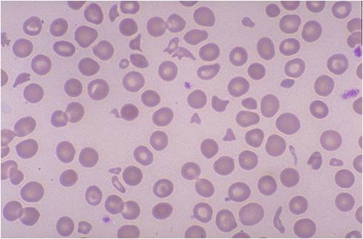
Microangiopathic hemolytic anemia
-
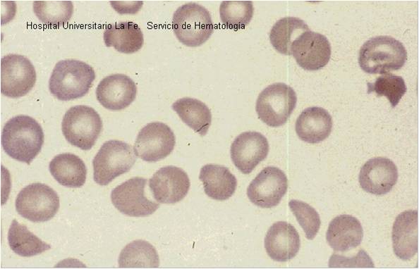
Microangiopathic hemolytic anemia

